R E V I E W
Open Access
Panniculitides of particular interest to the
rheumatologist
Thâmara Cristiane Alves Batista Morita
1,5*, Gabriela Franco Sturzeneker Trés
1, Maria Salomé Cajas García
2,
Ilana Halpern
1, Paulo Ricardo Criado
1,3and Jozelio Freire de Carvalho
4Abstract
The panniculitides remain as one of the most challenging areas for clinicians, as they comprise a heterogeneous group of inflammatory diseases involving the subcutaneous fat with potentially-shared clinical and
histopathological features. Clinically, most panniculitides present as red edematous nodules or plaques. Therefore, in addition to a detailed clinical history, a large scalpel biopsy of a recent-stage lesion with adequate representation of the subcutaneous tissue is essential to specific diagnosis and appropriate clinical management. Herein we review the panniculitides of particular interest to the rheumatologist.
Keywords:Cutaneous polyarteritis nodosa, Erythema nodosum, Infective panniculitis, Lupus panniculitis, Panniculitis, Subcutaneous tissue, Vasculitis
Background
The panniculitides comprise a heterogeneous group of in-flammatory diseases involving the subcutaneous fat. The basic unit of the subcutaneous fat is a cohesive collection of adipocytes called primary microlobule. An aggregation of primary microlobules, termed a secondary lobule, is surrounded by an easily discernible rim of connective tis-sue known as septum, which house the nerves, the lymph-atic vessels, and the arteries and veins of the subcutis. Every adipocyte in the subcutaneous fat is encircled by a capillary [1].
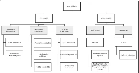
This knowledge is crucial, once classification of the pan-niculitides is far too complex and often contradictory. From an academic point of view, in this review we have chosen to follow the three-step approach for histopatho-logic diagnosis of panniculitides. The first step is to clas-sify them as mostly septal (Fig. 1) or lobular (Fig. 2) depending on where the inflammatory cell infiltrate is lo-cated. Secondly, is necessary to define if vasculitis is present or not. Finally, the third step is further character-izing the type of the inflammatory cell infiltrate and the presence and pattern of necrosis [2, 3]. Nevertheless,
different stages in the evolution of diseases and categories that are not exclusive may lead clinicians to confusion. Therefore, after that initial classification, efforts should be made to look for the additional histopathological findings that may be considered as clues to reach specific diagnoses.
The purpose of this review is to present a comprehen-sive clinic pathologic overview of the panniculitides of particular interest to the rheumatologist.
Main text
Primarily-septal panniculitides
Primarily-septal panniculitides without vasculitis
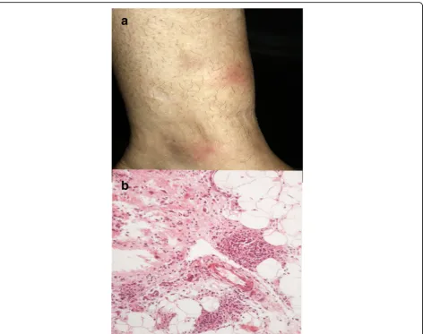
Erythema nodosum Erythema nodosum (EN) is the most common clinical form of panniculitis. In children, up to the age of 12 years, the female-to-male ratio is ap-proximately equal, whereas in adults it occurs three to six times more often in woman, usually in the second and third decades of life. EN typically manifest by the sudden onset of multiple painful inflammatory, cutane-ous and subcutanecutane-ous red nodules, which may assume a deep bruised appearance as they fade, and coalescence-forming plaques. The most common location are the an-terior and lateral surfaces of the lower limbs, although the extensor surfaces of the forearm, the thighs, and the trunk may also be affected (Fig.3a). Systemic symptoms
© The Author(s). 2019Open AccessThis article is distributed under the terms of the Creative Commons Attribution 4.0 International License (http://creativecommons.org/licenses/by/4.0/), which permits unrestricted use, distribution, and reproduction in any medium, provided you give appropriate credit to the original author(s) and the source, provide a link to the Creative Commons license, and indicate if changes were made. The Creative Commons Public Domain Dedication waiver (http://creativecommons.org/publicdomain/zero/1.0/) applies to the data made available in this article, unless otherwise stated.
* Correspondence:thamaramorita@usp.br
1Faculdade de Medicina FMUSP, Universidade de Sao Paulo, Sao Paulo, SP,
Brazil
5Private practice, Aracaju, SE, Brazil
Full list of author information is available at the end of the article
Advances in Rheumatology
Moritaet al. Advances in Rheumatology (2019) 59:35such as low-grade fever, fatigue, malaise, arthralgia, are present in more than 30% of the cases. The lesions never ulcerate and resolve spontaneously without leaving scars in approximately two to eight weeks [4, 5]. Currently, it is believed that EN migrans, subacute nodular migratory panniculitis, and chronic EN are clinical variants that may all be included within the spectrum of EN [6].
EN has been regarded a late type hypersensitivity reac-tion to several antigenic stimuli. The main etiologies are shown in Table 1 and may vary according to the geo-graphic region. Streptococcal infection is a frequent cause of this panniculitis worldwide, especially in children; other
potential infectious agents are represented by Epstein-Barr virus, Cytomegalovirus, Yersinia spp., Mycoplasma,
Chlamydia,HistoplasmaandCoccidioides. Due to the ele-vated prevalence of tuberculosis in Brazil,Mycobacterium tuberculosismay be the most important factor in EN in our country. Drugs such as nonsteroidal anti-inflammatory drugs (NSAIDs), oral contraceptives, “biological” thera-peutic agents (adalimumab, infliximab, imatinib, BRAF inhibitors, and immune checkpoint modulators) [7], and antibiotics, besides a wide group of conditions, which in-clude sarcoidosis, pregnancy, autoimmune disorders, malig-nancies (mostly Hodgkin’s disease), and enteropathies Fig. 1Primarily-septal panniculitides
(Crohn’s disease and ulcerative colitis) can lead to second-ary EN [8]. Meanwhile, nearly half of the cases are classified as having idiopathic EN [9,10].
EN is the paradigm of predominantly septal panniculitides with no vasculitis, although hemorrhages within the small vessels may occur. The composition of the inflammatory in-filtrate in the septa varies with the age of the lesion. In early lesions, sparse neutrophils are seen interstitially arranged and at the periphery of fat lobules, together with some foamy macrophages. Fully formed lesions are characterized by septal fibrosis, granulation tissue, lymphocytes and histio-cytes, many of them multinucleated [2,6]. Visualization of the so-called“Miescher radial granulomas”, nodular aggre-gations of histiocytes and neutrophils radially surrounding small blood vessels, is quite characteristic in the histopatho-logical evaluation (Fig.3b) [4]. Fat lobules are reduced mark-edly in size.
In contrast, erythema nodosum leprosum (ENL) is an immune-mediated common complication of lepromatous
leprosy, occurring in about 50% of these patients. The histology of ENL lesions shows an intense perivascular in-filtrate of neutrophils throughout the dermis and subcutis in a lobular pattern often associated with vasculitis [11]. However, macrophage granuloma without neutrophil infil-tration occur in up to 35% of the patients. Foamy histio-cytes containing large numbers of acid-fast bacilli, known as Virchow cells, are frequently seen in ENL lesions, as they are in lepromatous leprosy [12].
Rheumatoid nodule Almost one-third of the patients with rheumatoid arthritis exhibit skin lesions related to the disease, 98.3% of which are rheumatoid nodules (RNs). RNs develop in patients with long-standing rheumatoid arthritis (RA) and a higher disease activity score compared to pa-tients without skin lesions. Rheumatoid factor positivity is detected in 75% of them. These nodules are commonly lo-cated on the metacarpophalangeal and proximal interpha-langeal joints, on the olecranon process, and on the Fig. 3Erythema nodosum.aRed nodules anterior on the anterior aspect of the lower limb.bSeptal thickening with lymphohistiocytic infiltrate and formation of Miescher radial granulomas at the periphery of the subcutaneous fat lobule. H&E, 400x
proximal ulna; less frequently the palms, the back of the hands and, in bedridden patients, the occiput and ischium are affected. RNs are often asymptomatic, persistent, firm, and moveable in subcutaneous planes, characteristics that make them distinguishable from RNs mimickers, mostly tenosynovitis and bursitis nodules, that are softer, com-pressible and painful in the majority of the cases [13–16].
RNs have a distinctive histological appearance, resultant of an immune-mediated granulomatous process involving the deeper dermis and extending into the septa of the sub-cutaneous fat. They exhibit a well-defined palisading of elongated histiocytes around central collagen degener-ation, with an intensely eosinophilic accumulation of fibrin in the center of the granuloma. Extensive fibrosis is prom-inent in old lesions. Accelerated rheumatoid nodulosis, a phenomenon that has been observed not only in patients on methotrexate therapy, but also during treatment with azathioprine, leflunomide, infliximab, and etanercept re-veals the same histopathological findings of RNs [16–18]. Histopathologic differential diagnosis is made with sub-cutaneous granuloma annulare and necrobiosis lipoidica.
Deep morphea Morphea, or localized scleroderma, is a rare fibrosing disorder of the skin resulting from inflam-mation and deposition of collagen-rich extracellular
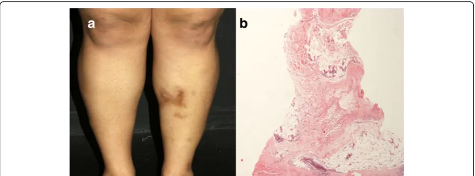
matrix [19]. When sclerotic changes extend beyond the dermis to involve the subcutaneous tissue, the fascia or the superficial muscle, we refer to deep variant of circum-scribed morphea (previously known as subcutaneous mor-phea or morphea profunda). In contrast, when there is exclusive deep fascia involvement, classification as eosino-philic fasciitis or Shulman’s syndrome is more appropriate [20]. Clinically, deep morphea appears in the form of indu-rated plaques or nodules, frequently located on the upper part of the trunk close to the midline and on the lower limbs (Fig.4a). The overlying skin may feel hardened and adherent to the underlying tissues; however, acute inflam-matory signs, such as edema and erythema, are rarely ob-served [21–23]. The presence of deep sclerosis, in some cases perineural, may account for the presence of pain [19]. Morphea and systemic sclerosis cannot be differentiated by histopathologic examination. In the early stages, deep morphea shows perivascular infiltration in the reticular dermis that is predominantly lymphoplasmacytic, al-though eosinophils are found in one-third of the speci-mens, with swollen endothelial cells and thickened collagen bundles (Fig.4b) [23]. Late morphea is character-ized by dense collagen sclerosis, which produces thickened septa and fat lobule obliteration. Lipomembranous changes can also be observed [20,24]. On the other hand,
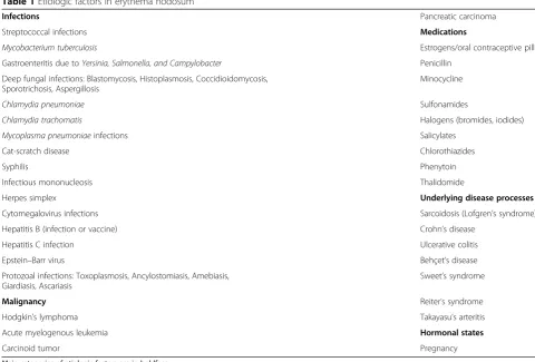
Table 1Etiologic factors in erythema nodosum
Infections Pancreatic carcinoma
Streptococcal infections Medications
Mycobacterium tuberculosis Estrogens/oral contraceptive pills
Gastroenteritis due toYersinia, Salmonella, and Campylobacter Penicillin
Deep fungal infections: Blastomycosis, Histoplasmosis, Coccidioidomycosis, Sporotrichosis, Aspergillosis
Minocycline
Chlamydia pneumoniae Sulfonamides
Chlamydia trachomatis Halogens (bromides, iodides)
Mycoplasma pneumoniaeinfections Salicylates
Cat-scratch disease Chlorothiazides
Syphilis Phenytoin
Infectious mononucleosis Thalidomide
Herpes simplex Underlying disease processes
Cytomegalovirus infections Sarcoidosis (Lofgren’s syndrome)
Hepatitis B (infection or vaccine) Crohn’s disease
Hepatitis C infection Ulcerative colitis
Epstein–Barr virus Behçet’s disease
Protozoal infections: Toxoplasmosis, Ancylostomiasis, Amebiasis, Giardiasis, Ascariasis
Sweet’s syndrome
Malignancy Reiter’s syndrome
Hodgkin’s lymphoma Takayasu’s arteritis
Acute myelogenous leukemia Hormonal states
Carcinoid tumor Pregnancy
fibrous nodule formation in the setting of both systemic scleroderma and morphea consist histologically of mid dermal regions of fibrous tissue and inflammatory cells with increased mucin and decreased elastic fibers, indis-tinguishable from keloids [25].
Primarily-septal panniculitides with vasculitis
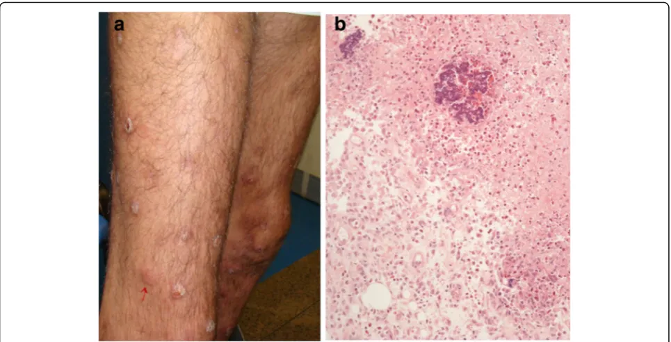
Cutaneous polyarteritis nodosa Polyarteritis nodosa (PAN) is a systemic necrotizing vasculitis that predomin-antly targets small and medium-sized muscular arteries. In adult patients, it is more common in male sex and the median age at presentation is 33 (19–71) years. Both genders are equally affected in children and the median age at disease onset is 11 (2–16) years [26, 27]. The pathway(s) leading to necrotizing inflammation of the blood vessels have not yet been completely unraveled. Immunological mechanisms seem to play a relevant role in the pathogenesis of PAN. Most of the cases are idio-pathic; nevertheless, several triggers have been identified. Hepatitis B virus infection (HBV) is the most common and it is well known that patients with HBV-related PAN have more severe disease, even though its occur-rence has largely decreased due to widespread vaccin-ation [27]. PAN can also be associated with malignancies and others infectious agents, such as group A β -hemolyticStreptococcus[28,29].
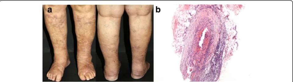
The most common manifestations are palpable purpura and reddish-purple subcutaneous nodules mainly located on the lower limbs; nevertheless, they may also include livedo racemosa,‘punched out’ necrotic ulcers, atrophie blanche, and ultimately gangrene of digits, especially in children (Fig. 5a). Cutaneous PAN (C-PAN), also known as cutaneous arteritis since the 2012 Revised International Chapel Hill Consensus Conference, is a special form of
the disease restricted to the skin. Despite frequent re-lapses, cutaneous arteritis has a more benign course, milder constitutional symptoms and extra-cutaneous manifestations, which include arthritis, muscle pain and peripheral neuropathy, limited to the same area with over-lying skin lesions [30,31].
Skin biopsy is extremely useful for a definitive diagnosis. Cutaneous arteritis consists of a necrotizing vasculitis af-fecting small and medium-sized arteries within deep der-mal or at the septa of the subcutaneous fat tissue. Fibrinoid degeneration involves all layers of the vascular wall, and internal elastic lamina disruption is detected (Fig.5b). The histological changes can be divided into four stages (acute, subacute, reparative and healed), with a transition from a predominantly neutrophilic to lympho-cytic infiltrate in the subacute and reparative stages. Then, vascular lumens become occluded by the fibrin thrombi with perivascular neovascularization in the healed stages [32, 33]. Direct immunofluorescence staining may show C3 and IgM deposits within vessels in the lesions [34]. Nodular vasculitis and thrombophlebitis are considered histopathologic differential diagnosis.
Primarily-lobular panniculitides
Primarily-lobular panniculitides without vasculitis
Gouty panniculitis Gouty panniculitis represents an ex-tremely rare manifestation of gout, characterized by the deposition of monosodium urate crystals in the lobular hypodermis. The pathophysiology is currently poorly understood and serum uric acid level need not to be ele-vated to its development [35]. Gouty panniculitis may pre-cede, appears concomitantly or years after the articular clinical expression of tophaceous gout. On physical exam-ination, patients present with indurated, erythematous, Fig. 4Deep morphea.aIndurated plaque on the lower limb.bThickening of the hypodermic septa by sclerotic collagen bundles, permeated by some lymphocytes and plasma cells, which extend to the periphery of the fat lobule. H&E, 100x
and irregular subcutaneous nodules or plaques, frequently painful, which can ulcerate and drain serous or opaque fluid. Lesions are located predominantly on the lower extremities, although they may be observed on upper ex-tremities, torso, buttocks and scalp [36–38]. Skin biopsy reveals amorphous eosinophilic deposits in the deep der-mis and subcutaneous tissue surrounded by a granuloma-tous reaction with macrophages and many multinucleated giant cells. Urate crystals causing lobular panniculitis show a needle-like shape and are doubly refractive under polar-ized light [39].
Post-steroid panniculitisThis is a very rare panniculitis mostly described in childhood, but also in early adult-hood, one to 10 days after cessation of systemic cortico-steroid therapy. It has been suggested that a sudden decrease or withdrawal of high doses of corticosteroids either oral or intravenous may cause an increase in the ratio of saturated to unsaturated fatty acids, leading to crystal formation. The nodules tend to appear in those areas prone to accumulation of fat during steroid ther-apy such as the cheeks, jawline, arms and trunk, al-though it has also been reported on the legs. Usually there are no general symptoms [40,41].
Characteristic histopathological findings consist of lobular panniculitis without vasculitis, with narrow strands needle-shaped clefts in radial arrangement within the cytoplasm of histiocytes, multinucleated giant cells, and the great majority of fat cells. The needle-shaped clefts represent negative images of crystals of tri-glycerides dissolved during tissue processing. These fea-tures are also seen in sclerema neonatorum and subcutaneous fat necrosis of the newborn. The lesions generally subside in weeks or months even without any treatment, leaving residual hyperpigmentation. If ulcer-ation occurs in late stage, resuming steroid therapy may be indicated and then a slower and gradual decrease of the dose should be programmed [40,41].
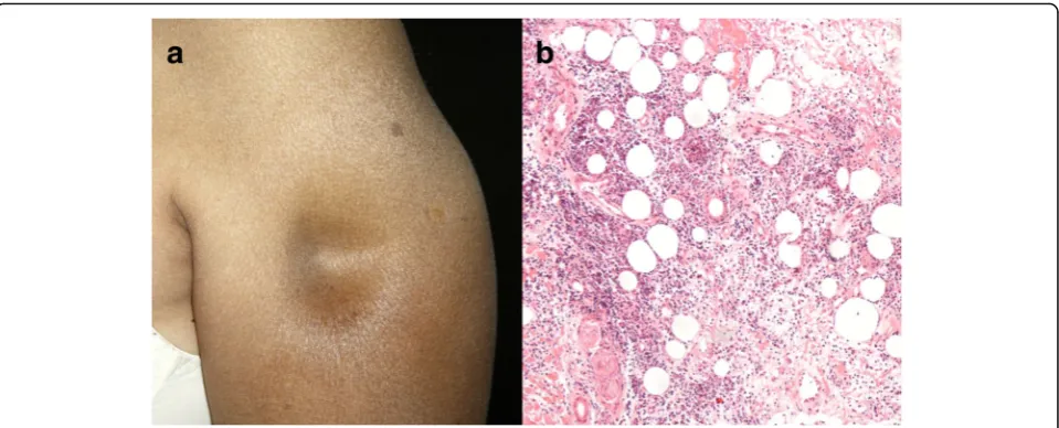
Lupus panniculitis Lupus panniculitis (LEP), also known as lupus erythematosus profundus, affects ap-proximately 2% of the patients diagnosed with lupus ery-thematosus [42]. It can also appears concomitantly with discoid lupus erythematosus or subacute cutaneous lupus erythematosus (on the overlying skin or else-where), as well as an isolated phenomenon [43]. The development of LEP after an injury to the area, such as HPV vaccination, preceding trauma and injection site of adalimumab - a human monoclonal antibody to tumor necrosis factor (TNF)-α - has been previ-ously described [44–47].
There is a predilection for the female sex. The mean age at diagnosis range from 28.5 to 32.9 years in different case series. In approximately two-thirds of the patients, the lesions are found on the head, mainly on the cheeks, and scalp. LEP involve predom-inantly the proximal part when affecting the limbs (Fig. 6a). The lateral aspect of the arms, the trunk, including the breasts, and the buttocks are commonly affected [43, 48, 49]. It presents clinically as deep subcutaneous nodules and erythematous to violaceous infiltrated plaques, which occasionally ulcerate and often resolve leaving depressed areas [42, 48]. A linear configuration of LEP has been sometimes reported, especially in children and adolescents [50]. Serologic abnormalities include antinuclear antibodies, usually at low titers, double-stranded-DNA and anti-ENA-antibodies [51].
immune-deposit in direct immunofluorescence is IgM, and C3 and IgG are seen in half of the cases [43,50].
The most troublesome differential diagnosis of LEP is subcutaneous panniculitis-like T-cell lymphoma, a sub-type of non-Hodgkin lymphoma presenting with nod-ules, solitary or multiple, or deeply seated plaques mainly involving the legs, the arms and the trunk, that leave areas of lipoatrophy after regression. Systemic symptoms, such as fever, fatigue and weight loss are common. Although lymphoid nodules with germinal centers within fat cells are not pathognomonic of LEP, they are especially useful since their occurrence is not observed in cases of subcutaneous lymphoma, whilst rimming of fat lobules by lymphocytes and vascular in-vasion by atypical lymphocytes have been described in both entities. Immunohistochemistry is even more help-ful in recognizing subcutaneous panniculitis-like T-cell lymphoma, revealing a cytotoxic T-cell phenotype con-sisting of CD3-positive, CD8-positive, CD4-negative lymphoid cells, coexpressing cytotoxic proteins gran-zyme B, TIA-1 and perforin [45, 49, 52]. Although rare, rheumatologists should be also aware of this malignancy, since there is one case report of subcutaneous panniculitis-like T-cell lymphoma in a patient receiving etanercept for rheumatoid arthritis [53].
Panniculitis in dermatomyositis Panniculitis is an usual finding in both juvenile and adult forms of dermatomyositis. It is characterized by a lobular infil-trate composed of lymphocytes and plasma cells. A certain degree of pseudomembranous changes, that refers to pseudocystic spaces lined with eosinophilic membranes in areas of fat necrosis, may be seen in a great number of panniculitides, besides those cases
related to dermatomyositis, including: sclerosing pan-niculitis, LEP, erythema induratum (EI), necrobiosis lipoidica, traumatic panniculitis, pancreatic panniculi-tis and subcutaneous sarcoidosis. There are reports of overlying dermal-epidermal vacuolar change and in-creased mucin deposition in the panniculitis associ-ated with dermatomyositis resembling LEP [54, 55].
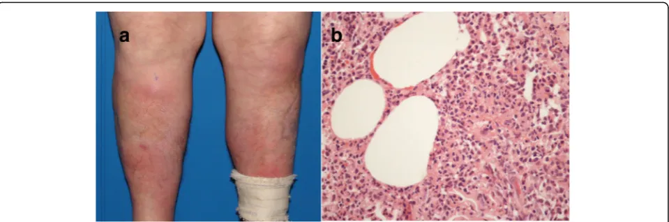
Neutrophilic lobular panniculitis The term neutro-philic (lobular) panniculitis refers to histopathological findings shared by a variety of cutaneous and systemic disorders, which includes pustular panniculitis associ-ated with RA, pancreatic panniculitis, α1-antitrypsin de-ficiency panniculitis, subcutaneous Sweet syndrome, and factitious panniculitis [56, 57]. The most common form of panniculitis occurring in the setting of RA is EN [13]. However, rare cases of neutrophilic lobular panniculitis have also been reported in these patients. Cutaneous eruption manifests as tender erythematous nodules on the lower legs in which eventual ulceration and dis-charge of purulent material allow clinical distinction from EN (Fig.7a). Histopathology in neutrophilic panni-culitis associated with RA shows fat necrosis and neu-trophilic dust in the subcutaneous tissue with surrounding fibrosis (Fig.7b) [58].
Vemurafenib and dabrafenib were recently approved for the treatment of patients with unresectable or meta-static melanoma harboring the BRAF V600E mutation. Several cases of BRAF inhibitors-induced panniculitis have been reported within 7 days to 16 months after starting the medication. Joint pain and fever were associ-ated with tender subcutaneous nodules on the upper or lower limbs, mostly the thighs, in 44 and 31% of the pa-tients, respectively [7]. Histopathological investigation Fig. 6Lupus panniculitis.aDepressed area involving the proximal part of the arm.bExtensive lobular infiltrate composed of lymphocytes. H&E, 200x
has shown mixed (both septal and lobular) panniculi-tis in some instances, but primarily lobular neutro-philic panniculitis usually predominates. Occasionally, small foci of fatty necrosis and some foamy macro-phage are seen. Only in a few cases an inflammatory infiltrate surrounding blood vessel walls and focal erythrocyte extravasation were revealed, as did non-necrotizing granulomas. Spontaneous resolution des-pite drug maintenance is possible; however, low-dose topical or systemic steroids and nonsteroidal anti-inflammatory drugs may be used for symptomatic treatment [7, 59]. Similarly, panniculitis has been re-ported as a rare side effect of specific inhibitors of tyrosine kinase activity ibrutinib, with marked activity in several B-cell malignant neoplasms, and dasatinib, used to treat chronic myeloid leukemia in chronic, blastic or accelerated phase that is resistant or in-tolerant to imatinib mesylate [60, 61].
Primarily-lobular panniculitides with vasculitis
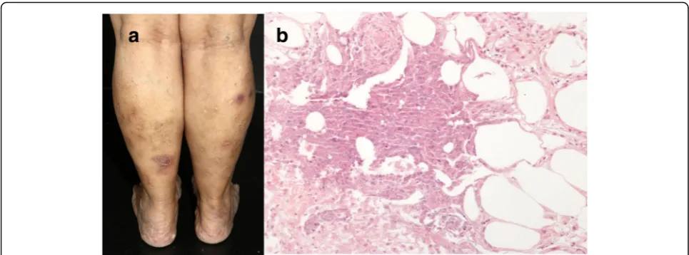
Erythema induratum Erythema induratum (EI) has a female predominance and typically affects patients with some degree of obesity and venous insufficiency of the lower extremities. The disease presents with relapsing episodes of painless violaceous nodules on the calves and shins, but also on the buttocks and lower trunk, mostly during the winter. Lesions have a tendency to central ulceration and resolve spontaneously within a few months, leaving post inflammatory hyperpigmenta-tion and occasionally atrophic pigmented scars (Fig.8a). There are no accompanying systemic symptoms [62,63]. EI occurs as hypersensitive immune reaction to
Mycobacterium tuberculosis. As others tuberculids, which include lichen scrofulosorum and papulonecrotic tuberculid, although extremely rarely EI can be induced by tuberculin skin test, as well as by Bacillus Calmette–Guérin vaccine
even in children. Ziehl-Neelsen staining for acid-fast bacilli and mycobacterial culture usually result nega-tive [64, 65]. However, there is one report in which
M. marinum was isolated [66]. The most striking fea-tures of EI are strongly positive tuberculin purified protein derivative (PPD) with an induration greater than 20 mm and clearance of skin lesions with anti-tuberculosis therapy, which occurs within 1–6 months (mean, 2.1 months). Once diagnosis is sus-pected a skin biopsy should be obtained for histologic examination and polymerase chain reaction analysis for mycobacterial DNA [67].
Histopathological analysis reveals a predominant lobular panniculitis, with mild to moderate inflamma-tion of the neighboring fibrous septa, which appear widened. At an early stage, the inflammatory infiltrate is mainly composed of neutrophils, with or without leukocytoclasis; whereas histiocytes and lipophages predominate in fully developed lesions. Some type of vascular damage is detected in 90% of the cases. The most common pattern identified is inflammation in-volving small venules of the center of the fat lobule; however, vasculitis involving large septal vessels can also be found, irrespectively of the stage of the lesion [68]. There are varying degrees of caseous and coagu-lative necrosis, and poorly developed granulomas (Fig. 8b) [69]. Although nowadays most authors use both terms indistinctly, nodular vasculitis usually re-fers to the nontuberculous variant of EI. There is one case report on a 28-year-old man who developed ery-thematous painful nodules on the lower legs, one year after starting etanercept for psoriasis. Physical exam-ination and histologic manifestations were compatible to nodular vasculitis. Since the screening for infec-tious and auto-immune conditions was negative, the development of this panniculitis was attributed to TNF-α inhibitor treatment [70].
Fig. 7Neutrophilic lobular panniculitis associated with rheumatoid arthritis.aErythematous nodules on the anterior aspect of the legs.b
Panniculitis with crystals following etanercept injection According to Llamas-Velasco and Requena [71], panniculitis with lipid crystallization within adipocytes may be seen in several disorders, including crystal-storing histio-cytosis, gouty panniculitis, post-steroid panniculitis, oxalo-sis and subcutaneous fungal infections by mucormycooxalo-sis, zygomycosis or aspergillosis. These authors reported the first case of panniculitis with crystals induced by etanercept subcutaneous injection in a 62-year-old woman with severe psoriasis who developed an erythematous, slightly painful nodule on the skin of the anterior abdominal wall. The bi-opsy demonstrated a mostly lobular panniculitis with lymphocytic infiltrate, venulitis, as well as granulomas with foreign-body-type giant cells. Small radially arranged crys-tals surrounded by histiocytes were present at the interface between deep reticular dermis and subcutis. These crystals failed to stain with periodic acid-Schiff (PAS) and were slightly refractile under polarized light.
Mixed (septal and lobular) panniculitides
Infective panniculitis Infective panniculitis (IP) may occur as a primary infection by direct inoculation, as an extension from an underlying source of infection or sec-ondarily via microbial hematogenous dissemination with subsequent infection of the subcutaneous tissue, induced by bacteria, mycobacteria, fungi, protozoa, or viruses. This type of panniculitis often manifests as multiple nodules predominantly found on the peripheral extremities, re-sembling EN (Fig.9a). Other locations of involvement are possible such as the upper extremities, the gluteal region and the abdominal wall. However, the clinical picture can vary from infiltrated nodules with discharge or not to nec-rotizing ulcers depending on the organism involved, the
route of infection, the host immune response, and the dur-ation of the lesion at the time of biopsy [72,73].
There are many bacterial etiologic agents of IP. The more common of these include Streptococcus pyogenes,
Staphylococcus aureus,Pseudomonasspp.,Mycobacterium
spp. (most of the cases of mycobacterial panniculitis being caused by nontuberculous mycobacteria), Actinomyces,
Nocardia spp., Borrelia burgdorferi, and Klebsiella spp. When it comes to fungal infections that involve the sub-cutaneous fat, they can be divided into two major types: (1) panniculitis in the setting of a disseminated fungal infection and (2) classical subcutaneous mycosis (with the most important being mycetoma, chromoblastomycosis, and sporotrichosis), introduced into the subcutaneous tis-sue from the environment via inoculation. Cytomegalo-virus has been rarely reported as a causative agent; immunocompromised patients were particularly affected, as in most of these IP [72, 73]. Similarly, there has been sporadic reports on parasitic organisms (Leishmania spp.;
Trypanosoma cruzi; and Gnathostoma spp.) causing pan-niculitis [74].
Diagnosis is quite challenging, not only because of a nonspecific clinical presentation, but also due to its histologic findings, which can be indistinguishable from several types of panniculitides. Bacterial IP should be strongly considered when extensive suppurative panni-culitis with perivascular, lobular, or mixed septal-lobular neutrophil-dominated infiltrate is present, which often extends into the dermis (Fig. 9b) [58, 72]. IP caused by atypical mycobacteria or deep fungal infection shows suppurative granulomas within the lobule, which consist of collections of neutrophils surrounded by epithelioid histiocytes [39,73]. Borreliosis have been documented to show a histological picture mimicking LEP and subcuta-neous panniculitis-like T-cell lymphoma, with a dense Fig. 8Erythema induratum.aErythematous nodules and atrophic scars in the legs.bCaseous necrosis in the lobules of the hypodermis.
H&E, 400x
lymphocytic infiltrate of the subcutaneous tissue with scat-tered plasma cells [20]. Remarkably, besides typical cyto-megalic inclusions localized predominantly within the endothelial cells of small vessels of the subcutaneous cellu-lar tissue, skin biopsy of noducellu-lar lesions in panniculitis as-sociated with cytomegalovirus infection may show septal involvement with abundant neutrophils, histiocytes, karyor-rhexis and phagocytosis of cellular debris [75].
Regardless of the microorganism, vascular damage in-cluding necrotizing small vessel vasculitis involving arteri-oles and venules and/ or a thrombotic microangiopathy in the deep reticular dermis and subcutaneous fat (in sec-ondary cutaneous infections) may be noteworthy [58,76]. A high degree of clinical awareness is needed, since additional histological studies are necessary to identify the causative agent in many cases, including special stains (Gram, periodic acid-Schiff, Ziehl-Neelsen, methenamine-silver, anti–bacillus Calmette-Guerin antibody); cultures and molecular diagnostic techniques, like polymerase chain reaction PCR, especially in patients with preserved immune response [39,72,73].
Erythema nodosum-like lesions in Behçet’s disease EN-like lesions occur in 22.5 to 45.5% of the patients di-agnosed with Behçet’s disease (BD) [77,78]. Nodular le-sions are mostly present on the lower legs, but upper extremities may also be involved. They are histopatho-logically similar to those of conventional EN in approxi-mately one-third of the cases, showing a mostly septal
panniculitis pattern, and an inflammatory infiltrate com-posed of neutrophils and various numbers of lympho-cytes and histiolympho-cytes in the thickened septa of the subcutis. However, biopsy specimens with features of a panniculitis lobular or mixed septal and lobular in pat-tern, with variable degrees of lymphohistiocytic and neu-trophilic infiltration, and clear evidence of vasculitis are even more common to be found [79,80].
Vessels involvement is usually extensive and not lim-ited to the areas of severe inflammation. It can be both of the venulitis or of the phlebitis type. The latter char-acteristically simulates C-PAN, as in superficial thrombophlebitis, and therefore must be clarified based on the investigation of the internal elastic lamina fiber by specific staining, such as Verhoeff van Gieson. Al-though the subcutaneous muscular veins in the lower legs usually have a compact concentric smooth muscle pattern with a round lumen and intimal elastic fiber pro-liferation due to the persistent hydrostatic pressure mimicking the characteristic features of arteries, the elastic fibers in the muscular layer are distributed be-tween the bundled smooth muscle in veins, whereas the elastic fibers are scantly distributed in the medial mus-cular layer in arteries [81]. A variable degree of fat ne-crosis is frequently observed [79,80].
Conclusion
different stages of development. Even in the same stage of evolution, location and type of inflammation may vary among different cases of the same panniculitis; whereas similar histopathologic findings may be observed in pan-niculitides of different etiologies, for instance in superfi-cial thrombophlebitis and C-PAN, or in EN and EI. In evaluating cases of panniculitidesis essential to know the associated changes outside the subcutis to establish a specific diagnosis.
Acknowledgements
The author’s wish to acknowledge Alexandre Vargas, photographer from the Dermatology Department of Universidade de São Paulo.
Authors’contributions
TCABM wrote the manuscript in consultation with GFST. GFST contributed to the writing of the manuscript. MSCG designed the tables and figures. IH provided histopathological images and aided in descripting
histopathological findings of each panniculitis. PRC contributed to the design of the manuscript, provided clinical images and supervised the work. JFC conceived the idea of the manuscript and supervised the work. All authors read and approved the final manuscript.
Funding
The authors received no financial support for the research, authorship, and/ or publication of this article.
Availability of data and materials
Data sharing is not applicable to this article as no datasets were generated or analyzed during the current study.
Ethics approval and consent to participate
All participants were volunteers and provided informed consent for the photographs included in this paper.
Consent for publication
Consent for publication of pictures was given by all participants.
Competing interests
The authors declare that they have no competing interests.
Author details
1Faculdade de Medicina FMUSP, Universidade de Sao Paulo, Sao Paulo, SP,
Brazil.2Instituto Dermatológico Professor Rubem David Azulay, Rio de Janeiro, RJ, Brazil.3Faculdade de Medicina do ABC FMABC, Santo Andre, SP, Brazil.4Instituto de Ciências da Saúde, Universidade Federal da Bahia (UFBA), Salvador, BA, Brazil.5Private practice, Aracaju, SE, Brazil.
Received: 31 August 2018 Accepted: 12 July 2019
References
1. Segura S, Requena L. Anatomy and histology of Normal Subcutaneous fat, necrosis of adipocytes, and classification of the panniculitides. Dermatol Clin. 2008;26:419–24.
2. Llamas Velasco M, et al. Clues in histopathological diagnosis of panniculitis. Am J Dermatopathol. 2018;40:155–67.
3. Cascajo CD, Borghi S, Weyers W. Panniculitis. Am. J. Dermatopathol. 2000;22: 530–49.
4. Chowaniec M, Starba A, Wiland P. Erythema nodosum - review of the literature. Reumatologia. 2016;54:79–82.
5. Requena L, Yus ES. Erythema Nodosum. Dermatol Clin. 2008;26:425–38. 6. Requena L, Yus ES. Panniculitis. Part I. Mostly septal panniculitis. J. Am. Acad.
Dermatol. 2001;45:163–83.
7. Mössner R, et al. Erythema nodosum-like lesions during BRAF inhibitor therapy: report on 16 new cases and review of the literature. J Eur Acad Dermatology Venereol. 2015;29:1797–806.
8. García-Porrúa C, et al. Erythema nodosum: etiologic and predictive factors in a defined population. Arthritis Rheum. 2000;43:584–92.
9. Mert A, et al. Erythema nodosum: an experience of 10 years. Scand J Infect Dis. 2004;36:424–7.
10. Psychos DN, Voulgari PV, Skopouli FN, Drosos AA, Moutsopoulos HM. Erythema nodosum: the underlying conditions. Clin Rheumatol. 2000;19:212–6. 11. Negera E, et al. Clinico-pathological features of erythema nodosum
leprosum: a case-control study at ALERT hospital, Ethiopia. PLoS Negl Trop Dis. 2017;11:1–13.
12. Sarita S, et al. A study on histological features of lepra reactions in patients attending the Dermatology Department of the Government Medical College, Calicut, Kerala, India. Lepr. Rev. 2013;84:51–64.
13. Ziemer M, Müller AK, Hein G, Oelzner P, Elsner P. Incidence and classification of cutaneous manifestations in rheumatoid arthritis. JDDG - J Ger Soc Dermatology. 2016;14:1237–46.
14. Ergun T, et al. Skin manifestations of rheumatoid arthritis: a study of 215 Turkish patients. Int J Dermatol. 2008;47:894–902.
15. Nakamura T, Inaba M, Yoshinaga T, Takaoka H, Iyama K. Nodules in patients with rheumatoid arthritis and methotrexate treatment. Mod Rheumatol. 2015;7595:1–2.
16. Tilstra JS, Lienesch DW. Rheumatoid Nodules. Dermatol Clin. 2015;33:361–71. 17. Wick MR. Granulomatous & histiocytic dermatitides. Semin Diagn Pathol.
2017;34:301–11.
18. Chua-Aguilera CJ, Moller B, Yawalkar N. Skin manifestations of rheumatoid arthritis, juvenile idiopathic arthritis, and Spondyloarthritides. Clin Rev Allergy Immunol. 2017.https://doi.org/10.1007/s12016-017-8632-5. 19. Walker D, Susa JS, Currimbhoy S, Jacobe H. Histopathological changes in
morphea and their clinical correlates: results from the Morphea in adults and children cohort V. J Am Acad Dermatol. 2017;76:1124–30.
20. Shiau CJ, Abi Daoud MS, Wong SM, Crawford RI. Lymphocytic panniculitis: an algorithmic approach to lymphocytes in subcutaneous tissue. J Clin Pathol. 2015;68:954–62.
21. Bielsa Marsol I. Update on the classification and treatment of localized scleroderma. Actas Dermo-Sifiliográficas (English Ed). 2013;104:654–66. 22. Toledano C, et al. Localized scleroderma: a series of 52 patients. Eur J Intern
Med. 2009;20:331–6.
23. Fett N, Werth VP. Update on morphea: Part I. Epidemiology, clinical presentation, and pathogenesis. J. Am. Acad. Dermatol. 2011;64:217–28. 24. Bielsa I, Ariza A. Deep Morphea. Semin Cutan Med Surg. 2007;26:90–5. 25. Evangelisto A, Werth V, Schumacher HR. What is that nodule?: a diagnostic
approach to evaluating subcutaneous and cutaneous nodules. J Clin Rheumatol. 2006;12:230–40.
26. Erden A, et al. Comparing polyarteritis nodosa in children and adults: a single center study. Int J Rheum Dis. 2017;20:1016–22.
27. Sharma A, et al. Polyarteritis nodosa in North India: clinical manifestations and outcomes. Int J Rheum Dis. 2017;20:390–7.
28. Fain O, et al. Vasculitides associated with malignancies: analysis of sixty patients. Arthritis Care Res. 2007;57:1473–80.
29. Fathalla BM, Miller L, Brady S, Schaller JG. Cutaneous polyarteritis nodosa in children. J Am Acad Dermatol. 2005;53:724–8.
30. Pagnoux C, et al. Clinical features and outcomes in 348 patients with polyarteritis nodosa: a systematic retrospective study of patients diagnosed between 1963 and 2005 and entered into the French vasculitis study group database. Arthritis Rheum. 2010;62:616–26.
31. Nakamura T, et al. Cutaneous polyarteritis nodosa: revisiting its definition and diagnostic criteria. Arch Dermatol Res. 2009;301:117–21.
32. Furukawa F. Cutaneous Polyarteritis Nodosa: an update. Ann Vasc Dis. 2012;5:282–8.
33. Morimoto A, Chen K-RR. Reappraisal of histopathology of cutaneous polyarteritis nodosa. J Cutan Pathol. 2016;43:1131–8.
34. Kawakami T, Yamazaki M, Mizoguchi M, Soma Y. High titer of anti-phosphatidylserine-prothrombin complex antibodies in patients with cutaneous polyarteritis nodosa. Arthritis Care Res. 2007;57:1507–13. 35. Dahiya A, Leach J, Levy H. Gouty panniculitis in a healthy male. J Am Acad
Dermatol. 2007;57:S52–4.
36. Weberschock T, Gholam P, Hartschuh W, Hartmann M. Gouty panniculitis in a 68-year-old man: case report and review of the literature. Int J Dermatol. 2010;49:410–3.
37. Ochoa CD, et al. Panniculitis: another clinical expression of gout. Rheumatol Int. 2011;31:831–5.
38. Forbess LJ, Fields TR. The broad Spectrum of urate crystal deposition: unusual presentations of gouty tophi. Semin Arthritis Rheum. 2012;42: 146–54.
39. Requena L, Yus ES. Panniculitis. Part II. Mostly lobular panniculitis. J. Am. Acad. Dermatol. 2001;45:325–61.
40. Torrelo A, Hernández A. Panniculitis in Children. Dermatol. Clin. 2008;26: 491–500.
41. Kim ST, et al. Post-steroid panniculitis in an adult. J Dermatol. 2008;35:786–8. 42. Tuffanelli DL. Lupus Erythematosus Panniculitis (Profundus). Arch. Dermatol.
1971;103:231.
43. Ng PP-L, Tan SH, Tan T. Lupus erythematosus panniculitis: a clinicopathologic study. Int J Dermatol. 2002;41:488–90.
44. Choi JY, Kim HS, Lee GY. Case of lupus erythematosus panniculitis triggered by human papillomavirus quadrivalent vaccine injection. J Dermatol. 2017; 44:1420–1.
45. Castrillón MA, Murrell DF. Lupus profundus limited to a site of trauma: case report and review of the literature. Int J Womens Dermatol. 2017;3:117–20. 46. Durand A-L, et al. Anti-tumour necrosis factorα-induced lupus
erythematosus panniculitis. J. Eur. Acad. Dermatol Venereol. 2017;31:e318–9. 47. Lee H, Kim DS, Chung KY. Adalimumab-induced lupus panniculitis. Lupus.
2014;23:1443–4.
48. Jacyk WK, Bhana KN. Lupus erythematosus profundus in black South Africans. 2006:717–21.https://doi.org/10.1111/j.1365-4632.2005.02770.x. 49. Park HS, Choi JW, Kim B, Cho KH. Lupus erythematosus panniculitis:
Clinicopathological, Immunophenotypic, and molecular studies. Am J Dermatopathol. 2010;32:24–30.
50. Elbendary A, Griffin J, Li S, Tlougan B, Junkins-Hopkins JM. Linear Sclerodermoid lupus erythematosus Profundus in a child. Am J Dermatopathol. 2016;38:904–9.
51. Kündig TM, Trüeb RM, Krasovec M. Lupus profundus/panniculitis. Dermatology. 1997;195:99–101.
52. Willemze R. Cutaneous lymphomas with a panniculitic presentation. Semin Diagn Pathol. 2017;34:36–43.
53. Michot C, et al. Subcutaneous panniculitis-like T-cell lymphoma in a patient receiving etanercept for rheumatoid arthritis. Br J Dermatol. 2009;160:889–90. 54. Braunstein I, Werth VP. Update on management of connective tissue
panniculitides. Dermatol Ther. 2012;25:173–82.
55. Polcari IC, Stein SL. Panniculitis in childhood. Dermatol Ther. 2010;23:356–67. 56. Cohen P, Subcutaneous R. Sweet’s syndrome: a variant of acute febrile
neutrophilic dermatosis that is included in the histopathologic differential diagnosis of neutrophilic panniculitis. J Am Acad Dermatol. 2005;52:927–8. 57. Nobeyama Y, Nakagawa H. Subcutaneous Sweet’s syndrome and
neutrophilic panniculitis. J Dermatol. 2014;41:861–2. 58. Chan MP. Neutrophilic panniculitis: algorithmic approach to a
heterogeneous group of disorders. Arch Pathol Lab Med. 2014;138:1337–43. 59. Choy B, Chou S, Anforth R, Fernández-Peñas P. Panniculitis in patients
treated with BRAF inhibitors: a case series. Am J Dermatopathol. 2014; 36:493–7.
60. Fabbro SK, Smith SM, Dubovsky JA, Gru AA, Jones JA. Panniculitis in patients undergoing treatment with the Bruton tyrosine kinase inhibitor ibrutinib for lymphoid leukemias. JAMA Oncol. 2015;1:684-6.
61. Assouline S, Laneuville P, Gambacorti-Passerini C. Panniculitis during dasatinib therapy for imatinib-resistant chronic myelogenous leukemia. N Engl J Med. 2006;354:2623–4.
62. Segura S, Pujol RM, Trindade F, Requena L. Vasculitis in erythema induratum of Bazin: A histopathologic study of 101 biopsy specimens from 86 patients. J Am Acad Dermatol. 2008;59(5):839–51.
63. Mascaró JM, Baselga E. Erythema induratum of Bazin. Dermatol Clin. 2008; 26(4):439–45.
64. Sekiguchi A, Motegi S, Ishikawa O. Erythema induratum of Bazin associated with bacillus Calmette-Guérin vaccination: implication of M1 macrophage infiltration and monocyte chemotactic protein-1 expression. J Dermatol. 2016;43(1):111–3.
65. Posada García C, et al. Erythema induratum of Bazin induced by tuberculin skin test. Int J Dermatol. 2015;54(11):1297–9.
66. Papathemeli D, Franke I, Bonnekoh B, Gollnick H, Ambach A. Explosive generalization of nodular vasculitis - Mycobacterium marinum challenges the paradigm. J Eur Acad Dermatol Venereol. 2016;30(12):e189–91. 67. Lighter J, Tse DB, Li Y, Borkowsky W. Erythema induratum of bazin in a
child: evidence for a cell-mediated hyper-response to Mycobacterium tuberculosis. Pediatr Infect Dis J. 2009;28(4):326–8.
68. Segura S, Pujol RM, Trindade F, Requena L. Vasculitis in erythema induratum of Bazin: a histopathologic study of 101 biopsy specimens from 86 patients. J Am Acad Dermatol. 2008;59:839–51.
69. Gilchrist H, Patterson JW. Erythema nodosum and erythema induratum (nodular vasculitis): diagnosis and management. Dermatol Ther. 2010;23(4): 320–7.
70. Park S, et al. Nodular vasculitis developed during etanercept treatment in a patient with psoriasis. J Am Acad Dermatol. 2014;70:AB177.
71. Llamas-Velasco M, Requena L. Panniculitis with crystals induced by etanercept subcutaneous injection. J Cutan Pathol. 2015;42:413–5. 72. Morrison LK, Rapini R, Willison CB, Tyring S. Infection and panniculitis.
Dermatol Ther. 2010;23:328–40.
73. Delgado-Jimenez Y, Fraga J, García-Díez A. Infective Panniculitis. Dermatol. Clin. 2008;26:471–80.
74. Norgan AP, Pritt BS. Parasitic infections of the skin and Subcutaneous tissues. Adv Anat Pathol. 2018;25:106–23.
75. Ballestero-Diez M, Alvarez-Ruiz SB, Aragues Montanes M, Fraga J. Septal panniculitis associated with cytomegalovirus infection. Histopathology. 2005;46:720–2.
76. Magro CM, Dyrsen ME, Crowson AN. Acute infectious id panniculitis/ panniculitic bacterid: a distinctive form of neutrophilic lobular panniculitis. J Cutan Pathol. 2008;35:941–6.
77. Davatchi F, et al. Behcet’s disease in Iran: analysis of 6500 cases. Int J Rheum Dis. 2010;13:367–73.
78. Tursen U, Gurler A, Boyvat A. Evaluation of clinical findings according to sex in 2313 Turkish patients with Behçet’s disease. Int J Dermatol. 2003;42:346–51. 79. Misago N, Tada Y, Koarada S, Narisawa Y. Erythema Nodosum-like lesions in
Behçet’s disease: a Clinicopathological study of 26 cases. Acta Derm Venereol. 2012;92:681–6.
80. Kim B, LeBoit PE. Histopathologic features of erythema nodosum-like lesions in Behcet disease: a comparison with erythema nodosum focusing on the role of vasculitis. Am J Dermatopathol. 2000;22:379–90.
81. Chen KR. The misdiagnosis of superficial thrombophlebitis as cutaneous polyarteritis nodosa: features of the internal elastic lamina and the compact concentric muscular layer as diagnostic pitfalls. Am J Dermatopathol. 2010; 32:688–93.
Publisher’s Note