Open Access
Research article
Five mucosal transcripts of interest in ulcerative colitis identified by
quantitative real-time PCR: a prospective study
Anders Eriksson*
1, Carl-Fredrik Flach
2, Anders Lindgren
3, Eva Kvifors
1and
Stefan Lange
4Address: 1Department of Internal Medicine, Gastroenterology Unit, Sahlgren's University Hospital/Östra, Göteborg, Sweden, 2Institute of
Biomedicine, Department of Microbiology and Immunology, Göteborg University, Gothenburg, Sweden, 3Department of Internal Medicine,
Borås Lasarett, Borås, Sweden and 4Institute of Biomedicine, Department of Clinical Bacteriology, Göteborg University, Gothenburg, Sweden
Email: Anders Eriksson* - [email protected]; Carl-Fredrik Flach - [email protected];
Anders Lindgren - [email protected]; Eva Kvifors - [email protected]; Stefan Lange - [email protected] * Corresponding author
Abstract
Background: The cause and pathophysiology of ulcerative colitis are both mainly unknown. We have previously used whole-genome microarray technique on biopsies obtained from patients with ulcerative colitis to identifiy 5 changed mucosal transcripts. The aim of this study was to compare mucosal expressions of these five transcripts in ulcerative colitis patients vs. controls, along with the transcript expression in relation to the clinical ulcerative colitis status.
Methods: Colonic mucosal specimens from rectum and caecum were taken at ambulatory colonoscopy from ulcerative colitis patients (n = 49) with defined inflammatory activity and disease extension, and from controls (n = 67) without inflammatory bowel disease. The five mucosal transcripts aldolase B, elafin, MST-1, simNIPhom and SLC6A14 were analyzed using quantitative real-time PCR.
Results: Significant transcript differences in the rectal mucosa for all five transcripts were demonstrated in ulcerative colitis patients compared to controls. The grade of transcript expression was related to the clinical disease activity.
Conclusion: The five gene transcripts were changed in patients with ulcerative colitis, and were related to the disease activity. The known biological function of some of the transcripts may contribute to the inflammatory features and indicate a possible role of microbes in ulcerative colitis. The findings may also contribute to our pathophysiological understanding of ulcerative colitis.
Background
Ulcerative colitis (UC) is a disorder characterized by chronic mucosal inflammation of the large intestine. It is frequently associated with various extraintestinal manifes-tations. The inflammation may be limited to the rectum (proctitis), but mucosal lesions often continue more
prox-imally (left-sided UC) or additionally embrace the trans-verse colon (extensive colitis) or the entire large bowel (pancolitis). The immune and cellular (non-immune) response is dysregulated in both the acute and the chronic phase of UC [1,2]. In Scandinavia, UC has been found to affect individuals of all ages, with an annual incidence of
Published: 12 August 2008
BMC Gastroenterology 2008, 8:34 doi:10.1186/1471-230X-8-34
Received: 28 June 2007 Accepted: 12 August 2008
This article is available from: http://www.biomedcentral.com/1471-230X/8/34 © 2008 Eriksson et al; licensee BioMed Central Ltd.
about 15 per 100 000 [3,4] and a prevalence of about 300 per 100 000 inhabitants [5].
The pathogenesis and pathophysiology of UC are still under investigation [6]. We can tentatively say that the cause and onset of the disease is polygenic with environ-mental interaction; that is, there is a genetic predisposi-tion [7-9] in combinapredisposi-tion with eliciting environmental factors which may precipitate the phenotype of UC [10]. In addition, interaction between the colonic epithelium and microbiological flora as well as a disintegrated mucosal barrier function may be important factors in the onset and development of UC [6]. The use of microarray technique analyses on mucosal specimens obtained from both patients with established UC and controls has allowed identification of candidate genes, which are valu-able in research on UC pathogenesis. However, these UC candidate genes must be carefully selected, since recent evaluations of microarray data have revealed considerable divergence after examination of similar tissues [11-13]. Such divergent results are commonly presented in studies using pooled patient samples. In the present study how-ever, the transcripts selected are based on our earlier indi-vidual whole-genome microarray screening and quantitative real-time PCR (RT-PCR) in patients with UC [14], where five changed genes/transcripts were identified; aldolase B, elafin, MST-1, simNIPhom (similar to NIP homolog), and SLC6A14. The pathophysiological proper-ties of SimNIPhom have not yet been clarified, but the other transcript products have potential importance in secretion [15,16], anti-microbiological activity [17], and cell-mediated immune response [18].
The primary aim of the present study was to define differ-ences in the mucosal expression of five selected tran-scripts, retrieved from two different colonic locations in UC, by using a quantitative RT-PCR technique. We also aimed to evaluate the influence of ongoing anti-inflam-matory treatment as well as the importance of the colonic UC extension and the severity class.
Methods
Patients and tissue specimens
Before the colonoscopy procedure, consecutive male and female subjects (UC patients and controls, >18 y) were recruited to the present study. The UC diagnosis was based on the medical history, endoscopic findings, histo-logical examination, laboratory tests, and the clinical dis-ease presentation. The extent of UC and the clinical activity were classified in accordance with the Montreal Classification [19]. In brief, the colonic inflammatory involvement is defined as extension (letter E) combined with a number between 1–3 (E1 denotes proctitis, E2 left-sided UC, and E3 extensive colitis). In addition, the clini-cal severity grade (letter S) is defined. The S-score ranges
from clinical remission (S0) to severe UC (S3). Mucosal biopsies were obtained from the rectum (10–15 cm prox-imal from anal verge) and caecum from all participants.
No uses of corticosteroids, aminosalicylates, or immuno-suppressants were registered in the control group, while 9 patients (18%) in the UC group were treated systemically with corticosteroids (prednisolone 10–20 mg). Twenty-seven UC patients (55%) were treated systemically with aminosalicylates (mesalazine 1.600–2.400 mg/24 h). Among these, two patients had additional ongoing ther-apy with aminosalicylate (mesalazine 500 mg QD) ene-mas and three patients were treated with corticosteroid enemas (prednisolone 37.5 mg QD or BID). Seven patients had stable (>3 months) ongoing immunosup-pressant treatment (Azathioprine, 1.8–2.2 mg/kg bw).
The remaining demographic and clinical data are pre-sented in Figure 1.
RNA isolation
The biopsy specimens were immediately stored in RNA-later solution for isolation of RNA. The RNA- RNA-later-pre-served biopsies were homogenized in a lysis buffer from the GenElute Mammalian Total RNA kit (Sigma, St. Louis, MO.) and total RNA was isolated according to the manu-facturer's instructions. The RNA concentration was meas-ured spectrophotometrically.
Quantification by real-time polymerase chain reaction (RT-PCR)
Two μg of total RNA from each sample were converted into cDNA. The cDNA synthesis was performed as described previously [20]. Oligonucleotide primers pur-chased from MWG-BIOTECH AG (Ebersberg, Germany) were used for the relative quantification (ABI-7500 sys-tem, software version 1.3) (Table 1). Glyceraldehyde-3-phosphate dehydrogenase (GAPDH) was used as a refer-ence gene in all experiments. The expression level in each sample was compared with a calibrator by using the ΔΔC T-formula (ΔCT(calibrator) - ΔCT(sample)).
Statistical analysis
Descriptive statistics and the Wilcoxon signed rank test (SAS, Statview®) were used. Median values are presented.
Ethics
The study was approved by the local research ethical com-mittee. All patients were given oral and written informa-tion before entering the study. Informed consent was obtained from all patients and controls.
Results
colitis/pan-colitis 9.2 years). Neither age nor gender was matched between the UC group and the control group.
In order to evaluate any differences in transcript expres-sions within the control group (n = 67) with respect to background diagnoses (anaemia, diverticulosis, irritable bowel disease and polyposis), statistical analysis of each background diagnosis were compared to the remaining group of controls. No significant differences (p > 0.05) were detected for any of these diagnoses.
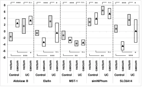
Significantly higher transcript expressions of aldolase B and SimNIPhom and significantly lower transcript expres-sions of elafin, MST-1, and SLC6A14 were found in caecal biopsies in comparison to rectal biopsies from the control group (Figure 2). The only significant differences between rectal and caecal transcript expressions in UC patients were the decreased transcript expressions of elafin and SLC6A14 in caecal biopsies in comparison to rectal biop-sies.
Demographic and clinical data from the control and UC group respectively
Figure 1
Demographic and clinical data from the control and UC group respectively.
Analyzed cases, n=116
Controls, n=67
Demographic data
Men: mean age;
68 (31-86), n=31
Women: mean age;
62 (19-88), n=36
Smoking habits
Current smokers;
n=9
Non-smokers;
n=46
Ex-smoker; n=12
Reasons for colonoscopy
1Unexplained anemia;
n=21
2Abdominal pain;
n=24
3Suspect diverticulosis; n=21
4Post volvolus control; n=1
Colonoscopy diagnosis
1
Normal endoscopic findings; n=5
Single adenomas;
n=16
2
IBS;
n=24
3
Diverticulosis;
n=21
5Status post volvolus;
n=1
Ulcerative colitis, n=49
Demographic data
Men: mean age;
49 (22-86), n=31
Women: mean age;
41 (21-71), n=18
Smoking habits
Current smokers;
n=4
Non-smokers; n=30
Ex-smoker; n=15
Montreal classification
(number of patients)
E1 E2 E3 Sum
S0
2 2 17 21
S1
2 3 5 10
S2
2 2 3 7
S3
1 2 8 11
Comparison of rectal biopsies from controls (n = 67) with rectal biopsies from UC patients with inflammatory activ-ity in accordance with Montreal classifications S1–S3 (n = 28) showed significant elevations (p < 0.05) in UC patients of all transcript expressions with the exception of MST-1, which showed significantly (p < 0.05) decreased expression in UC patients. The same analysis of caecal biopsies from controls and patients with S1–S3 UC (n = 16) showed significantly elevated transcript expressions of aldolase B and SLC6A14 only. Distal biopsies from con-trols, compared with UC patients without inflammatory
activity (S0), showed increased transcript expression in aldolase B only (median -1.62 vs. 1.0, p = 0,012). All other transcript analyses from both locations showed no signif-icant differences (p > 0.05).
All transcript analysis with respect to UC extension showed that left-sided (E2) and total colitis (E3) differed significantly from controls (p < 0.05); this was not the case for proctitis (E1).
Table 1: Primers used for real-time polymerase chain reaction.
Gene Forward primer Reverse primer
Aldolase B 5'-aaggctgcaaacaaggaggcaacc-3' 5'-tgaagagcgactgggtggaagcag-3'
Elafin 5'-tgtgaaggctcttgcgggatgg-3' 5'-agggcagcagggacttaggaccag-3'
SimNIPhom 5'-cgccagacagctaggggagtgaag-3' 5'-gcatttctgatattttgtgaccacgcac-3'
SLC6A14 5'-gctgcttggttttgtttctccttggtc-3' 5'-gcaattaaaatgccccatccagcac-3'
MST-1 5'-aaccaggagtgtaacatcaagcaccgag-3' 5'-cagttgtgggtaaagcaggcaagtgg-3'
GAPDH 5'-gagcaccaggtggtctcctctgacttc-3' 5'-gccaaattcgttgtcataccaggaaatg-3'
RT-PCR result (ΔΔCt(=ΔCttarget-ΔCtcalibrator)) for controls (filled dots) and UC patients (▲) presented as median values and
25th and 75th percentil (bars) Figure 2
RT-PCR result (ΔΔCt(=ΔCttarget-ΔCtcalibrator)) for controls (filled dots) and UC patients (▲) presented as median values and 25th and 75th percentil (bars). * = p < 0.05, ** = p < 0.01, *** = p < 0.001, n.s. = non significant.
Statistical analysis concerning the influence of anti-inflammatory treatment on the transcript expressions within the UC cohort showed no statistical differences (p > 0.05) when comparing UC patients with ongoing corti-costeroids (n = 9) or azathioprine (n = 7) respectively with the remaining UC patients. However, the 27 patients treated with mesalazine show a significant increase in aldolase B (median 0.48 vs. 3.02, p = 0.035) in compari-son to the remaining UC patients.
Discussion
Genetic predisposition, psychological stress, nutritional and environmental influences, intestinal pathogens and disturbed intestinal barrier function have all been propo-seas pathogenetic factors in UC [6]. However, current knowledge about the pathogenesis and pathophysiology of UC [21] is incomplete. Moreover, with the exception of a few general serological inflammatory activity biomark-ers, even less information is available regarding mucosa-associated transcript changes and their potential pathoge-netic and pathophysiological role in UC [22]. This lack of knowledge may sometimes lead to uncertainty in diagno-sis, judgement of prognosis and clinical management of UC patients.
On the basis of exsisting knowledge of the biological func-tions of the transcripts investigated in this study, it is rea-sonable to believe that the demonstrated alterations might be related to predisposition and/or the pathophys-iological response in UC.
It is intriguing that aldolase B and SLC6A14 were up-reg-ulated in rectal as well as caecal mucosa in UC compared to controls. Aldolase B is known to be mainly expressed in the intestinal villus cells and it has a central role in the gly-colytic pathway. It also participates in regulation of intes-tinal secretion [16]. Since SLC6A14 is also known to encode a Na+/Cl- driven amino acid transporter B(0+)
[15], the up-regulation of aldolase B and SLC6A14 might be a common pathophysiological response, aimed at counteracting the exaggerated loss of fluid seen in UC. Theoretically, the up-regulation of these two transcripts could be a local response to the increased feacal/fluid stream, where bioactive molecules comprise ability to reg-ulate transcript expression. Additionally, since the inflam-matory activity and load of fluid over time is usually most pronounced in the distal part of the colon, the registered changes in aldolase B and SLC6A14 may reflect long-term inflammatory activity. Our finding that aldolase B from distal biopsies is significantly elevated during the remis-sion phase (S0) indicates that the regulation of this tran-script not only is secondary to the inflammatory activity.
The involvement of the microflora and its importance in the onset, development and preservation of UC has been
discussed [6]. The SLC6A14 transcript expression is there-fore also interesting in this respect, since it is involved in the host's antibacterial response [21]. In addition, the defensine-like epithelium associated antimicrobial mole-cule elafin, antagonizing human neutrophil elastase pre-venting tissue injury via inhibition of excessive release of proteolytic enzymes from inflammatory cells is interest-ing in this context [18]. The present results confirm an elafin transcript enhancement in caecal as well as rectal biopsies from patients with UC. Thus, the combined ele-vation of elafin and SLC6A14 may contribute to an ampli-fied defence reaction aimed to restoration and maintenance of the mucosal integrity. This finding may indicate a pathogenetic role of the microflora in UC.
MST-1 was included in the present study due to its altera-tions in UC, as shown in our previous experiment [14], although it was excluded from that publication due to deviation in its control group. MST-1 is known to be capa-ble of inhibiting cell-mediated immune responses via down-regulation of IL-12 production and subsequently inhibition of macrophage activation [23]. Consequently, the observed down-regulation of MST-1 in rectal speci-mens may contribute to an enhanced cellular immune response in UC. A reasonable explanation of the concom-itant decreased MST-1 transcript expression and increased aldolase B, SLC6A14, and elafin transcript expression is that the changes describe a pathophysiological response to a more pronounced inflammatory and, possibly, an exaggerated microbial load in at least the rectal part of the colon mucosa.
The fifth identified significantly up-regulated transcript (in rectum only) SimNIPhom (similar to the numb-inter-acting homolog), encodes a hypothetical protein, at present of unknown pathophysiological importance.
Our results supports that specimens from the rectal mucosa are more suitable for further analysis of the selected transcripts, due to the more predictable inflam-matory involvement in the rectum and its availability for direct inspection and easy biopsy sampling.
Our data can not answer whether the observed changes in expressions of the five selected transcripts may be present in e.g. other inflammatory, infectious or autoimmune conditions since this study uniquely focused on UC patients compared to non-inflamed controls.
Conclusion
Publish with BioMed Central and every scientist can read your work free of charge "BioMed Central will be the most significant development for disseminating the results of biomedical researc h in our lifetime."
Sir Paul Nurse, Cancer Research UK
Your research papers will be:
available free of charge to the entire biomedical community
peer reviewed and published immediately upon acceptance
cited in PubMed and archived on PubMed Central
yours — you keep the copyright
Submit your manuscript here:
http://www.biomedcentral.com/info/publishing_adv.asp
BioMedcentral
Competing interests
The authors declare that they have no competing interests.
Authors' contributions
AE designed the study, preformed sampling of biopsies, analyzed the data, and prepared the manuscript. C-FF ana-lyzed the data. AL preformed sampling of biopsies. EK coordinated the study. SL designed the study, analyzed the data and prepared the manuscript. All authors read and approved final manuscript.
Acknowledgements
This work was supported by grants from the Swedish federal government under the LUA/ALF agreement, (grant no. 7157). The RT-PCR analysis was performed by Index Pharmaceuticals, Stockholm, Sweden. We thank Pro-fessor Sven Wallerstedt for critical reading of the manuscript.
References
1. Fiocchi C: Inflammatory bowel disease: etiology and patho-genesis. Gastroenterology 1998, 115:182-205.
2. Macdonald TT, Monteleone G: Immunity, inflammation, and allergy in the gut. Science 2005, 307:1920-1925.
3. Ekbom A, Helmick C, Zack M, Adami HO: The epidemiology of inflammatory bowel disease: a large, population-based study in Sweden. Gastroenterology 1991, 100:350-358.
4. Moum B, Vatn MH, Ekbom A, Aadland E, Fausa O, Lygren I, Sauar J, Schulz T, Stray N: Incidence of ulcerative colitis and indetermi-nate colitis in four counties of southeastern Norway, 1990– 93. A prospective population-based study. The Inflamma-tory Bowel South-Eastern Norway (IBSEN) Study Group of Gastroenterologists. Scand J Gastroenterol 1996, 31:362-366. 5. Jacobsen BA, Fallingborg J, Rasmussen HH, Nielsen KR, Drewes AM,
Puho E, Nielsen GL, Sorensen HT: Increase in incidence and prevalence of inflammatory bowel disease in northern Den-mark: a population-based study, 1978–2002. Eur J Gastroenterol Hepatol 2006, 18:601-6.
6. Kucharzik T, Maaser C, Lugering A, Kagnoff M, Mayer L, Targan S, Domschke W: Recent understanding of IBD pathogenesis: implications for future therapies. Inflamm Bowel Dis 2006,
12:1068-83.
7. Satsangi J, Parkes M, Louis E, Hashimoto L, Kato N, Welsh K, Ter-williger JD, Lathrop GM, Bell JI, Jewell DP: Two stage genome-wide search in inflammatory bowel disease provides evi-dence for susceptibility loci on chromosomes 3, 7 and 12. Nat Genet 1996, 14:199-202.
8. Schwab M, Schaeffeler E, Marx C, Fromm MF, Kaskas B, Metzler J, Stange E, Herfarth H, Schoelmerich J, Gregor M, Walker S, Cascorbi I, Roots I, Brinkmann U, Zanger UM, Eichelbaum M: Association between the C3435T MDR1 gene polymorphism and suscep-tibility for ulcerative colitis. Gastroenterology 2003, 124:26-33. 9. Lawrance IC, Fiocchi C, Chakravarti S: Ulcerative colitis and
Crohn's disease: distinctive gene expression profiles and novel susceptibility candidate genes. Hum Mol Genet 2001,
10:445-456. [9]
10. Roussomoustakaki M, Satsangi J, Welsh K, Louis E, Fanning G, Targan S, Landers C, Jewell DP: Genetic markers may predict disease behavior in patients with ulcerative colitis. Gastroenterology
1997, 112:1845-53.
11. Mah N, Thelin A, Lu T, Nikolaus S, Kuhbacher T, Gurbuz Y, Eickhoff H, Kloppel G, Lehrach H, Mellgard B, Costello CM, Schreiber S: A comparison of oligonucleotide and cDNA-based microarray systems. Physiol Genomics 2004, 16:361-370.
12. Tan PK, Downey TJ, Spitznagel EL Jr, Xu P, Fu D, Dimitrov DS, Lem-picki RA, Raaka BM, Cam MC: Evaluation of gene expression measurements from commercial microarray platforms.
Nucleic Acids Res 2003, 31:5676-5684.
13. Dieckgraefe BK, Stenson WF, Korzenik JR, Swanson PE, Harrington CA: Analyzis of mucosal gene expression in inflammatory bowel disease by parallel oligonucleotide arrays. Physiol Genomics 2000, 4:1-11.
14. Flach CF, Eriksson A, Jennische E, Lange S, Gunnerek C, Lönnroth I:
Detection of elafin as a candidate marker for ulcerative col-itis by whole-genome microarray screening. Inflamm Bowel Dis
2006, 12:837-847.
15. Sloan JL, Mager S: Cloning and functional expression of a human Na(+) and Cl(-)-dependent neutral and cationic amino acid transporter B(0+). J Biol Chem 1999,
274:23740-23745.
16. Wachsmuth ED: Differentiation of epithelial cells in human jejunum: localization and quantification of aminopeptidase, alkaline phosphatase and aldolase isozymes in tissue sec-tions. Histochemistry 1976, 48:101-109.
17. Simpson AJ, Maxwell AI, Govan JR, Haslett C, Sallenave JM: Elafin (elastase-specific inhibitor) has anti-microbial activity against gram-positive and gram-negative respiratory patho-gens. FEBS Lett 1999, 452:309-313.
18. Schmid M, Fellermann K, Fritz P, Wiedow O, Stange EF, Wehkamp J:
Attenuated induction of epithelial and leukocyte serine anti-proteases elafin and secretory leukocyte protease inhibitor in Crohn's disease. J Leukoc Biol 2007, 81:907-915.
19. Silverberg MS, Satsangi J, Ahmad T, Arnott ID, Bernstein CN, Brant SR, Caprilli R, Colombel JF, Gasche C, Geboes K, Jewell DP, Karban A, Loftus EV Jr, Pena AS, Ridell RH, Sachar DB, Schreiber S, Steinhart AH, Targan SR, Vermeire S, Warren BF: Toward an integrated clinical, molecular and serologic classification of inflamma-tory bowel disease: Report from a working Party of the 2005 Montreal World congress of gastroenterology. Can J Gastroen-terology 2005, 19(suppl A):5-36.
20. Flach CF, Lange S, Jennische E, Lönnroth I: Cholera toxin induces expression of ion channels and carriers in rat small intestinal mucosa. FEBS Lett 2004, 561:122-126.
21. Sartor RB: Mechanisms of disease: pathogenesis of Crohn's disease and ulcerative colitis. Nat Clin Pract Gastroenterol Hepatol
2006, 3:390-407.
22. Desai D, Faubion WA, Sandborn WJ: Review article: biological activity markers in inflammatory bowel disease. Aliment Phar-macol Ther 2007, 25(3):247-55.
23. Morrison AC, Wilson CB, Ray M, Correll PH: Macrophage-stimu-lating protein, the ligand for the stem cell-derived tyrosine kinase/RON receptor tyrosine kinase, inhibits IL-12 produc-tion by primary peritoneal macrophages stimulated with IFN-gamma and lipopolysaccharide. J Immunol172:1825-1832. 2004, Feb 1
Pre-publication history
The pre-publication history for this paper can be accessed here: