ARTICLE
Fetal Tricuspid Valve Size and Growth as Predictors
of Outcome in Pulmonary Atresia With Intact
Ventricular Septum
Joshua W. Salvin, MDa,b, Doff B. McElhinney, MDa,b, Steven D. Colan, MDa,b, Kimberlee Gauvreau, ScDa, Pedro J. del Nido, MDc,d,
Kathy J. Jenkins, MD, MPHa,b, James E. Lock, MDa,b, Wayne Tworetzky, MDa,b
Departments ofaCardiology andcCardiac Surgery, Children’s Hospital Boston, Boston, Massachusetts; Departments ofbPediatrics anddSurgery, Harvard Medical School, Boston, Massachusetts
The authors have indicated they have no financial relationships relevant to this article to disclose.
ABSTRACT
OBJECTIVE.Pulmonary atresia with intact ventricular septum is a complex congenital cardiovascular anomaly that frequently requires single ventricle palliation. Fetal diagnosis of pulmonary atresia with intact ventricular septum is common, but the natural history of pulmonary atresia with intact ventricular septum diagnosed in midgestation, predictors of neonatal anatomy, and predictors of biventricular repair have not been determined. The objective of this study was to determine whether the size and rate of growth of the fetal tricuspid valve predict neonatal anatomy and biventricular repair.
DESIGN AND RESULTS.Twenty-three fetuses diagnosed with pulmonary atresia with intact ventricular septum between 1990 and 2004 were studied. Of 13 fetuses with a midgestation fetal tricuspid valvezscore ⱕ⫺3, 1 achieved biventricular repair, compared with 5 of 5 with a tricuspid valve zscore ⬎⫺3. Of 13 fetuses with a midgestation fetal tricuspid valvezscoreⱕ⫺3, 8 were diagnosed postnatally with a right ventricular dependent coronary circulation, compared with none with a tricuspid valvezscore⬎⫺3. Midgestation and late gestation fetal tricuspid valvez
scores correlated with neonatal tricuspid valvezscore. The average rate of tricuspid valve growth between mid- and late fetal echocardiograms was significantly lower in patients who did not achieve biventricular repair than in those who did (0.012
⫾0.008 cm per week vs 0.028⫾0.014 cm per week).
CONCLUSIONS.Fetal tricuspid valve z score and rate of growth predict postnatal outcome in pulmonary atresia with intact ventricular septum. These findings may have important implications for prenatal counseling and selection of patients for fetal pulmonary valve dilation.
www.pediatrics.org/cgi/doi/10.1542/ peds.2006-0428
doi:10.1542/peds.2006-0428
Key Words
echocardiography, congenital heart defects, pulmonary heart disease, pulmonary atresia
Abbreviations
PA/IVS—pulmonary atresia with intact ventricular septum
RV—right ventricle
RVDCC—right ventricule-dependent coronary circulation
TV—tricuspid valve
EGA— estimated gestational age
P
ULMONARY ATRESIA WITHintact ventricular septum (PA/IVS) is a morphologically heterogeneous lesion characterized by variable right ventricle (RV) hypoplasia and abnormal communications between the RV and cor-onary arteries, with an RV-dependent corcor-onary circula-tion (RVDCC) in the most severe cases. Single ventricle palliation is required in a substantial proportion of pa-tients.1–6Current neonatal management strategies beginwith establishment of flow through the hypoplastic RV, with the aim of promoting RV growth and achieving an RV of sufficient size and function to support the pulmo-nary circulation. Several previous reports suggest that a small tricuspid valve (TV) and the presence of RVDCC in the neonate are predictors of failure to achieve a biven-tricular circulation.1,3,4Given the increasing prenatal
de-tection rate of PA/IVS, the complexity of this defect, and the range of possible outcomes, it becomes important to be able to predict in utero which patients will ultimately achieve a biventricular repair. In addition, with the emerging prospect of in utero valvuloplasty7,8 for PA/
IVS, prediction of postnatal outcome at the time of in utero diagnosis may facilitate appropriate patient selec-tion for fetal intervenselec-tion. The primary aim of this study was to determine whether TV size and the rate of TV growth in midgestation fetuses with PA/IVS are indica-tors of the ultimate capacity of the right heart to support a biventricular circulation. The secondary purpose of this study was to determine whether fetal TV size and rate of growth can predict TV size at birth and/or the presence of an RVDCC. We hypothesized that a fetal TVzscore of
ⱕ⫺3, along with slow in utero TV growth, will predict failure to achieve a biventricular repair, small TV at birth, and presence of RVDCC.
METHODS
Patient Selection
The echocardiography database was queried for all fetal studies with a diagnostic code for PA/IVS. Selection was restricted to 1990 –2004, because the neonatal manage-ment of PA/IVS at our center was consistent during this time period. Live-born fetuses that subsequently had all postnatal interventions (catheter-based and surgical) at Children’s Hospital were selected for full analysis. Fe-tuses that did not go on to live birth and feFe-tuses in which a fetal intervention was attempted were used only for the initial demographic description and were excluded from further analysis. Fetuses with associated Ebstein’s anomaly of the TV were excluded, because there was no consistent management strategy for this subset of pa-tients during the study time period. The study was ap-proved by the institutional review committee at Chil-dren’s Hospital Boston.
Echocardiographic Data
Two-dimensional echocardiograms were reviewed on all of the fetuses and neonates. Archived analog videotapes
were digitized into a database using a commercially available product (EchoTrace, Marcus Laboratories, Bos-ton, MA). Studies completed after 2003 were reviewed in their original digital format. The TV was imaged in all of the fetal studies, and annulus size was felt to be a reproducible value without technical limitations. A sin-gle fetal echocardiographer (W. T.) blinded to patient outcome measured the TV annulus diameter from the 4-chamber view in all of the fetal echocardiograms. The TV was measured between the hinge points of the TV leaflets with the valve open in diastole. The annulus diameter was expressed as a z score to adjust for the estimated gestational age ([EGA] estimated from stan-dard femur length and head circumference measure-ments) of the fetus, based on unpublished normative data from our institution derived from fetuses without heart disease according to the following formulas: TVp (mean TV diameter for gestational age)⫽0.04071 * EGA⫺0.33017 TVsd (standard deviation of the regres-sion)⫽0.00417 * (EGA⫺29.83000)⫹(0.010976 * {1.0128
⫹[(EGA⫺29.83)2]/2735})0.5.The same blinded reviewer
measured TV annulus diameter in the 4-chamber view of the first neonatal echocardiogram. Annulus diameters measured on fetal and neonatal echocardiograms were expressed aszscores to adjust for body surface area using the formula: TVzscore⫽TVz⫽(TV⫺TVp)/TVsd.
Figure 1 demonstrates the normative mean TV an-nulus size in millimeters measured across various times in gestation.zscores are derived using the SD around the mean. Other institutions have used similar algorithms for quantitative analysis of fetal cardiac structures.9
Fetal TVzscores were dichotomized intoⱕ⫺3 and⬎⫺3 groups based on the findings of Hanley et al3and the
natural division of patients in our series. To account for variability in EGA at the time of fetal echocardiogram, studies for each fetus were grouped into 1 of 2 windows based on the EGA at the time of the study. The midges-tation window was defined as 20 to 29 weeks’ EGA. The late gestation window was defined as 30 to 39 weeks’ EGA. If 2 echocardiograms were completed within 1 window, measurements from the earlier EGA were used for analysis.
The rate of fetal TV growth was calculated by sub-tracting the TV annulus diameter (cm) at the first fetal echo from the TV annulus diameter (cm) at the final fetal echo and dividing this value by the number of weeks between these 2 studies. This number was ex-pressed as the rate of fetal TV growth in centimeters per week.
Catheterization, Coronary Angiography, and Management Strategy
PA/IVS have been described previously.10,11A coronary
artery was considered RV dependent if it demonstrated a communicating RV fistula and it was atretic, stenotic, or interrupted proximal to the RV to coronary artery com-munication, which could lead to ischemia if the RV was to be decompressed. RVDCC was defined as coronary circulation in which there is atresia of 1 or both coronary ostia or obstruction within ⬎1 major coronary artery (right, left main, left anterior descending, circumflex, and/or posterior descending coronary artery). At Chil-dren’s Hospital Boston, the initial management strategy for newborns with PA/IVS is based on the presence or absence of RVDCC and rarely takes into account RV or TV size. When RVDCC is documented by coronary an-giography, a systemic-to-pulmonary artery shunt is placed with the goal of single ventricle palliation. In neonates with non-RVDCC, RV decompression is at-tempted based on the size of the RV and TV. This is accomplished either by transcatheter balloon valvotomy or surgical RV outflow tract augmentation with or with-out systemic-to-pulmonary artery shunt. Cardiac cathe-terization is then performed at 6 months of age to test the adequacy of the TV and RV. The systemic-to-pulmo-nary artery shunt and interatrial communication may be closed at that time, assuming the RV is able to support the pulmonary circulation. A bidirectional superior cavo-pulmonary anastomosis is performed in patients in whom the RV is unable to support the pulmonary circulation, and then the cardiac catheterization may be repeated at 1 to 3 years of age to determine suitability for a “one and one-half ventricle” repair or Fontan operation.
Outcomes
Outcomes assessed included achievement of a biven-tricular repair, TV size at birth, and the presence of RVDCC. A biventricular repair was defined as an RV supporting the pulmonary circulation, a systemic arterial
saturation of ⬎90%, and either no interatrial commu-nication or a left-to-right shunt at any remaining atrial communication. Patients with a cavopulmonary shunt (with or without a patent RV outflow tract) were con-sidered not to have a biventricular circulation.
Data Analysis
Comparison of continuous and categorical data between or within groups was performed using the appropriate parametric or nonparametric tests for comparing un-paired or un-paired samples, respectively. Because definitive biventricular repair is often a staged process in patients with PA/IVS, achievement of biventricular circulation is a time-dependent variable and, therefore, was assessed both as a discrete outcome and by assessment of time to biventricular repair. The time to achievement of biven-tricular repair was determined by review of catheteriza-tion and echocardiographic and pulse oximetry data, using the Kaplan-Meier method, with comparison be-tween groups using the log-rank test. For Kaplan-Meier analysis, patients were censored at the time of death, at the time of definitive single ventricle palliation (ie, Fon-tan completion, bidirectional Glenn without a patent pulmonary outflow tract), at the time of commitment to a single ventricle management strategy (ie, at the time of diagnosis of RVDCC, an anatomic criteria that is consid-ered at our center to preclude biventricular repair), or at the time of most recent follow-up if a definitive biven-tricular circulation had not been achieved but remained a possibility (ie, systemic-to-pulmonary shunt or bidirec-tional Glenn with a patent pulmonary outflow tract). Continuous data are presented as mean⫾SD.
RESULTS
Demographics and Diagnostic Data
Between 1990 and 2004, 36 fetuses were diagnosed with PA/IVS, resulting in 25 live births. Of the 11 fetuses not
FIGURE 1
carried to term, there were 10 elective terminations and 1 fetal demise. Of 25 liveborn fetuses, 2 underwent fetal interventional procedures and were not included in this study. Thus, the study group included 23 patients. (Fig 2) The EGA at diagnosis for the original 36 fetuses was 23.4 ⫾ 5.6 weeks. Fetuses not carried to term were diagnosed significantly earlier in gestation than those that were (20.4⫾3.1 vs 24.5⫾5.7 weeks;P⫽.04).
Within the study group, the median number of fetal echocardiograms was 2 (range: 1–3). For fetuses with⬎1 study (n⫽18), the mean age at the final fetal echocar-diogram was 30.6⫾3.6 weeks’ EGA. The mean duration of time from first fetal echo to last fetal echo was 9.8⫾ 3.2 weeks. Eighteen patients hadⱖ1 study in midgesta-tion, and 20 patients had ⱖ1 study in late gestation. Fifteen patients had an echocardiogram in both the mid-and late gestational age windows.
Outcomes
Among 23 live born patients, there were 7 with a biven-tricular circulation and 16 with a single ventricle pallia-tion. Twenty one were alive at a median follow-up of 5.4 years (10 months to 13 years), and the 2 deaths occurred in neonates with severe RV hypoplasia and RVDCC who died after a systemic-to-pulmonary arterial shunt. Ten patients were diagnosed postnatally with RVDCC. One patient had coarctation of the aorta requiring surgical repair, and none had significant extracardiac abnormal-ities. Among the 14 living patients with a single ventricle circulation, 9 had undergone a Fontan procedure, and 5 had a bidirectional superior cavopulmonary anastomo-sis. Of the 5 patients with a bidirectional cavopulmonary anastomosis, 2 had documented RVDCC and were
awaiting a Fontan operation, and the other 3 had a patent RV outflow tract and an open atrial septum at most recent follow-up (Fig 2). Fetuses with TV zscore
ⱕ⫺3 on the midgestation echocardiogram were signifi-cantly less likely to undergo biventricular repair (P ⫽
.001) than patients with azscore⬎⫺3 and significantly more likely to be diagnosed with RVDCC (P ⫽ .036). Fetuses with a TV z score ⱕ⫺3 on the late gestation echocardiogram were similarly less likely to undergo biventricular repair (P⬍.001) and more likely to have an RVDCC (P⫽.051) than patients with azscore⬎⫺3. In both the mid- and late gestation groups, a TVzscore
ⱕ⫺3 suggested decreased freedom from biventricular repair (P⫽.059 andP⫽.014, respectively).
In utero TV growth was significantly slower in pa-tients who ultimately required single ventricle palliation (P⫽.006) and in patients who developed RVDCC (P⫽
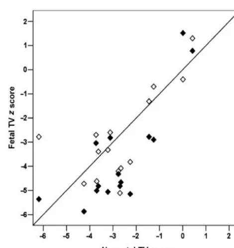
.033; Table 1) than in those who did not. Among the 15 patients with measurements in both mid- and late ges-tation windows, there was a distinct separation between biventricular and single ventricle outcome groups when tracking TV growth from mid- to late gestation (Fig 3). Both mid- (R2⫽0.48) and late (R2⫽0.74) gestation TV zscores correlated with neonatal TVzscores (Fig 4).
DISCUSSION
Among fetuses diagnosed with PA/IVS at our center since 1990, both the fetal TVzscore and the in utero rate of TV growth were associated with postnatal outcome. Fetuses with a TVzscore ⱕ⫺3 on either mid- or late-gestation fetal echocardiograms were significantly less likely to achieve a biventricular repair than those with a
FIGURE 2
Fetal PA/IVS demographics and postnatal outcomes, 1990 –2004. The study group contained 23 patients. Of the 5 Glenn shunts, 2 patients have RVDCC and, thus, are expected to continue along a single ventricle pathway. Two deaths occurred, both after BTS, in ne-onates with severe RV hypoplasia and RVDCC. BTS in-dicates Blalock-Taussig shunt.
TABLE 1 Biventricular Versus Single Ventricle Outcome and RVDCC Versus Non-RVDCC in Mid- and Late Gestation by TVzScore Group
Variable Biventricular
Repair
Single Ventricle
P RVDCC Non-RVDCC P
Mid-TVzscore
ⱕ⫺3 (n⫽13) 1 12 .001 8 5 .036
⬎⫺3 (n⫽5) 5 0 0 5
Late TVzscore
ⱕ⫺3 (n⫽14) 1 13 ⬍.001 7 7 .051
⬎⫺3 (n⫽6) 6 0 0 6
TV z score ⬎⫺3 and were also more likely to have a RVDCC. In contrast, all of the fetuses with a TVzscore
⬎⫺3 on either the mid- or late fetal echocardiogram achieved a biventricular repair. A fetal TVzscoreⱕ⫺3 on the midgestational fetal echocardiogram was associ-ated with postnatal diagnosis of RVDCC, and none of the fetuses with a TVzscore⬎⫺3 on mid- or late gestation fetal echocardiograms had RVDCC. In addition, the rate of TV growth in utero was more than twice as slow in the single ventricle patients than in those who achieved a biventricular repair or were born with a non-RVDCC. These findings may have an important impact on the prenatal and postnatal management of the fetus with PA/IVS. The frequency of fetal diagnosis of congenital heart disease is rising steadily, with screening practices affecting worldwide prevalence and types of congenital heart disease, including PA/IVS.12At our institution, the
frequency of fetal diagnosis of PA/IVS has followed this trend, with prenatal detection rising from 40% of all PA/IVS births between 1986 and 2000 to ⬎65% after 2000. With this increase in prenatal diagnosis, prognos-tic data may be helpful for prenatal decision-making and perinatal planning. Several studies have correlated neo-natal echocardiographic features with angiographic evi-dence of RVDCC. For example, Garcia et al13concluded
that abnormal neonatal coronary flow patterns are sug-gestive of fistulas; Giglia et al11suggested that RV volume
and TVzscores are significantly smaller in neonates with RVDCC; and Satou et al14demonstrated that a neonatal
TVzscore ofⱕ⫺2.5 has an 80% positive predictive value to predict RVDCC. Despite the use of neonatal echocardi-ography for predicting the presence of RVDCC, correlating fetal data are lacking. Fetal echocardiographic features of ventriculocoronary connections have been described15
but may not be predictive of true RVDCC. Fetal indicators
of RVDCC are important for 2 primary reasons. First, at our institution, coronary artery anatomy is the primary deter-minant of postnatal management. In newborns with PA/ IVS, decompression of the RV is thought to be essential in promoting growth of the RV and achievement of biven-tricular repair. Although RV fistulas are common in pa-tients with PA/IVS3,16and not considered a
contraindica-FIGURE 4
Scatterplots demonstrating relationship between early (〫) and late (⽧) window fetal TVzscores and neonatal TVzscores. ⁄, unity. By linear regression, the relationship be-tween early fetal and neonatal TVzscores is described by the equation: Neonatal TVz
score⫽(0.61⫻Early fetal RVzscore)⫺0.97 (R2⫽0.48). The relationship between late fetal and neonatal TVzscores is described by the equation: Neonatal TVzscore⫽(0.70 ⫻Late fetal RVzscore)⫺0.28 (R2⫽0.74).
FIGURE 3
tion to RV decompression, the presence of RVDCC is a contraindication.10At our institution, coronary anatomy is
documented in all neonates by angiography. If there is evidence of RVDCC, there is no subsequent attempt to decompress the RV, and the neonate progresses toward staged single ventricle palliation. Second, among patients with PA/IVS, outcomes are worse in those with an RVDCC than in those without. Intermediate follow-up of patients with an RVDCC demonstrated 83% 5-year survival, with the surviving patients at ongoing risk for ischemia.17
Pre-diction of an RVDCC in midgestation may aid in prenatal counseling.
PA/IVS is a heterogeneous disease, and not all institu-tions base their initial management strategy on the pres-ence of RVDCC. Recent reports have demonstrated a tiered grading system for RV hypoplasia, which is predictive of failure to achieve a biventricular repair.18Others describe
specific RV morphology in an attempt to predict the need for single ventricle palliation.19Although our data are
de-rived from the management strategy consistently used at a single institution over the past 15 years, the strong corre-lations between fetal TVzscore and both neonatal TV size (a surrogate for RV size) and the presence of RVDCC are likely to be useful regardless of the approach to the man-agement of PA/IVS. In addition, the use of serial fetal echocardiograms to document the rate of in utero growth may aid in parental counseling at centers that rely on RV size as a criterion for palliation. The ability to predict which midgestation fetuses with PA/IVS are likely to have RVDCC and/or to require single ventricle palliation may aid in appropriate patient selection for advanced fetal ther-apy. There have been several reports of balloon dilation of the pulmonary valve in the fetus, which is based on the theory that decompression of the hypoplastic and hyper-tensive RV in utero will promote antegrade flow across the TV and PV, thus encouraging RV growth and preventing the development of an RVDCC.7,8,20Although much more
experience is necessary to substantiate the feasibility and efficacy of fetal balloon pulmonary valvuloplasty, appropriate fetal selection and timing of this proce-dure may ultimately improve the ability of a fetus with PA/IVS to achieve a biventricular repair postna-tally. Additional anatomic factors will almost certainly influence the success of fetal balloon pulmonary val-vuloplasty, and careful evaluation of fetal TV size and growth by serial echocardiography may be the first step toward optimal patient selection for fetal intervention.
REFERENCES
1. Ashburn DA, Blackstone EH, Wells WJ, et al. Determinants of mortality and type of repair in neonates with pulmonary atre-sia and intact ventricular septum.J Thorac Cardiovasc Surg2004; 127:1000 –1008
2. Dyamenahalli U, McCrindle BW, McDonald C, et al. Pulmo-nary atresia with intact ventricular septum: management of, and outcomes for, a cohort of 210 consecutive patients.Cardiol Young.2004;14;299 –308
3. Hanley FL, Sade RM, Blackstone EH, Kirklin JW, Freedom RM, Nanda NC. Outcomes in neonatal pulmonary atresia with in-tact ventricular septum. A multiinstitutional study. J Thorac Cardiovasc Surg1993;105:406 – 427
4. Rychik J, Levy H, Gaynor JW, DeCampli WM, Spray TL. Out-come after operations for pulmonary atresia with intact ven-tricular septum.J Thorac Cardiovasc Surg.1998;116:924 –931 5. Jahangiri M, Zurakowski D, Bichell D, Mayer JE, del Nido PJ,
Jonas RA. Improved results with selective management in pulmonary atresia with intact ventricular septum. J Thorac Cardiovasc Surg.1999;118:1046 –1055
6. Daubeney PE, Wang D, Delany DJ, et al. Pulmonary atresia with intact ventricular septum: predictors of early and medi-um-term outcome in a population-based study.J Thorac Car-diovasc Surg.2005;130:1071
7. Arzt W, Tulzer G, Aigner M, Mair R, Hafner E. Invasive intrauter-ine treatment of pulmonary atresia/intact ventricular septum with heart failure.Ultrasound Obstet Gynecol.2003;21;186 –188 8. Tulzer G, Arzt W, Franklin RC, Loughna PV, Mair R, Gardiner
HM. Fetal pulmonary valvuloplasty for critical pulmonary steno-sis or atresia with intact septum.Lancet.2002;360;1567–1568 9. Schneider C, McCrindle BW, Carvalho JS, Hornberger LK,
McCarthy KP, Daubeney PE. Development of Z-scores for fetal cardiac dimensions from echocardiography.Ultrasound Obstet Gynecol.2005;26;599 – 605
10. Giglia TM, Mandell VS, Connor AR, Mayer JE, Jr, Lock JE. Diagnosis and management of right ventricle-dependent cor-onary circulation in pulmcor-onary atresia with intact ventricular septum.Circulation.1992;86:1516 –1528
11. Giglia TM, Jenkins KJ, Matitiau A, et al. Influence of right heart size on outcome in pulmonary atresia with intact ven-tricular septum.Circulation.1993;88:2248 –2256
12. Bull C. Current and potential impact of fetal diagnosis on prevalence and spectrum of serious congenital heart disease at term in the UK. British Paediatric Cardiac Association.Lancet
1999;35:1242–1247
13. Garcia JA, Zellers TM, Weinstein EM, Mahony L. Usefulness of Doppler echocardiography in diagnosing right ventricular cor-onary arterial communications in patients with pulmcor-onary atresia and intact ventricular septum and comparison with angiography.Am J Cardiol.1998;81:103–104
14. Satou GM, Perry SB, Gauvreau K, Geva T. Echocardiographic predictors of coronary artery pathology in pulmonary atresia with intact ventricular septum.Am J Cardiol.2000;85;1319 –1324 15. Maeno YV, Boutin C, Hornberger LK, et al. Prenatal diagnosis
of right ventricular outflow tract obstruction with intact ven-tricular septum, and detection of ventriculocoronary connec-tions.Heart.1999;81:661– 668
16. Fyfe DA, Edwards WD, Driscoll DJ. Myocardial ischemia in patients with pulmonary atresia and intact ventricular septum.
J Am Coll Cardiol.1986;8:402– 406
17. Powell AJ, Mayer JE, Lang P, Lock JE. Outcome in infants with pulmonary atresia, intact ventricular septum, and right ventri-cle-dependent coronary circulation. Am J Cardiol 2000;86: 1272–1274, A9
18. Odim J, Laks H, Plunkett MD, Tung TC. Successful manage-ment of patients with pulmonary atresia with intact ventricular septum using a three tier grading system for right ventricular hypoplasia.Ann Thorac Surg.2006;81;678 – 684
19. Yoshimura N, Yamaguchi M, Ohashi H, et al. Pulmonary atresia with intact ventricular septum: strategy based on right ventricular morphology.J Thorac Cardiovasc Surg.2003;126;1417–1426 20. Kohl T, Sharland G, Allan LD, et al. World experience of
percutaneous ultrasound-guided balloon valvuloplasty in hu-man fetuses with severe aortic valve obstruction.Am J Cardiol.
DOI: 10.1542/peds.2006-0428
2006;118;e415
Pediatrics
J. del Nido, Kathy J. Jenkins, James E. Lock and Wayne Tworetzky
Joshua W. Salvin, Doff B. McElhinney, Steven D. Colan, Kimberlee Gauvreau, Pedro
Atresia With Intact Ventricular Septum
Fetal Tricuspid Valve Size and Growth as Predictors of Outcome in Pulmonary
Services
Updated Information &
http://pediatrics.aappublications.org/content/118/2/e415 including high resolution figures, can be found at:
References
http://pediatrics.aappublications.org/content/118/2/e415#BIBL This article cites 20 articles, 3 of which you can access for free at:
Subspecialty Collections
http://www.aappublications.org/cgi/collection/cardiology_sub Cardiology
following collection(s):
This article, along with others on similar topics, appears in the
Permissions & Licensing
http://www.aappublications.org/site/misc/Permissions.xhtml in its entirety can be found online at:
Information about reproducing this article in parts (figures, tables) or
Reprints
DOI: 10.1542/peds.2006-0428
2006;118;e415
Pediatrics
J. del Nido, Kathy J. Jenkins, James E. Lock and Wayne Tworetzky
Joshua W. Salvin, Doff B. McElhinney, Steven D. Colan, Kimberlee Gauvreau, Pedro
Atresia With Intact Ventricular Septum
Fetal Tricuspid Valve Size and Growth as Predictors of Outcome in Pulmonary
http://pediatrics.aappublications.org/content/118/2/e415
located on the World Wide Web at:
The online version of this article, along with updated information and services, is
by the American Academy of Pediatrics. All rights reserved. Print ISSN: 1073-0397.