Synthesis and characterization of different
structured ZnO nanomaterial through
Polyethylene Glycol along with
antibacterial activity study by direct
attachment to the E.coli Dh5
α
and S.aureus
ATCC 25923 bacterial cell membrane
P BHADRA*
Department of Metallurgical and Material Engineering, Jadavpur University, Kolkata 700032, India First author E-mail: [email protected]
M.K.MITRA
Department of Metallurgical and Material Engineering, Jadavpur University, Kolkata 700032, India
Second author E-mail: [email protected] G.C.DAS
Department of Metallurgical and Material Engineering, Jadavpur University, Kolkata 700032, India Third author E-mail: [email protected]
R .DEY
Department of Metallurgical and Material Engineering, Jadavpur University, Kolkata 700032, India Fourth author E-mail: [email protected]
S. MUKHERJEE
School of Material Science and Technology, Jadavpur University, Kolkata 700032, India Fifth author E-.mail: [email protected]
Abstract
The development of new resistant strains of bacteria to current antibiotics has become a serious problem in the human health; therefore, there is a strong incentive to develop new bactericides. To overcome this problem Nanotechnology is expected to open new avenues to fight and prevent disease using atomic scale tailoring of materials. Here we achieve the considerable control over ZnO nanomaterials shape comprises volume percentage control of the polymer PEG. In this article, first time we present cuboid like structure to hexagonal disc to columnar structure and finally most challenging tetrapod like structure at room temperature. The products were characterized by several sophisticated instrumental techniques. Zone of inhibition study showed the enhanced biocidal activity of ZnO nanotetrapods compared with other nanostructures in repeated experiments. It demonstrated that the bactericidal efficacy of ZnO nanostructures changes with changing nanomaterials structure. Apart from this, we also investigated cell internalization process through SAED pattern.
Keywords: ZnO nanostructure; PEG; Internalisation.
1. Introduction
meV) and novel surface chemical properties [1]. The synthesis of low-dimensional ZnO nanostructures, for example nanorods [2] , nanobelts [3] and nanoplatelets [4] has been reported recently. There is Various Synthesis Route of ZnO Nanomaterial Sol– gel technique [5], Micro emulsion synthesis [6], Mechanochemical processing [7], Spray pyrolysis and drying [8], Thermal decomposition of organic precursor [9], RF plasma synthesis [10], Supercritical-water processing [11], Self-assembling [12], Hydrothermal processing [13], Vapor transport process [14], Sonochemical or microwave-assisted synthesis [15], Direct precipitation [16] and Homogeneous precipitation [17] etc. Different ZnO nanostructures have been prepared through the sol–gel or solution process under different experimental conditions. The preparation parameters chosen are thus a very important factor when designing ZnO for different applications. In order to rationally control the synthesized ZnO nanostructures and understand the roles of the experimental parameters, the growth mechanism should be fully investigated. Here we proposed the growth of different structured ZnO nanomaterial in the presence of PEG. Changing of volume percentage of PEG can determine the structural differentiation of ZnO nanomaterial.
The polyethylene structure having OH groups and this group is the main binding agent with whom ZnO nanoparticles can attach. Different volume percentage of PEG is main responsible for producing several structures. Zinc oxide (ZnO) is currently being investigated as an antibacterial agent in both microscale and nanoscale formulations. Earlier results have indicated that ZnO nanoparticles show antibacterial activity apparently greater than for microparticles [18]. While the exact mechanisms of the antibacterial action have not yet been clearly elucidated, suggested mechanisms include, the role of reactive oxygen species (ROS) generated on the surface of the particles, zinc ion release, membrane dysfunction [19], and nanoparticle internalization [20]. Nevertheless, the excellent study by Sawai et al. [21] clearly showed that ROS concentrations increased with the ZnO content of slurries makes this mechanism worthy of further detailed evaluations. With regard to the role of cell membrane versus cell internalization, transmission electron microscopy study showed that many particles of 10–14 nm ZnO were internalized after overnight exposure with subsequent cell membrane damage. The effect of particle size on internalization of ZnO is not known. Another aspect of relevance to completed studies is the role of the medium in which the exposures are carried out. ZnO can be processed through diethylene glycol (DEG) or aqueous routes. DEG can cause damage to bacterial membranes which complicates the interpretation of the role of ZnO. Here in our report the antibacterial activity has been changed due to the structural effect. The objective of this study is to thoroughly examine the significance of PEG assisted ZnO nanomaterials on bacterial cell wall destruction and the current status of these nanomaterials after getting entered into the bacterial cell. Actually In this study, we should investigate the probable mechanism of antibacterial property of these ZnO nanomaterials. Generally, the antimicrobial mechanism of chemical agents like PEG assisted ZnO nanomaterials can be understand by studying the specific binding on the surface of the agent with the microorganism and the consequent mode of action against those bacteria.
2. Experimental
2.1. Materials and Methods
2.2. Synthesis of PEG assisted ZnO nanomaterial
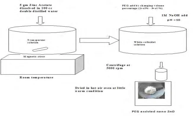
PEG assisted different structured ZnO nanomaterials were synthesized by co-precipitation technique. The general procedure involves addition of 5 gm Zinc acetate dehydrate to a 100 cc of double distilled water in the presence of Polyetheleneglycol. Five different volume percentage of PEG (1vol%, 3 vol%, 5 vol%, 7 vol% and 9 vol%) were used to synthesize different structures of ZnO. The pH of the solution was maintained at 10 by drop wise addition of 1M NaOH. After adding the precipitating agent a white colloidal solution was formed. After vigorously stirring, the white precipitation was collected after one day by centrifuging the samples at 3000 rpm and washed several times by water and ethanol and finally dried in hot air oven at slight warm condition. By changing the volume percentage of this capping agent i.e. PEG, separately five different samples were prepared. The schematic diagram of the synthesis process is given in FIG. 1. as a flow chart.
2.3. Bacterial cultures and evaluation of antibacterial activities
For antibacterial experiments, E. coli Dh5α and S. aureus ATCC 25923 was selected as the target organism. All materials were sterilized in an autoclave before the experiments. Nutrient broth and nutrient agar were used as sources for culturing bacteria. Both E.coli and S.aureus bacteria were incubated at 37C, under continuous shaking in a rotary platform at 120 rpm. At the exponential phase bacteria were harvested by centrifugation at 4000g for 10 min at 4C followed by washing twice with 10mM phosphate buffer saline (PBS, pH 7.2).The bacteria were suspended in PBS. Separately, 0.02 gm of PEG assisted ZnO nanomaterials for 3, 7 and 9 volume percentage samples were mixed in the bacterial medium & were incubated under same condition. At the exponential phase, the bacteria with nanomaterials were harvested identically. Finally the bacteria attached to PEG assisted ZnO samples were dried in a vacuum drier.
Apart from this, antibacterial activities of ZnO nanostructures were further studied by measuring the zone of inhibition. The Petri plates used in the tests were prepared using a nutrient agar medium. Separately both the bacteria were sprayed evenly on top of the plates using a sterile glass rod. After allowing the bacteria to dry (within 5–10 min) 100 µl test solutions of PEG assisted ZnO nanostructures of 0.02 g/ml concentration (different particle structures) were dropped within a cup. The zone of inhibition was measured after 18 hr incubation.
3. Results and Discussion
3.1. XRD analysis of PEG assisted ZnO nanomaterials
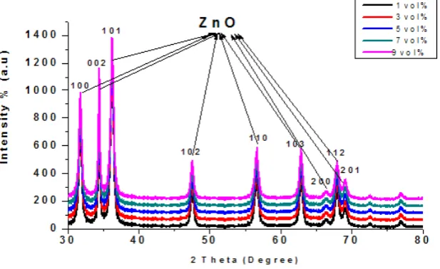
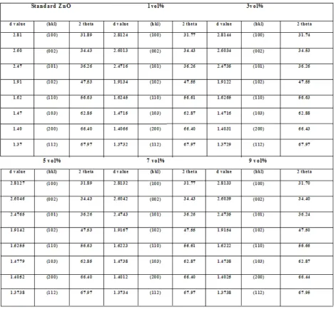
FIG. 2 shows the whole XRD plots of PEG assisted ZnO nanomaterials. The sharp intense peaks of ZnO confirms the good crystalline nature of ZnO and the peak originates from (100), (002), (101), (102), (110), (103), (200), (112) and (201) plane which reflects the formation of hexagonal ZnO. No peaks for Zn or other impurities were detected in the spectrum, revealing the phase purity of the products. The d values were
computed and compared with standard d values of ZnO (JCPDS No( 036-1451)) resulting in excellent matching for all embedded samples. The computed d values of all samples and standard d values of ZnO have been summarized in Table 1.
3.2. UV-VIS Spectroscopy analysis of PEG assisted ZnO nanostructures
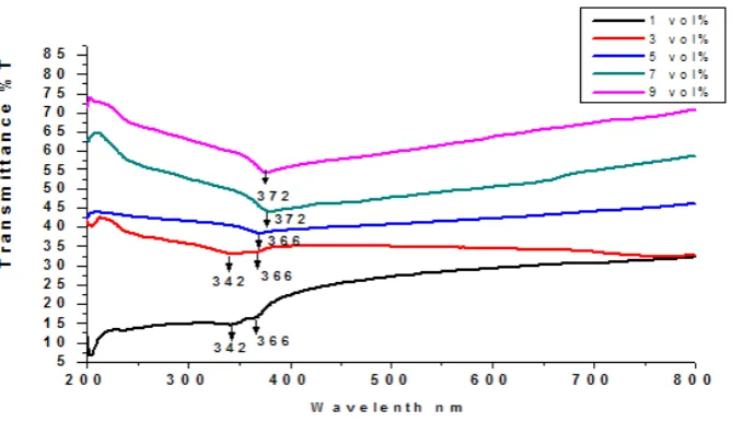
The transmission of 1 vol% PEG assisted ZnO nanomaterial is about 342 & 366 nm, and it is unchanged its transmission after getting attach with 3 vol% PEG. The transmission further shifts towards right direction after capping with 5 vol%, 7 vol% and 9 vol% PEG and the transmission fall is in the region of 366nm, 372nm and 372nm respectively. The transmission fall towards red shifted after getting attached with 5 vol% capping agent and the value reveals at 366 nm in wavelength. Finally it further shifts towards red region and this may be due to the growth of nanomaterials to coarser size. The transmission of 7 vol% sample shows at 372 nm and it remains unchanged up to extend of 9 vol% or more (FIG. 3.).
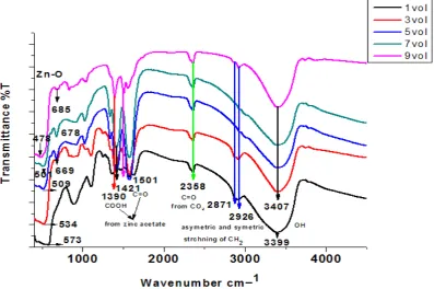
3.3. FTIR Spectroscopy analysis of PEG assisted ZnO nanostructures
The connection between ZnO with PEG has been studied by the FTIR measurement. It provided evidences of the modification of hydroxyl groups on the surface of ZnO. The spectra consist of several vibrational bands. The vibrations due to the carboxyl group obtains at 1390 cm−1 whereas the stretching mode of vibration corresponding to C=O centres at 1421cm−1. These two bands arise from Zinc acetate dehydrate precursor [22]. The stretching mode of vibration (ν4) corresponding to C=C originates at 1501 cm−1 [23], and the alkyl group obtains in the range between 2847 to 2930 cm−1. The bands at 2871 cm−1 and 2926 cm−1 corresponds asymmetric and symmetric strechning vibration of CH2 group respectively [24, 25]. The band at 3399 cm−1 and
3407 cm−1 attributes to the O–H mode of vibration, i.e. due to the hydroxyl group originates from PEG. The bands obtains in the range between 478 cm−1 to 685 cm−1 corresponds to the stretching of Zn–O–Zn [26]. The most interesting observation shows as the structure of ZnO nanomaterial changes from cuboid like morphology to tetrapod like structure. The band assigns to the stretching of Zn–O–Zn has been changed. Initially the cuboid like structure shows 573 cm-1 corresponds to the Zn–O–Zn band. After the structure forms in plate like morphology it revelas 534 cm-1 , which changes from previous assigned band i.e. 573 cm-1.Whenever the capping content further increases, it retains the structure up to the 7vol% but the obtained structure assembles thin plate for 5vol%, columnar for 7vol% and tetrapod for 9vol% PEG capping. Here the assigned band at 534 cm-1, which has been observed in 3 vol% sample,disappears and two new bands have generated at around 509 cm-1 and 669 cm-1 for assembled thin plate material (5 vol%), whereas in case of columnar material two bands have appeared at around 501 cm-1 & 678 cm-1 and 478 cm-1 & 685 cm-1 for tetrapod material. This investigation attributes to the change in particle size and morphology [27, 28, 29]. The deformation of cuboid like structure towards thin plate like structure and so on was may be responsible for generating new bands. The FTIR analysis of all PEG assisted ZnO nanomaterials have been shown in FIG. 4.
3.4. Morphological analysis by FESEM study
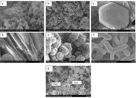
The nanomaterials morphology has been studied by FESEM analysis. The volume percentage of capping content varies from 1-9. Initially 1vol% PEG assisted ZnO nanomaterial shows cuboid like structure with 5-100 nm width and 100-300 nm length whereas the structure modifies into hexagonal plate or sheet like morphology with 25-50 nm thickness after getting attach with 3vol% PEG. With the increase of PEG to 5 vol%, the thin plate assembles, which has been clearly shown from the FIG. 5(d), confirms the stacking of several thin plates with the individual thickness of approximately 30 nm. This micrograph shows the side section of the hexagonal plate. The microscopic analysis reveals further stacking of several hexagonal plates with the increase of PEG to 7 vol% and finally leads to columnar structure. This column made up of several hexagonal plates, which measures 60 nm in thickness and 200nm – 300nm in length. On increasing PEG vol% to 9, the columnar ZnO changes its structure into tetrapod like morphology with several varieties of tetrapod pattern. At the same time different sizes of tetrapods have been formed. Further it shows that 9vol% PEG forms two types of tetrapod structure, where one consist blunt tip & another consist sharp tip end with the variation of size. The blunt tip tetrapod structure is 250-300 nm in diameter whereas the obtained sharp tip tetrapod structure is 20-50 nm in diameter. The sharp tip clearly observes through FESEM picture. Finally 9vol% PEG assisted ZnO sample confirms the formation of mixed tetrapod like structure. FIG. 5 shows the FESEM images of PEG assisted different structured ZnO nanomaterials.
FIG. 5. FESEM micrograph, a) 1vol% PEG assisted ZnO nanomaterial shows cuboid like morphology with 5-100 nm width and 100-300 nm length, b) Hexagonal plate or sheet like morphology with 50 nm thickness after getting attach with 3vol% PEG, c) Assembled thin plate
was formed after getting attach with 5vol% PEG, d) Side view of assembled thin plates, e) 7vol% PEG assisted ZnO sample this all hexagonal plates assembled in a control manner and finally formed column like structure , f) In case of 9vol% PEG the columnar ZnO totally changed its structure into tetrapod like morphology and g) 9vol% PEG had insisted to form two type of tetrapod structure one was
blunt tip another one was sharp tip
3.5. Morphological analysis by HRTEM study
indicating the preferential growth of tetrapod arms along the [002] direction. SAED pattern has been taken from one of the pods which confirm that the product is a perfect crystaline wurtzite ZnO.
FIG.6. HRTEM micrograph, a) 3vol% PEG assisted hexagonal ZnO nanoplate/nanosheet, b) Latice frindges 7vol% PEG assisted hexagonal ZnO nanoplate stacked with each other and finally formed columnar structure, c) Upper View of columnar structure, e) Side view of columnar structure, d) f) Latice frindges, g) 9vol% PEG had insisted to form two type of tetrapod structure one was blunt tip another one
was sharp tip. Here the tip of the sharp tip tetrapod structure confirmed the tip diameter was 25 nm. h) Latice frindges & SAED pattern from a single pod
3.6. Growth mechanism
The role of PEG in controlling the shape and size of the ZnO nanomaterials has been presented here. PEG is soluble in water and generally utilized in the surface modification of inorganic nanoparticles. The general formula of PEG is HO– (CH2–CH2–O–)n–H. The alcohol group is situated at the chain ends, and the chain is formed by joining the ethylene oxide group. The use of compounds of this type is particularly advantageous, since it forms a bond to the growth unit of the ZnO nuclei. The present work is related to a surface-modified nanoscale zinc oxide, where the width as well as length of the ZnO hexagonal nanoplate can be controlled by the involvement of PEG in the reaction procedure. In the presence of PEG, the Zn2+ ion from zinc acetate dihydrates is more easily adsorbed on the oxygen site of the C–O–C chain. The interaction of an ionic zinc specie and PEG led to the formation of the PEG coils into solvated Zn(II)–PEG chains. Thus the polymer chains containing Zn2+ neutralize with hydroxide ions to produce the PEG-Zn(OH)2 complex. The zinc hydroxide has
been initially fixed in a polymer network. With subsequent heating by slightly warm condition, the PEG-Zn(OH)2 complex dehydrates to form ZnO nuclei. Due to the natural tendency of ZnO for polar crystal growth
along [0001] direction, the oriented ZnO nuclei attaches to form cuboid like structures. The long-chain polymer PEG acts as a mediator for ZnO hexagonal plate like structures to grow and attain a definite shape. Hence, the presence of PEG reveals an advantage for the growth of hexagonal plates on the active sites of the PEG-Zn(OH)2 complex. A random network of nanosheets/nanoplates has been obtained without a clear center or
3.7. Optical property study by Photoluminescence spectroscopy analysis
FIG. 7. PL analysis, a) Photoluminescence study of 1vol% PEG, b) 3vol% and c) 5vol% PEG assisted ZnO thin nanoplate and assembled nanoplate, d) 7vol% and e) 9vol% PEG assisted ZnO columnar structure and mixed tetrapod structure
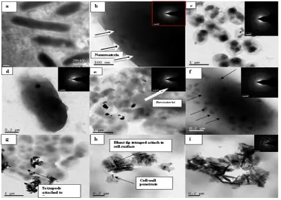
3.8. Study of attachment of PEG assisted ZnO nanostructures with both E.coli and S.aureus bacterial cell by TEM
The ability of the different structured ZnO nanomaterial to rupture bacterial cells had been tested by the TEM analysis. Here three different structure of ZnO nanomaterial was taken. The TEM image of a typical E. coli
pattern confirms the presence of ZnO nanomaterial inside the amorphous E.coli cell, indicates the internalization process. The peptydoglycan layer is much thicker in case of S.aureus. The spherical structure of this bacterial cell changes after getting attached with PEG assisted ZnO hexagonal thin nanoplates (FIG. 8(c)). Cell internalization process has been observed for gram positive bacteria i.e S.aureus but some amount of agglomeration tendency on the cell surface has been noticed. The nanoplate does not penetrate the cell membrane barrier easily due to the thicker membrane of S.aureus bacteria.
In case of 7vol% PEG assisted ZnO, the E.coli bacterial cell has been fully damaged and an uneven cellular structure has been observed. Here the outermost cell membrane layer destructs and the cytoplasm is no longer uniform. The samples have been found inside the bacterial cell which clearly reveals by the TEM image FIG. 8(d), but the cellular internalization is more prominent compared to that of cellular internalization due to the hexagonal nanoplate structure. This investigation is also applicable for S.aureus
(FIG.8(e)).
Increasing to 9vol% PEG on ZnO both the E.coli and S.aureus bacterial cells have been found fully damaged and the outermost cell membrane layer has been destroyed. Here two types of tetrapods have been observed, blunt tip ended tetrapods are larger in size and due to this it attaches on the cell surface of the bacteria only. Whereas the tip diameter of the sharp tip ended nanoterapods are smaller in size which penetrates easily into the bacterial cell. The sharp tip has been clearly revealed through TEM image. The dotted ring has been shown from the all SAED patterns which confirms the presence of ZnO nanocrystal inside the amorphous E.coli and S.aureus cell, indicates the internalization process.
3.9. Study of attachment of PEG assisted ZnO nanostructures with both E.coli and S.aureus bacterial cell by FTIR
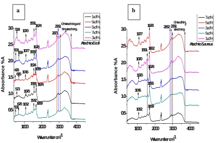
The comparative FTIR spectral analysis of untreated and treated E.coli, S.aureus with the sample has been shown in FIG. 9. After treatment with PEG assisted different structured ZnO nanomaterials the bands relative to those observed in untreated cells has been shifted to lower value. The obvious shift of these bands suggests that polysaccharides and proteins of the bacterial cells are responsible in the attachment which may cause the ultimate destruction of bacterial cell. After treatment with mixed tetrapod sample the bands at 1084 cm -1, 1678 cm -1 and 1583 cm -1 observes in untreated E.coli has been shifted to lower values 1046 cm -1, 1638 cm -1 and 1534 cm -1. In case of columnar structured sample the bands shifts to lower values 1062 cm -1, 1669 cm -1 and 1542 cm -1. Similarly for thin plate structured samples the shifting of the bands leads to lower values 1070 cm -1, 1674 cm -1 and 1551 cm -1. Whereas for cuboid shaped samples the bands have been shifted to lower values
FIG. 8. TEM micrograph, a) Intact E.coli cell; b), c), E.coli & S.aureus cell internalization by 3vol% hexagonal thin plate, d) E.coli cell internalization, e) S.aureus cell internalization due to 7vol% assembled thin plate, f) E.coli cell
1070 cm -1, 1674 cm -1 and 1558 cm -1 and for assembled thin plate shaped samples it shifts towards lower values 1077 cm -1, 1685cm -1 and 1565 cm -1. From This FTIR data it reveals the interaction of these nanomaterials with E.coli bacteria and at the same time it suggests that the attachment depends on the structure of the material [34, 35, 36, 37].
In case of S.aureus the all mentioned bands which is assigned by P=O stretching in peptidoglycan layer, O-H and NH (the functional group in polysaccharides and proteins) strechning and Amide I, Amide II stretchning shifts to lower value but this shifting is not as much blue shift as like as in the E.coli. The FTIR analysis concludes the interaction of these nanoparticles with E.coli is much stronger than the S.aureus. Figure 9 has been shown the FTIR spectroscopy analysis.
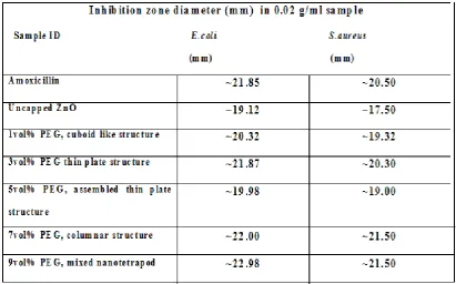
3.10. Antibacterial activity test through zone of inhibition study; Kirby-Bauer test (Cup plate method)
The antibacterial activity of uncapped ZnO nanomaterial, PEG assisted five different ZnO nanostructures with amoxicillin have been carried out for both E.coli Dh5α and S.aureus ATCC 25923. The antibacterial effect has been analyzed by the study of inhibition zone in agar plate with respect to known antibiotic Amoxicillin. The experimental result reveals the antibacterial effect on the basis of inhibition zone due to the presence of known antibiotic (amoxicillin) and different structured ZnO nanomaterial respectively. From the table it confirms that larger inhibition zone has been created by mixed ZnO nanotetrapods among the all. All tests have been repeated three times after culture incubation at 37°C overnight and the average zone diameter has been given in Table 2.
1000 2000 3000 4000 0.5 1.0 1.5 2.0 2.5 3.0 2871 509 669 573 534 501678 685 478 1678 1565 1674 1551 1674 1558 1669 1542 1638 1534 1070 1062 1046 1077 Abs o rb an ce %A
Wavenumber cm-1
1vol% 9vol% 5vol% 7vol% 3vol% 1070
OH strechning and NH2strechning
2951
Attach to E.coli
a
1000 2000 3000 4000
0.5 1.0 1.5 2.0 2.5
3.0 1678 2951
1662 1678 1669 1685 1077 1070 1070 1062 A b sorbance %A
Wavenumber cm-1
7vol% 5vol% 3vol% 9vol% 1vol% 1046 1551
OH and NH2
strechning
2862
Attach to S.aureus
b
FIG. 9. FTIR study, a) PEG assisted different structured ZnO nanomaterials attach to
E.coli bacteria, b) PEG assisted different structured ZnO nanomaterials attach to S.aureus
4. Conclusion
Here in this study it has been synthesized different structured ZnO nanomaterials by changing the volume percentage of the capping agent. These different structures of ZnO nanomaterials and their morphological details have been studied by SEM and HRTEM analysis. The hydroxyl functional groups attached to the ZnO nanomaterials has been successfully investigated by FTIR analysis. It reveals the bonding between ZnO & polymer network. This PEG encapsulation helps the attachment to the bacterial cell. The X-ray diffraction (XRD) pattern confirms the presence of single crystalline ZnO nanomaterials. No impurity occurs due to the presence of PEG. The antibacterial efficacy along with the probable mechanism study has been undertaken for the E.coli Dh5 and S.aureus ATCC 25923 bacteria. All nanomaterial reveals good antibacterial result for the both bacteria but the mixed tetrapod nanomaterial shows better result among the all. According to the TEM analysis it has been visualized directly the morphological changes resulting in the microorganisms upon contact with PEG capped ZnO nanomaterials. Cellular internalization occurs in all samples for both E.coli and S.aureus
bacteria. The images clearly reveal the presence of PEG capped ZnO nanostructures inside the bacterial cell with subsequent release of cell cytoplasm. The most dramatic changes have been occurred in mixed nanotetrapod like structure. Due to the sharp tip end the nanoterapods which are smaller in size, can easily get penetrated into the bacterial cell wall whereas the blunt tip nanotetrapods, larger in size has been agglomerated and attached on the cell surface of the bacteria.
Acknowledgments
We acknowledge the use of characterizing instruments XRD, TEM, FESEM, PL, UV-VIS and FTIR procured through UPE programme of UGC. This work was financially supported by the UGC meritorious scheme. We would like to express our special gratitude to Dr Amalesh Samanta, from Dept of Pharmaceutical Technology, Jadavpur University for supplying the bacterial strain.
References
[1] H, Cao.; J.Y, Xu.; D.Z, Zhang.; S.H, Chang.; S.T, Ho.; E.W, Seelig.; X, Liu.; R.P.H, Chang. Phys. Rev. Lett., 2000, 84, 5584. [2] Lili, Wu.; Youshi, Wu.; Wei, LU. Physica E., 2005, 28, 76–82.
[3] X. Y, Kong.; Z. L, Wang. Nano Lett., 2003, 3, 12, 1625.
[4] Yongcai, Qiu.; Wei, Chen.; Shihe, Yang. J. Mater. Chem., 2010, 20, 1001-1006.
[5] D, Mondelaers.; G, Vanhoyland.; H, Van den Rul.; J.D, Haen.; M.K, Van Bael.; J, Mullens.; L.C, Van Poucke. Mater. Res. Bull., 2002, 37, 901.
[6] M, Singhal.; V, Chhabra.; P, Kang.; D.O, Shah. Mater. Res. Bull., 1997, 32, 239. [7] T, Tsuzuki.; P.G, McCormick. Scripta Mater., 2001, 44, 1731.
[8] K, Okuyama.; I.W, Lenggoro. Chem. Eng. Sci., 2003, 58, 537.
[10] T, Sato.; T, Tanigaki.; H, Suzuki.; Y, Saito.; O, Kido.; Y, Kimura.; C, Kaito.; A, Takeda.; S, Kaneko. J. Cryst. Growth., 2003, 255, 313.
[11] R, Viswanathan.; G.D, Lilly.; W.F, Gale.; R.B, Gupta. Ind Eng Chem Res., 2003, 42, 5535. [12] Y.W, Koh.; M, Lin.; C.K, Tan.; Y.L, Foo.; K.P, Loh. J. Phys.Chem B., 2004, 108, 11419. [13] B, Liu.; H.C, Zeng. J. Am. Chem. Soc., 2003 125, 4430.
[14] W.D, Yu.; X.M, Li.; X.D, Gao. Cryst. Growth Des., 2005 5, 1, 151. [15] X.-L, Hu.; Y.-J, Zhu.; S.-W, Wang. Mater. Chem. Phys., 2004, 88, 421. [16] J.M, Wang.; L, Gao. Inorg Chem Commun., 2003, 6, 877.
[17] J.H, Kim.; W.C, Choi.; H.Y, Kim.; Y, Kang.; Y.-K, Park. Powder Technol., 2005, 153, 166. [18] O, Yamamoto. Int. J. Inorg. Mater., 2001, 3, 643.
[19] R, Brayner.; R, Ferrari-Iliou.; N, Brivois.; S, Djediat.; M.F, Benedetti.; F, Fievet. Nano Lett., 2006, 6, 866–870. [20] L, Zhang.; Y, Jiang.; Y, Ding.; M, Povey.; D, York. J. Nanopart. Res., 2007, 9, 479–89.
[21] J, Sawai . J Microbiol Methods., 2003, 54, 177–182
[22] S. Bell, Nelson .;a) David R, Tallant.; Rebecca Raymond.; Timothy, J. Boyle. J. Mater. Res., 2008, 23, 2. [23] P S, Calefi.; A O, Ribeiro.; A M, Pires.; O A, Serra. J. Alloys Compounds., 2002, 344, 285.
[24] P, Cerai.; G.D, Guerra.; M, Tricoli. Macromolecular Chemistry., 1996, 197, 3367.
[25] O. A. C, Monterio.; C, Airoldi. International Journal of Biological Macromolecules., 1999, 26, 119-128. [26] Andres, VergesM.; A, Mifsud.; C.J, Serna. J. Chem. Soc., 1990, 86, 959.
[27] Song, Rui.;* Liu, Ying.; He, Linghao. Solid State Sciences., 2008, 10, 1563-1567. [28] M, Ristic.; S, Music.; M, Ivanda.; S, Popovic. J. Alloys Compd., 2005, 397, L1. [29] S, Music.; D, Dragcevic.; S, Popovic. J. Alloys Compd., 2007, 2, 429. [30] J, Dobryszycki.; S, Biallozor. Corros. Sci., 2001, 43, 1309.
[31] W.Z, Wang.; G.H, Wang.; X.S, Wang.; Y.J, Zhan.; Y.K, Liu.; C.L, Zheng. Adv. Mater., 2002, 14, 67. [32] Z.Q, Li.; Y.J, Xiong.; Y, Xie. Inorg. Chem., 2003, 42, 8105.
[33] J.P, Richters.; T, Voss.; L, Wishmwier.; L, Ruckmann.; Gutowski. J Appl. Phys. Lett., 2008, 92, 11103. [34] Sheng.; H, Yu.; C, Wang. Appl. Microbiol. Biot., 2006, 73, 204–210.
[35] S, Lin.; G.D, Rayson. Eviron. Sci.Technol., 1998, 32, 1488.
[36] L.R, Drake.; S, Lin.; G.D, Rayson. Eviron. Sci.Technol., 1996, 30, 110.