Sleepwalking and Sleep Terrors in Prepubertal Children:
What Triggers Them?
Christian Guilleminault, MD, Biol D; Luciana Palombini, MD; Rafael Pelayo, MD; and Ronald D. Chervin, MD, MS
ABSTRACT. Objectives. To evaluate the clinical pre-sentation and polysomnography of prepubertal children with repetitive sleep terrors and sleepwalking, to com-pare them with a control group, and to evaluate the treatment of associated sleep disorders.
Methods. Patients with complaint of sleep terrors with or without sleepwalking were studied retrospec-tively. A control group was also recruited. Each subject received a standardized evaluation, which included the following: 1) Pediatric Sleep Questionnaire; 2) interview regarding child’s medical and sociofamilial history, orth-odontic history, schooling, psychological difficulties, medication intake, and family history of medical and sleep disorders; 3) general pediatric physical examination and neurologic, otolaryngological, and craniofacial exam-ination by a specialist; 4) obtaining medical history on variables relevant to early life sleep disorders; 5) poly-somnography, which included electroencephalogram (EEG; C3/A2, Fp1/T1, T1/O1, O1/C3, C4/A1, Fp2/T2, T2/ O2, O2/C4), chin and leg electromyelogram, right and left electro-oculogram, and electrocardiogram (modified V2 lead); respiration was monitored with a nasal cannula/ pressure transducer system, mouth thermistor, chest and abdominal bands, pulse oximeter, and neck microphone; respiratory effort was monitored with calibrated esoph-ageal manometry; variables were collected on a comput-erized sleep system; and 6) available family members with a positive history of sleep terrors and sleepwalking received clinical evaluations similar to those used for index cases; they also underwent ambulatory monitoring with an Edentrace system, which monitors heart rate, body position, oro-nasal flow, chest impedance, breath-ing noises (neck microphone), and pulse oximetry. Move-ments are deduced from artifact, and leg moveMove-ments may be recorded on one channel if the equipment is preset for such recording. Subjects used logs to record “lights out” time, “lights on” time, nocturnal awakenings, and other events that occurred during the night. All original and follow-up recordings were rescored by 2 of 4 randomly selected specialists who were blind to subject identity. Mann-Whitney Utest was used for group comparison. Nonparametric2test was used to compare percentages of symptoms in symptomatic children versus control children.
Results. Eighty-four children (5 with sleep terrors and 79 with both sleep terrors and sleepwalking) and 36 normal control children formed the studied population. All subjects were Tanner stage 1 (prepubertal). None of
the control children had any parasomnias. Fifty-one (61%) of 84 children with parasomnia had a diagnosis of an additional sleep disorder: 49 with sleep-disordered breathing (SDB) and 2 with restless leg syndrome (RLS). Twenty-nine of the children with both parasomnia and SDB had a positive family history of parasomnias, and 24 of the 29 also had a positive family history of SDB. Of the 51 children with associated sleep disorders, 45 were treated. Forty-three of 49 children with SDB were treated with tonsillectomy, adenoidectomy, and/or turbinate re-vision, and 2 of 2 children with RLS were treated with Pramipexole, a dopamine agonist, at bedtime. Treatment of the precipitating sleep disorder eliminated parasom-nias in all 45 children. In all 43 children who received surgery, polysomnography performed 3 to 4 months later indicated the disappearance of SDB. The recordings also showed an absence of confusional arousals. The number of EEG arousals significantly decreased from a mean of 9ⴞ2.6 EEG arousals>3 seconds/hour during total sleep time to 3ⴞ1.5. The number of EEG arousals>3 seconds during the first sleep cycle of slow wave sleep (stage 3– 4 non–rapid eye movement sleep) decreased from 4ⴞ1.4 to 1ⴞ 0.2. In all surgically treated cases, parents also re-ported subsequent absence of the parasomnia. The 2 symptomatic children who were treated with Pramipexole had a complete absence of confusional arousals on the follow-up recording and reported no parasomnia since treatment. The periodic limb move-ment syndrome arousal index (number of EEG arousals associated with periodic limb movement/hour) decreased from 11 and 16 to 0 and 0.2, respectively. Parasomnia persisted in the 6 children who were untreated for SDB. Surgeons had refused to perform surgery on these chil-dren because of lack of data on the relationship between parasomnia and SDB-related tonsil and adenoid enlarge-ment.
Conclusion. Children with chronic parasomnias may often also present SDB or, to a lesser extent, RLS. Fur-thermore, the disappearance of the parasomnias after the treatment of the SDB or RLS periodic limb movement syndrome suggests that the latter may trigger the former. The high frequency of SDB in family members of chil-dren with parasomnia provided additional evidence that SDB may manifest as parasomnias in children. Children with parasomnias are not systematically monitored dur-ing sleep, although past studies have suggested that pa-tients with sleep terrors or sleepwalking have an ele-vated level of brief EEG arousals. When children receive polysomnographies, discrete patterns (eg, nasal flow lim-itation, abnormal respiratory effort, bursts of highor slow␣ EEG frequencies) should be sought; apneas are rarely found in children. Children’s respiration during sleep should be monitored with nasal cannula/pressure transducer system and/or esophageal manometry, which are more sensitive than the thermistors or thermocouples From the Stanford University Sleep Disorders Clinic, Stanford, California.
Received for publication Jun 17, 2002; accepted Sep 11, 2002.
currently used in many laboratories. The clear, prompt improvement of severe parasomnia in children who are treated for SDB, as defined here, provides important evidence that subtle SDB can have substantial health-related significance. Also noteworthy is the report of familial presence of parasomnia. Studies of twin cohorts and families with sleep terror and sleepwalking suggest genetic involvement of parasomnias. RLS and SDB have been shown to have familial recurrence. RLS has been shown to have genetic involvement. It remains to be investigated whether a genetic factor directly influences sleep terror and sleepwalking or instead influences other disorders that fragment sleep and lead to confusional arousals. Additional studies are needed to investigate the association between SDB and non–rapid eye movement parasomnias in the general population. Pediatrics
2003;111:e17–e25. URL: http://www.pediatrics.org/cgi/ content/full/111/1/e17; sleepwalking, sleep terrors, sleep disordered breathing, restless leg syndrome, periodic limb movement syndrome, familial aggregation, prepubertal.
ABBREVIATIONS. NREM, non–rapid eye movement; SWS, slow wave sleep; EEG, electroencephalogram; SDB, sleep-disordered breathing; RLS, restless leg syndrome; AHI, apnea-hypopnea in-dex; RDI, respiratory disturbance inin-dex; PLMS, periodic limb movement syndrome.
S
leep terrors and sleepwalking, 2 common child-hood parasomnias, are arousal disorders that arise from deep non–rapid eye movement (NREM) sleep. In a landmark study, Klackenberg1,2followed for 20 years⬎200 children who were born in Stockholm and documented these parasomnias and their pattern of occurrence and potential associ-ation with psychopathology. He described the evo-lution, in some cases, of atypical, rare nocturnal be-haviors into clinical syndromes in need of treatment: sleepwalking can lead to self-inflicted injuries or, in teenagers, involuntary aggression toward others.
Sleep terrors and sleepwalking are states of confu-sion and partial arousal that emerge during the first third of the night when children exit slow wave sleep (SWS; ie, stages 3 and 4 of NREM sleep). Patients rarely remember the events in detail, but if actively probed after 4 years of age, they often report vague memories of having to act—run away, escape, or defend themselves—against monsters, animals, snakes, spiders, ants, intruders, or other threats. Children may report feeling complete isolation and fear. Parents often describe terrified facial expres-sions, mumbling, shouting, and inability to be con-soled.
Despite widespread prevalence of these disorders and the recognition that they may arise from incom-plete arousal, their pathophysiology is not well un-derstood. Recent polysomnographic recordings of these events have shown that they are associated with 2 abnormalities during the first sleep cycle: abnormally low ⌬ electroencephalogram (EEG) power and frequent, brief, nonbehavioral EEG-de-fined arousals.3,4 One study also showed that most
abnormal behaviors were preceded by a short-lived increase in⌬EEG frequency, a pattern that can also reflect physiologic activation.5 However, none of
these studies of sleep terrors and sleepwalking has
identified a cause for frequent arousals or decreased
⌬EEG power in the first sleep cycle.
To explore what might precipitate sleep terrors and sleepwalking in children, we performed a retro-spective analysis of clinical and polysomnographic data from 84 prepubertal children, aged 2 and 11 years, who were referred for these behavioral prob-lems during sleep. We compared their findings with those of 36 normal children who were recruited from the community.
METHODS Subject Recruitment
All parents of the children who were seen and monitored in the sleep clinic were asked to sign a consent form approved by the institution for use of clinical data and polysomnographic record-ings for research purposes. Parents of community children also signed an informed consent to participate in this research.
Inclusion Criteria
Eighty-four children (39 girls) met the following inclusion cri-teria: 1) diagnosis of sleep terrors or sleepwalking; 2) availability of complete chart and nocturnal polysomnographic data from a minimum of 8.5 hours of recording; 3) sleep study performed within the past 4 years; 4) clinical follow-up obtained for at least 12 months, and 5) if treated, follow-up nocturnal polysomnogra-phy had been obtained, at a mean of 3 months after treatment. All children who were seen consecutively and met the above criteria were included.
Thirty-six prepubertal children (19 girls) with parental report of at least 8.5 hours of nightly sleep, absence of known daytime consequences of sleep disorders (eg, daytime sleepiness, cata-plexy, hyperactivity, morning headache, mouth breathing), and normal health as reported by physicians over systematic visits were recruited from the community by advertisement to serve as control subjects for specific research protocols. Parents signed informed consents for each protocol. These children also had complete charts and underwent similar 8.5-hour polysomno-graphic recordings.
Protocol
Each child underwent the same standardized evaluation pro-tocol.
1. Parents completed a pediatric sleep questionnaire created in the late 1980s and derived from the questionnaire by Brouillette et al.6
2. Parents and children were interviewed about the children’s medical and sociofamilial history, orthodontic history, schooling, psychological difficulties, medication intake, and family history of medical and sleep disorders. Each category of responses was collected on a standardized form.
3. The general pediatric evaluation included a neurologic, oto-laryngological, and craniofacial examination by a specialist. Spe-cific findings were categorized as present or absent (eg, enlarged inferior turbinate7,8) or graded according to common convention
(eg, tonsil size, 0 for absent to 4⫹for “kissing”).
4. Medical histories included systematic assessment of variables relevant to early life sleep disorders, such as allergies and asthma. Family histories included inquiries about parasomnias and other sleep disorders, and affected relatives were examined when avail-able.
5. Polysomnography included EEG (C3/A2, Fp1/T1, T1/O1, O1/C3, C4/A1, Fp2/T2, T2/O2, O2/C4), chin and leg electromy-elogram, right and left electro-oculogram, and electrocardiogram (modified V2 lead). Respiration was monitored with a nasal can-nula/pressure transducer system (initially using a Medex [Dublin, OH] pressure transducer, thereafter Protec [Woodinville, WA]), mouth thermistor, chest and abdominal bands, pulse oximeter, and neck microphone. Respiratory effort was monitored with calibrated esophageal manometry. Variables were collected on a computerized sleep system (Sandman, Nellcor Puritan Bennett [Melville] Ltd, Ottawa, Ontario, Canada).
were sent to otolaryngologists for surgical therapy assessments, and patients with restless leg syndrome (RLS) were treated phar-macologically. The same variables as those monitored at baseline were monitored in a follow-up polysomnogram.
For the purposes of the current research, all recordings were rescored by 4 knowledgeable scorers masked to subject identity and status. A randomly chosen primary scorer identified EEG arousals, awakenings, and respiratory events on the basis of pre-defined criteria. A second scorer reviewed this initial scoring. When there was a discrepancy for any event, a third scorer re-viewed the questionable event and the majority score was used. A similar masked approach was used for the scoring of the post-treatment polysomnograms. A preestablished cutoff point of an apnea-hypopnea index (AHI) of 1 or more and a respiratory disturbance index (RDI; defined as index of apnea, hypopnea, event with flow limitation, abnormal breathing efforts, and seg-ment of tachypnea) of 2 or more event per hour of sleep had been previously established as abnormal in our center on the basis of clinical data.
Available family members with a positive history of sleep terrors and sleepwalking received clinical evaluations similar to those used for index cases. They also underwent ambulatory monitoring with an Edentrace system (Nellcor Puritan Bennett [Melville] Ltd, Ottawa, Ontario, Canada), which monitors heart rate, body position, oro-nasal flow, chest impedance, breathing noises (neck microphone), and pulse-oximetry. Movements are deduced from artifact, and leg movements may be recorded on 1 channel if the equipment is preset for such recording.9Subjects
used logs to record “lights out” time, “lights on” time, nocturnal awakenings, and other events that occurred during the night. Presence of arousals and their distribution and repetitiveness and presence of apnea, hypopnea, and oxygen saturation drops were scored from this ambulatory recording.
Polysomnographic Definitions
Before scoring, the definitions presented in Table 1 were estab-lished on the basis of previous clinical information.
Statistical Analysis
Mann-WhitneyUtest was used for group comparison. Percent-ages were compared using the nonparametric2test.
RESULTS Subjects
The 84 symptomatic children had a mean age of 6.85 ⫾ 3.6 years (range: 2.1–11.1 years). Their body mass index was between the 52nd and 83rd percen-tiles for boys and the 56th and 78th percentile for girls, based on age.19None of them was overweight.
There were 39 girls with a mean age of 6.7 ⫾ 3.0 (range: 2.4 –10.8) and 45 boys with a mean age of 7.1⫾3.6 (range: 2.1–11.1). Five children (mean age: 3.28), including 2 girls, had reports of sleep terrors only. All other children reported both sleep terrors and sleepwalking. Sleep terrors preceded sleepwalk-ing, occurred between episodes of sleepwalksleepwalk-ing, or occurred simultaneously with sleepwalking. The 36 control children had a mean age of 7.35⫾3.5 (range: 2.3–10.9) years. There were 19 control girls with a mean age of 6.7 ⫾ 3.1 (range: 2.6 –9.7) and 17 boys with a mean age of 8 ⫾ 2.9 (range: 2.3–10.9). The symptomatic and control children groups were not significantly different in age or gender, but the con-trol group contained a higher proportion of girls (52.8%) than did the symptomatic group (46.2%;P⫽
.52 [not significant]). All subjects were Tanner stage 1 (prepubertal20). None of the controls had any
para-somnias.
Pediatric Questionnaire
This questionnaire indicated an absence of the fol-lowing in the symptomatic children: narcolepsy symptoms, abnormal daytime napping, and chronic drug intake. It also confirmed the presence of fre-quent parasomnia events in the last 6 months.
TABLE 1. Polysomnographic Definitions of SDB
Term Definition
Apnea Absence of airflow at nose and mouth for longer than 2 breaths, independent of desaturation or change in EEG.
Hypopnea Reduction by at least 50% in nasal flow signal amplitude10,11for a minimum of 2
breaths. Often but not always associated with snoring.
Abnormal respiratory effort Reduction in nasal flow of⬍50% with flattening of nasal cannula signal10,11and
decrease in the mouth signal (thermistor). Often seen with snoring and increased effort shown on Pes signal defined as
⫹Pes Crescendo12 Sequence of 4 or more breaths that show increasingly negative peak end
inspiratory pressure. May be seen with flow limitation.
⫹Continuous sustained effort13 Discrete flow limitation on nasal cannula/pressure transducer signal, with
“flattening” of the breath signal curve for at least 4 successive breaths. Repetitive abnormally negative peak end inspiratory pressures, ending at same negative inspiratory pressure without a crescendo pattern.
Pes reversal12 Termination of abnormal increase in respiratory effort with abrupt switch to a
less negative peak end inspiratory pressure. Respiratory event related
arousals (RERAs)
As defined by the American Academy of Sleep Medicine14
Tachypnea Increase in respiratory rate, above that seen during quiet unobstructed breathing, by minimum of 3 breaths/minute in NREM sleep or 4 breaths/minute in REM sleep, for 30 seconds or more. No changes in oxygen saturation, Pes, or EEG were required.
Arousals and other EEG changes Arousals defined according to the American Sleep Disorders Association Atlas.15
In addition, a breathing event may be associated with an abrupt burst of high-amplitude slow waves in the slowor fast␦range (2.5–4.5 Hz), not lasting more than 5 seconds. These high-amplitude slow waves are similar to the EEG burst described in part of the phase 1 ofCyclical Alternating Pattern16or by
Black et al17in patients with upper airway resistance syndrome.
Sleep/wake scoring Same as in Rechtschaffen and Kales international manual18
History of the Parasomnias in the Symptomatic Children
All children with parasomnia had recurrent and chronic sleep terror/sleepwalking, occurring for anywhere between 4 months and 7.4 years. The re-ported frequency of events was at least once per week for ages 2 to 4 years, once per 15 days for ages 4 to 7 years, and more variable for ages 7 years and up. Events typically recurred in bursts of several successive nights followed by up to several weeks without any events. A recurrence of the problems within the past 2 weeks preceded most consultations.
Sleep Terror/Sleepwalking
Table 2 presents the clinical symptoms and the positive health history of the children. A total of 49 (58.3%) of 84 patients presented with symptoms sug-gestive of SDB. An additional 2 presented with symptoms of leg movements during the day or dur-ing sleep, caused by RLS (n ⫽1) and RLS/periodic limb movement syndrome (PLMS; n⫽ 1). Pediatri-cians did not suspect depression in any of the chil-dren; none of them was referred to a psychiatrist. Fourteen symptomatic children had current school difficulties, including difficulty arising in the morn-ing (n ⫽ 6), report of inattention/hyperactivity in class (n ⫽ 4), and a decrease in or poor long-term school performance (n⫽ 4).
As for family history (Table 3), 29 children had a positive history of sleep terror and sleepwalking. Eight subjects had a positive history in 1 parent and 1 sibling, 12 subjects had a positive history in 1
parent, and 9 subjects had a positive history in 1 (8 children) or 2 siblings. The number of family mem-bers who reported having a positive history of sleep-walking/sleep terror was 38; all of them were avail-able for clinical evaluations.
When clinical evaluation and family history were combined, there was a positive family history of RLS/PLMS in 2 families (1 subject had both a parent and a sibling and the other had only 1 parent with positive history of RLS/PLMS). Among the 38 family members with a positive history for sleep terrors or sleepwalking, 24 had positive findings for SDB, such as history of regular snoring, history of nocturnal asthma, and presence of signs or symptoms related to SDB.21Family members reported some of the
fol-lowing symptoms or presented some of the follow-ing signs: daytime fatigue or daytime sleepiness, morning headache, regular nocturnal sweating, agi-tated and disrupted sleep, mouth breathing with or without nocturnal fluid intake, bruxism, history of wisdom teeth extraction early in life, enlarged infe-rior nasal turbinates, septum deviation, enlargement of soft palatal tissue, enlarged tonsils, high and nar-row palate, and a score of⬎70 on the morphometric model for obstructive sleep apnea.21The presence of
both signs and symptoms was necessary to be con-sidered positive for the findings. SDB was also found in family members of subjects who had SDB and sleepwalking and no history of sleepwalking or sleep terror themselves. Among the 29 symptomatic chil-TABLE 2. Clinical Symptoms in Studied Children
Clinical Symptom Symptomatic
Children N(%)
Control Children
N(%)
2
PValue
Noisy breathing/habitual snoring 51 (60.7) 0 (0.0) ⬍.00001
History of repetitive earaches 36 (42.8) 1 (2.8) ⬍.0001
History of repetitive upper airway infection 28 (33.3) 3 (8.3) ⬍.004
Nocturnal asthma 5 (6.0) 0 (0.0) ⬍.13 (NS)
Respiratory allergies 19 (22.6) 2 (5.6) ⬍.024
Gastroesophageal reflux 2 (2.4) 0 (0.0) ⬍.35 (NS)
Enuresis 3 (3.6) 0 (0.0) ⬍.25 (NS)
Restless sleep 61 (72.6) 0 (0.0) ⬍.00001
Abnormal nocturnal sleep 32 (38.1) 0 (0.0) ⬍.00002
Morning sleepiness/daytime fatigue 24 (28.6) 0 (0.0) ⬍.00034
School difficulties 14 (16.6) 1 (2.8) ⬍.035
Restless leg/leg movement during sleep 2 (2.4) 0 (0.0) ⬍.35 (NS)
Mouth breathing noted by parents 28 (33.3) 1 (2.8) ⬍.00034
Bruxism 14 (16.6) 0 (0.0) ⬍.0092
Orthodontic consultation 10 (11.9) 3 (8.3) ⬍.56 (NS)
NS indicates not significant.
TABLE 3. Data on Positive Family History
Clinical Symptom No. of Siblings
No. of Parents
Sleep terror/sleepwalking 18 (2*) 20
Regular snoring 29 36
Asthma 6 9
Wisdom tooth extraction (early in life) – 27
Bruxism 7 13
Orthodontic consultation 10 3
RLS/PLMS 1 2
* Number of siblings with a history ofisolatedsleep terrors.
TABLE 4. ENT and Maxillomandibular Clinical Evaluation
No. of Symptomatic Children
No. of Control Children
Tonsils
0⫺2⫹ 36 35
3⫹ 28 1
4⫹ 20 0
High, narrow hard palate 12 0
Overjet⬍4 mm* 4 0
Orthodontic treatment 10 3
Elongated uvula 11 1
Enlarged inferior turbinate 27 2
dren with family histories positive for these para-somnias, 24 (83%) also had a positive family history of SDB. Among the 55 symptomatic children without family histories of parasomnias, 14 (25%) nonetheless had family histories suggestive of SDB.
Clinical and Ears, Nose, and Throat Evaluations None of the children was obese or had growth problems.19The sleep evaluations that included
neu-rologic and psychiatric clinical assessments per-formed by board-certified specialists were benign. In particular, they revealed no evidence of complex partial seizure or migraines, major anxiety, or de-pressive disorders. Seventeen children reported fre-quent nightmares. The need to escape, fight, or avoid frightening situations were themes of dreams and nightmares in 21 children aged 7 to 11 years. The results of tonsil evaluations are presented in Table 4. Inferior turbinates7,8 and/or tonsils were scored as
clearly enlarged in 58 children, and only 3 were in the control group.
Baseline Polysomnography
All symptomatic children presented evidence of nocturnal sleep disruption during polysomnogra-phy. The events were, at minimum, confusional arousal with sitting up in bed, moving in bed, or sleep talking with coherent or incoherent sentences (n ⫽ 40). Recordings showed sleep terror (n ⫽ 12) and confusional arousal with attempts at walking out of bed with varying degrees of aggression (n⫽
32). All events were seen in the first third of the night, during SWS (stage 3 or 4 NREM). Subjects returned to sleep quickly and had amnesia of the event in the morning. The longest monitored confu-sional arousal lasted 4 minutes and 38 seconds; the shortest lasted 47 seconds.
Polysomnography indicated presence of periodic leg movements in 2 children (PLMS index: 11 and 16) and SDB in 49 children. The children with SDB were subdivided on the basis of their polysomnographic findings. Twenty-three children had tachypnea and flow limitation at nasal cannula, with Pes Crescendo and sustained breathing effort sequences. No apneic events were noted in their recordings; AHI (related to presence of hypopnea) was 0.7 ⫾ 0.5 events/ hour,22,23 but RDI was calculated to be 4.8 ⫾ 1.2
events/hour. In 26 cases, obstructive hypopneas and rare apneas were noted: the mean AHI was 1.6⫾0.6 events/hour, and the RDI was 6⫾1.8 events/hour. The 49 children with SDB/parasomnias had a mean RDI of 5.43⫾1.5. The mean number of EEG arousals per hour of sleep was 9⫾2.6.
Polysomnography of the control group indicated a mean AHI of 0.3⫾0.07 and a mean RDI of 0.5⫾0.3 events/hour. The mean number of EEG arousals⬎3 seconds per hour of sleep was 2.7⫾ 1.9 events per hour (P ⫽ .0001). Both AHI and RDI were signifi-cantly different (P⫽.05 andP⫽.0001, respectively) between the symptomatic children with SDB and control groups. None of the children presented with severe SDB, and oxygen saturation never fell below 94% from a baseline of 99% (Figs 1 and 2).
In summary, at entry, 49 children presented with a
history, clinical evaluation, and polysomnographic recording of SDB, and 24 of these children came from families in which at least 1 other family member had both positive family history of parasomnia and SDB. Two additional children had a history and polysom-nogram indicative of RLS/PLMS. Thus, the sleep terror or sleepwalking of 51 (61%) of 84 children occurred in association with other primary sleep dis-orders that potentially could trigger the parasom-nias.
Follow-up
All children with recognized SDB were sent to a local pediatric otolaryngologist for treatment. Tonsil-lectomy with or without adenoidectomy and with or without turbinate treatment was performed in 43 children. The 2 children whose diagnosis was PLMS and RLS were prescribed a 0.125-mg (1 child) and 0.25-mg (other child) dosage of Pramipexole, a do-pamine agonist, at bedtime. Ears, nose, and throat surgeons declined to perform surgery on 6 subjects because of a lack of data on the relationship between parasomnia and SDB-related tonsil and adenoid en-largement.
Thirty-three children with neither of these 2 health problems were considered to have “primary para-somnia.” All parents of sleepwalkers were provided with instructions on how to avoid accidents associ-ated with sleepwalking and sleep hygiene recom-mendations. A pediatric psychologist saw parents and children to evaluate possible conflictual situa-tions that may have been associated with the para-somnias. She sought factors that could increase the risk of sleepwalking or sleep terror and explored the parasomnia’s effect on familial interaction. After this evaluation, family counseling was recommended for 6 children.
No medication for parasomnia was prescribed in primary parasomnia, but parents were asked to keep sleep logs of parasomnia events. Sleep clinic follow-ups were scheduled approximately every 2 months for the following 6 months. All children who under-went surgical intervention for SDB had a follow-up nocturnal polysomnography between 12 and 18 weeks after the surgery. Children who were receiv-ing Pramipexole had a follow-up monitorreceiv-ing 5 to 12 weeks after the start of the treatment.
Six to 7 months after the initial visit, reevaluation included the same clinical workup as at entry, review of the sleep logs obtained at the previous follow-up visits, and review of the nocturnal polysomnograms. Children with SDB at entry but without otolaryngo-logical treatment also had a new polysomnogram at that time. This assessment reviewed initial com-plaints and several specific features, such as Tanner stage, craniofacial development, and other sleep symptoms.
Comparison of Polysomnograms Before and After Surgery
9⫾2.6 EEG arousalsⱖ3 seconds/hour during total sleep time to 3⫾1.5 (P⫽.0001). The number of EEG arousals ⱖ3 seconds during the first sleep cycle of SWS (stage 3– 4 NREM sleep) went from 4 ⫾ 1.4 to 1 ⫾ 0.2 (P ⫽ .01). In all surgically treated cases, parents and sleep logs also indicated absence of the parasomnia.
Comparison of Initial Versus 6-Month Follow-up Polysomnogram in Untreated Patients With SDB
There was persistence of abnormal SDB without significant RDI changes in 6 children. The mean arousal index was 8.3⫾3 (not significant). There was persistence of at least 1 confusional arousal in each child during the recording night. (Sleep log indicated persistence of parasomnia at a comparable frequency and severity.)
Comparison of Children Before and After Pramipexole Treatment
The 2 children who were treated with Pramipexole had a complete cessation of confusional arousal and report of parasomnia at the time of follow-up record-ing. The PLMS arousal index (number of EEG
arous-als associated with periodic limb movement/hour) went from 11 and 16 to 0 and 0.2, respectively.
Summary
In summary, at the 6-month follow-up (mean: 15 weeks; range: 13–19 weeks after surgery), none of the children who were treated for their other sleep dis-order presented with sleepwalking or night terror. Five of the children had progressed from Tanner stage 1 to stage 2. The children who were treated for RLS/PLMS had mean treatment duration of 22 weeks and a complete disappearance of parasomnia (1 child was in Tanner stage 2). The 6 children with untreated SDB and sleepwalking were unchanged. Among the 33 children without evidence of SDB or RLS/PLMS at entry, parents of 24 wanted to con-sider drug treatment for parasomnias that had per-sisted with unchanged frequency. Of the 9 children who did not receive drug treatment, 1 child was in Tanner stage 220and was reported by parents to have
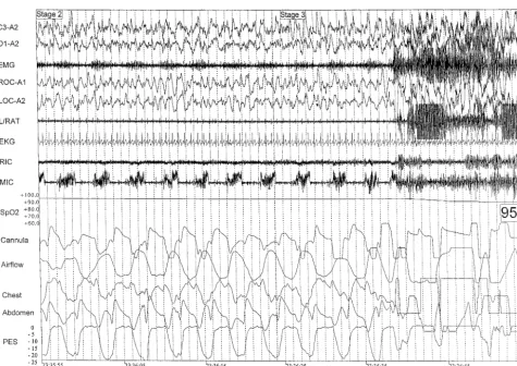
had no parasomnia for the last 13 weeks. Two young children switched from having sleep terrors to con-fusional arousals and sleepwalking; their parents re-ported a clear decrease in the frequency of parasom-Fig 1. Polysomnography of onset of sleepwalking in an 8-year-old child. On the right side of figure are the movement artifacts associated with the arousal and beginning of the parasomnias. The preceding recording segment (left side of figure) shows the abnormal breathing that occurred during SWS. The abnormal breathing can be seen on the nasal cannula/pressure transducer recording (Cannula). There is a flow limitation and very negative peak end inspiratory esophageal pressure (Pes) that resolve with the beginning of the arousal with a reversal of the abnormal respiratory effort (PES signal) and start of the sleepwalking. The patient snores continuously (microphone [MIC] channel) and is a mouth breather with a very good signal obtained from the mouth thermistor (Airflow channel). The child is in stage 3 NREM sleep just before the onset of the event. The drop in the pulse oximeter channel (Spo2) on the right of the figure is related
nia. Three of the 6 children for whom family counseling had been recommended had a disappear-ance of symptoms for 6, 10, and 14 weeks, respec-tively; a great reduction in frequency was reported in the fourth child, and no change occurred in the last 2.
DISCUSSION
This study of 84 children who were referred for repetitive sleep terrors or sleepwalking and 36 nor-mal children shows that other primary sleep disor-ders—SDB and, to a lesser extent, RLS/PLMS—may be comorbidities in these parasomnias when they are chronic. Furthermore, consistent resolution of these parasomnias after treatment for the underlying pri-mary sleep disorders suggests that in prepubertal children, SDB or RLS/PLMS may trigger or cause sleep terrors and sleepwalking. Among studied chil-dren with sleep terrors and sleepwalking, the high frequency of family members with SDB and, to a lesser extent, RLS/PLMS (disorders with strong fa-milial components) provides additional evidence that sleep disorders that are known to trigger arous-als could manifest as these parasomnias in some children. These findings provide new insight into the pathophysiology of certain NREM parasomnias and
have implications for clinical practice, where most children with parasomnias are not evaluated for any other underlying sleep disorders.
Sleep terror and sleepwalking episodes are dis-turbing to parents. Depending on the degree of con-fusion, bedroom location, furniture, and strength of the subject, sleepwalking may lead to accidents and self-injury. As shown by Klackenberg,1,2up to 50% of
children may experience 1 event during childhood, and these children, with a rare or isolated event, are not among those reported here. Our patient popula-tion is biased toward more severe, recurrent symp-toms that disturb family life. Our study provides little information on the frequency of SDB or PLMS/ RLS in patients who have parasomnia and are seen at general pediatrics clinics. However, one third of our patients was self-referred: the recurrent parasomnia was disturbing the family life, and the associated sleep disorder was discovered at the sleep clinic. SDB and RLS were unsuspected by parents. In view of the frequency with which SDB was found in our pa-tients, questions about signs and symptoms of SDB and RLS may be important in clinical practice when recurring sleep terrors or sleepwalking is reported.
Although past studies suggested that brief EEG Fig 2. Onset of sleep terror in a 3-year-old boy. The child is in stage 4 NREM sleep with high amplitude slow waves. On the right of the figure, movement artifacts begin. The chin electromyelogram changes abruptly. Before the start of the event, the child presents flow limitation seen on the nasal cannula, with a “flattening” at the top of the “Cannula” signal. Esophageal pressure signal (Pes), indicative of respiratory effort, is abnormally negative at end inspiration, reaching here 20 cm H2O. With the beginning of the confusional arousal,
arousals are increased and early night ⌬power de-creased, in patients with sleep terrors or sleepwalk-ing,3,4 several reasons could explain why SDB and
RLS/PLMS have not been implicated previously as a common cause of these changes in sleep architecture. Children with parasomnias, even if recurrent, are not systematically monitored during sleep. Breathing events during sleep in children rarely are apneas, and one must look for more discrete patterns. Arous-als ⬍15 seconds in duration are not systematically tabulated, are difficult to recognize visually, and are not mentioned in any sleep scoring atlases published before 1992. However, these short-lived EEG distur-bances, indicated by bursts of highor slow␣EEG frequencies in the central EEG leads (depending on the age of the child), should have underlying causes. Our study shows that at least 2 can be identified: SDB and RLS/PLMS. The possibility remains that we missed other causes of sleep fragmentation in some children. We did not perform, for example, esopha-geal pH measurement during sleep, although we had no history to suggest esophageal reflux.
Two other points deserve emphasis. One impor-tant aspect of childhood SDB is that obstructive sleep apnea is an uncommon feature in polysomnography: nasal flow limitation, abnormal respiratory effort, and bursts of tachypnea during sleep are more fre-quently noted. For facilitating recognition of these patterns, children’s respiration during sleep should be monitored with equipment such as nasal cannu-la/pressure transducer systems10,11 or esophageal
manometry, which are more sensitive than the ther-mistors or thermocouples currently used in many laboratories. One common limitation in the interpre-tation of pediatric sleep studies is the lack of suffi-cient data that link health-related outcomes with spe-cific polysomnographic findings. Previous authors have pointed out that breathing abnormalities more subtle than those commonly found in adult SDB may have significance in children,22 but these assertions
most often have been based on the rarity of overt, adult-defined apneic events in normal children.23
The current study used highly sensitive equipment, liberal definitions of apneic events, and inclusive definitions of SDB. Without outcome data, the high frequency of SDB in our sample (58%) might have been considered inflated, and clinical relevance would have been questionable. However, the clear, prompt improvement of severe parasomnias in chil-dren who were treated for SDB—as currently de-fined—provides important outcome-based evidence that SDB that is more subtle than that commonly recognized to be abnormal can have substantial health-related significance.
Also noteworthy is the report of a familial pres-ence of parasomnia. The investigation of twin co-horts and families with sleep terror and sleepwalk-ing has led to the suggestion of a genetic factor in parasomnias.24,25 The RLS has been shown to have
familial recurrence and genetic involvement, partic-ularly in early-onset cases. Familial aggregation also has been demonstrated in SDB. Thus, the question raised is whether a genetic factor directly influences sleep terror and sleepwalking or instead influences
other disorders that fragment sleep and lead to con-fusional arousals. Among our patients, 2 individuals had a positive family history of RLS. The ambulatory Edentrace unit with which we tested relatives is not the state of the art for recognizing mild SDB. How-ever, we can affirm that chronic snoring and some symptoms and signs of SDB were present in siblings and parents of patients who were reported to have had sleepwalking and sleep terrors. Additional stud-ies are needed to address the association between SDB and these NREM parasomnias in the general population.
ACKNOWLEDGMENT
Christian Guilleminault is the recipient of an Academic Award from the Sleep Disorders Center from the National Heart, Lung, and Blood Institute from the National Institutes of Health.
REFERENCES
1. Klackenberg G. A prospective longitudinal study of children. Acta Paediatr Scand.1971;224(suppl):1–239
2. Klackenberg G. Somnambulism in childhood: prevalence, course and behavioral correlation.Acta Paediatr Scand.1982;71:495– 499
3. Gaudreau H, Joncas S, Zadra A, Montplaisir J. Dynamics of slow-wave activity during NREM sleep of sleepwalkers and control subjects.Sleep. 2000;23:755–760
4. Guilleminault C, Poyares D, Abat F, Palombini L. Sleep and wakeful-ness in somnambulism, a spectral analysis study. J Psychosom Res. 2001;51:411– 416
5. De Gennaro L, Ferrara M, Bertini M. The spontaneous K-complex during stage 2 sleep: is it the ‘‘forerunner’ of delta waves?Neurosci Lett. 2000;291:41– 43
6. Brouillette RT, Hanson D, David R, et al. A diagnostic approach to suspected obstructive sleep apnea in children.J Pediatr.1984;105:10 –14 7. Wood RP II, Jafek BW, Eberhard R. Nasal obstruction. In: Bailey BJ, ed. Head and Neck Surgery-Otolaryngology.Philadelphia, PA: JB Lippincott Company; 1993:309
8. Gluckman JL. Nonallergic rhinitis. In: Donald PJ, Gluckman JL, Rice DH, eds.The Sinuses.New York, NY: Raven Press Ltd; 1995:151 9. Labanowski M, Schmidt-Nowara W, Guilleminault C. Sleep and
muscular disease: frequency of sleep disordered breathing in a neuro-muscular disease clinic population.Neurology.1996;47:1173–1180 10. Hosselet JJ, Norman RG, Ayappa I, Rapoport D. Detection of flow
limitation with nasal cannula/pressure transducer system.Am J Respir Crit Care Med.1998;157:1461–1467
11. Ayap I, Norman RG, Krieger AC, Rosen A, O’Malley R, Rapoport D. Noninvasive detection of respiratory effort-related arousals (RERAs) by a nasal cannula/pressure transducer system.Sleep.2000;23:763–771 12. Guilleminault C, Kim YD, Stoohs RA. Upper airway resistance
syn-drome.Oral Maxilofac Surg Clin North Am.1995;7:243–246
13. Guilleminault C, Poyares D, Palombini L, Koester U, Pelin Z, Black J. Variability of respiratory effort in relation to sleep stages in normal controls and upper airway resistance syndrome patients. Sleep Med. 2001;2:397– 406
14. American Academy of Sleep Medicine Task Force: Sleep related breath-ing disorders in adults: recommendations for syndrome definition and measurements techniques in clinical research.Sleep.1999;22:667– 689 15. American Sleep Disorders Association Atlas Task Force: EEG arousals:
scoring rules and examples.Sleep.1992;15:173–184
16. Terzano MG, Parrino L, Chervin R, et al. Atlas, rules and recording techniques for the scoring of the cyclical alternating pattern-CAP-in human sleep.Sleep Med.2001;2:537–554
17. Black J, Guilleminault C, Colrain I, Carillo O. Upper airway resistance syndrome: central EEG power and changes in breathing effort.Am J Respir Crit Care Med.2000;162:406 – 411
18. Rechtschaffen A, Kales A.A Manual of Standardized Terminology: Tech-niques and Scoring Systems for Sleep Stages of Human Subjects. Los Ange-les, CA: UCLA Brain Information Service/Brain Research Institute; 1968 19. Hammer LD, Kraemer HC, Wilson DM, Ritter PL, Dornbusch SM. Standardized percentiles curves of body mass index for children and adolescents.Am J Dis Child.1991;145:259 –263
22. Carroll JL, Loughlin GM. Diagnostic criteria for obstructive sleep apnea syndrome in children.Pediatr Pulmonol.1992;14:71–74
23. Marcus CL, Omlin KJ, Basinski DJ, et al. Normal polysomnographic values for children and adolescents. Am Rev Respir Dis. 1992;146: 1235–1239
24. Kales A, Soldatos CR, Bixler EO, et al. Hereditary factors in sleepwalk-ing and night terrors.Br J Psychiatry.1980;137:111–118
DOI: 10.1542/peds.111.1.e17
2003;111;e17
Pediatrics
Christian Guilleminault, Luciana Palombini, Rafael Pelayo and Ronald D. Chervin
Sleepwalking and Sleep Terrors in Prepubertal Children: What Triggers Them?
Services
Updated Information &
http://pediatrics.aappublications.org/content/111/1/e17
including high resolution figures, can be found at:
References
http://pediatrics.aappublications.org/content/111/1/e17#BIBL
This article cites 21 articles, 2 of which you can access for free at:
Subspecialty Collections
http://www.aappublications.org/cgi/collection/neurology_sub
Neurology
ub
http://www.aappublications.org/cgi/collection/psychosocial_issues_s
Psychosocial Issues
milestones_sub
http://www.aappublications.org/cgi/collection/growth:development_
Growth/Development Milestones
al_issues_sub
http://www.aappublications.org/cgi/collection/development:behavior
Developmental/Behavioral Pediatrics
following collection(s):
This article, along with others on similar topics, appears in the
Permissions & Licensing
http://www.aappublications.org/site/misc/Permissions.xhtml
in its entirety can be found online at:
Information about reproducing this article in parts (figures, tables) or
Reprints
http://www.aappublications.org/site/misc/reprints.xhtml
DOI: 10.1542/peds.111.1.e17
2003;111;e17
Pediatrics
Christian Guilleminault, Luciana Palombini, Rafael Pelayo and Ronald D. Chervin
Sleepwalking and Sleep Terrors in Prepubertal Children: What Triggers Them?
http://pediatrics.aappublications.org/content/111/1/e17
located on the World Wide Web at:
The online version of this article, along with updated information and services, is
by the American Academy of Pediatrics. All rights reserved. Print ISSN: 1073-0397.