Research Article
Antifungal, antibacterial and insecticidal
potential of Chara schweinitzii (A. Braun)
Kützing in Charsadda, Pakistan
Unab Begum
1, Uzair Ahmad
2and Imtiaz Ahmad
11. Department of Botany, Bacha Khan University, 24420- Charsadda, Pakistan.
2. Department of Plant Protection, The University of Agriculture, 25120- Peshawar, Pakistan
*Corresponding author’s email: [email protected] Citation
Unab Begum, Uzair Ahmad and Imtiaz Ahmad. Antifungal, antibacterial and insecticidal potential of Chara
schweinitzii(A. Braun) Kützing in Charsadda, Pakistan. Pure and Applied Biology. Vol. 6, Issue 1, pp87-96. http://dx.doi.org/10.19045/bspab.2017.60001
Received: 02/11/2016 Revised: 27/12/2016 Accepted: 03/01/2017 Online First: 05/01/2017
Abstract
Various organic solvents were experimented for the screening out of the natural activities of Chara schweinitzii and crude ethanolic extracts. In vitro potential like antifungal activities, antibacterial activities, insecticidal activities and some extracts (n-hexane, chloroform, ethyl acetate, methanol and ethanol) were tested into the experiment. The results of our study showed some major response and vital antifungal activity by the C. schweinitzii for the tested fungal species. Disc diffusion and agar well diffusion method was used to test the extract. The experiment was observed, where methanol extracts were inhibited the growth of the used bacteria. Completed tests of C.schweinitzii were carefully inspected against the resulted bacterial strains. The impregnated filter paper method process was used for investigation of the insecticidal potential of the algal extracts. The scope of our study is to understand the importance to natural resources (bioactive compounds) for pharmaceutical industry as well as our study is based on the premier findings in Nisatta region, district Charsadda.
Keywords: Antibacterial;Antifungal; C.schweinitzii; Insecticidal
Introduction
Algae are thought to be a vital character in our society by providing us various bioactive compounds. They create different active metabolites, which are important in pharmaceutical manufacturing [1]. The occurrence of calcium and magnesium carbonate in submerged muddy and watery bottom of the pools and ponds of Charsadda region is in galore. The presence of heavy water in the tested region is also in abundance, which is an excellent characteristic of the Charsadda region for collection of the said specie [2]. The aquatic
great ecological value and highly important. As they are covered with calcium carbonate deposits, they deposit lot of calcium in the bottom of lake, etc., and after a considerable time the whole lake or pond is filled up with calcareous deposits [2]. Many aquatic structures are majorly based on algae, where algae provide them major food sources. C. schweinitzii is found in fresh water and belongs to the class; Charophyta. It is cosmopolitan specie and is found worldwide. Chara has some local names which are, “Sand grass, stonewort and musk Grass”. Some of them grow in shallow water while most of them grow in deep water from, 4 cm to 20 m. The chara sticks to the bushy bottom of water, and are not found in oxygenated water or hard water. Their color and size ranges from green to light-gray and 1 mm - 5 cm in length, respectively. The branchlets make the whorl of musk grass, which are grouped at continuous linkages. They are easily been differentiated from other species of chara as cortex is absent and are monoecious. The above portion of the water surface covers it which makes the chara highly tolerable and protective into the environment. The need and focus of our study has been made that within the genus chara the condition of monoecism or dioecism is of limited taxonomic significance (especially in the tested region), and that our study reflects only minor variation, and is good sufficient to improve the literature for the genus chara [9]. The key objective of our study was to uncover the buried natural means, which are used in pharmaceutical industry for the benefit of the human beings.
Materials and methods Collection sites
The experiment was performed at the
The test specie was collected from Nisatta, Charsadda region from fresh shallow water through gloved hands from various small and large freshwater bodies like; canals, small streams and ponds. Our study is based on the methods of Khalid et al. [8].
Algal material
The experiment was started in September 2014-15 and the algal material was initiated to collect. The material was then brought to the lab, where attached dust, animal dung, external parasitic organisms, aerophytes, litter, insects, algae, spirogyra and sand particles were removed by gloved hands and washed thoroughly with the clean tap water. The major mass of the material was shade dried under the laboratory conditions. After a day, the breakdown of bulky molecules of certain thermo labile natural products initiated. The dried mass was then cut into tiny pieces and weighed on the balance. Antifungal activities
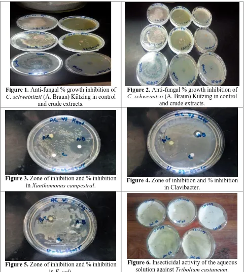
Our study included seven fungal species (Figures 1, 2 and 7) which were used to check the antifungal biological activities (Trichophyton longifusus, T. harzianum, Trichoderma hamatum, Rhizoctonia solani, P. oedochilum, Pythium aphanidermatum, Microsporum canis, Fusarium moniliforme, F. oxysporum and Aspergillus flavipes). The agar well diffusion method was used to test the antifungal activity for the above mentioned fungal species. The method for various tests and algal extraction process has been discussed earlier [8].
Antibacterial activities
used as a positive control while methanol as a negative control. The incubation of the antibacterial assay plates was done at 37 ○C for 24 hours and finally the diameters of the zone of inhibition were measured in mm. Insecticidal activities
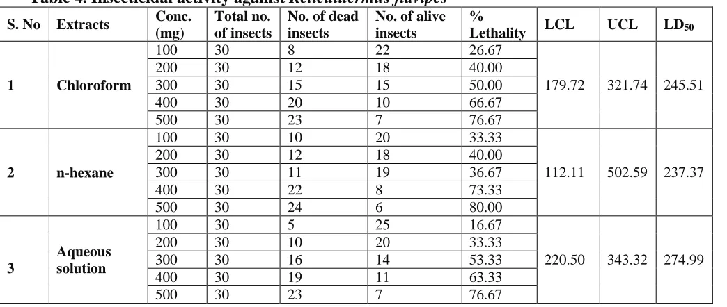
The impregnated filter paper process was used for investigating the insecticidal potential (Figures 6 and 9) of crude algal extracts [11]. The test was passed in petri plates (9 cm diameter) delivered with dual folds of the filter papers. To prepare the test sample, 200mg algal extract (from each extract) was dissolved in 3ml of methanol solvent. The sensitivity of the experiment was considered and the selection of the insect was done. The well insects (same size) were considered for the experiment.
The sizes of the petri plates were measured and the filter papers were cut of the same size. Double folds of the filter papers were kept inside the sterilized and clean petri plates. The tested sample was transferred on the filter paper. It was then freed so that the filter paper absorbs the solvent. The collected insects (healthy) were put into the petri plate with the help of the clean brush, which were further incubated at 27 ○C into the incubator. The similar method was conducted for every selected insect species into the experiment. For the positive control and negative control, the permethrin and DMSO were used. The checking of the extracts petri plates were observed after a day of incubation and percent mortality for
every used extract was find out by the formula:
Percent mortality = 100 - Number of alive insects in test / Number of alive insects in control × 100 [8].
The insects which were used assess the insecticidal potential of the methanol extracts of algal species are:
Termite and
Tribolium castaneum. Results
The crude ethanolic extract and various organic solvent fractions (ethanolic extracts) were brought into use. Different in vitro activities like; insecticidal activities, antibacterial activities and antifungal activities were conducted.
Table 1. Growth % inhibition of C. schweinitzii (A. Braun) Kützing against selected fungi strains
Solvent Test fungi
Antifungal activity of sample (mm)
Antifungal activity of control (mm)
% Inhibition
Chloroform
T. harzianum 21 32 65.62
Pythium sp. 19 32 59.37
Penicillium sp. 15 32 46.88
Fusarium solani 19 32 59.38
Microsporum canis 13 32 40.62
Aspergillus flavipes 11 32 34.38
Aspergillus niger 13 32 40.62
n-hexane
T. harzianum 17 32 53.5
Pythium sp. 21 32 65.75
Penicillium sp. 20 32 62.5
Fusarium solani 13 32 40.62
Microsporum canis 13 32 40.75
Aspergillus flavipes 10 32 31.25
Aspergillus niger 11 32 34.38
Crude methanol extract
T. harzianum 21 32 65.62
Pythium sp. 19 32 59.25
Penicillium sp. 15 32 46.88
Fusarium solani 17 32 53.12
Microsporum canis 11 32 34.38
Aspergillus flavipes 12 32 37.5
Aspergillus niger 12 32 37.5
Ethyl acetate
T. harzianum 20 32 62.5
Pythium sp. 19 32 59.75
Penicillium sp. 15 32 46.87
Fusarium solani 11 32 34.62
Microsporum canis 14 32 43.75
Aspergillus flavipes 10 32 31.25
Aspergillus niger 11 32 34.38
Figure 1. Anti-fungal % growth inhibition of C. schweinitzii (A. Braun) Kützing in control
and crude extracts.
Figure 2. Anti-fungal % growth inhibition of C. schweinitzii (A. Braun) Kützing in control
and crude extracts.
Figure 3. Zone of inhibition and % inhibition
in Xanthomonas campestral. Figure 4. Zone of inhibition and % inhibition in Clavibacter.
Figure 5. Zone of inhibition and % inhibition in E. coli.
Figure 7. Antifungal activity of chloroform, n-hexane, crude and ethyl acetate against the
selected strains of chara.
Figure 8. Antibacterial % inhibition of E. coli, X. campestral and clavibacter against the
selected strains of chara.
Figure 9. Insecticidal activity of n-hexane, chloroform and aqueous solution against T.
castaneum and R. flavipes.
The greatest activity was revealed by the chloroform extract for T. harzianum, with the zone of inhibition (21 mm). The moderate activity was shown by Pythium sp. with (19 mm), followed by Penicillium sp. (15 mm), Fusarium solani (19 mm), Microsporum canis (13 mm) and Aspergillus niger (13 mm). While the least activity was revealed by Aspergillus flavipes with the zone of inhibition (11 mm) (Table 1).
mm), Penicillium sp. (21 mm), Fusarium solani (20 mm), Microsporum canis (13 mm) and Aspergillus niger (13 mm), while the least potential was revealed by Aspergillus flavipes with the zone of inhibition (10 mm).
zone of inhibition (10 mm). Our study is in line with the experiment of Mahadevi et al.
[9].
The antibacterial examinations to find out the extracts of C. schweinitzii were tested against various bacterial strains (Table 2). The zone of inhibition for clavibacter revealed (17 mm), followed by ethyl acetate (21 mm), chloroform (22 mm), while the least was revealed against n-hexane (20 mm). The zone of inhibition for Xanthomonas campestral revealed (0.47 mm), followed by ethyl acetate (0.51 mm), chloroform (0.53 mm), while the least was showed against n-hexane (0.32 mm). The zone of inhibition for E. coli crude was revealed (0.40 mm), followed by ethyl acetate (0.50 mm), chloroform (0.60 mm),
while the least was revealed against n-hexane with the zone of inhibition (0.30 mm).
The percent inhibition in crude for clavibactor was recorded (50%), followed by ethyl acetate (48.75%), chloroform (52.5%), while the least percent inhibition was recorded for n-hexane (46.25%). The percent inhibition for Xanthomonas campestral in crude extract revealed (48.45%), followed by ethyl acetate (52.57%), chloroform (54.63%), while for n-hexane it revealed (32.98%). The percent inhibition of E. coli for crude extract revealed (44.4%), followed by ethyl acetate (55.5%), chloroform (66.6%) while in n-hexane it revealed (33.3%).
Table 2. Zone of inhibition and % inhibition of antibacterial bioassay of the test insects
Treatments Test species
Clavibacter Xanthomonas campestral E. coli
Zone of
inhibition (cm) %
Inhibition
Zone of
inhibition (cm) %
Inhibition
Zone of
inhibition (cm) %
Inhibition
-ve control 0 - 0 - 0 -
+ve control 0.8±0.10 - 0.97 ± 0.25 - 0.90 ± 0.01 -
Crude 0.40±0.10 50 0.47 ± 0.06 48.45 0.40 ± 0.01 44.4
Ethyl acetate 0.39±0.01 48.75 0.51± 0.08 52.57 0.50±0.01 55.5
Chloroform 0.42±0.12 52.5 0.53±0.07 54.63 0.60±0.02 66.6
n-hexane 0.37±0.10 46.25 0.32 ± 0.01 32.98 0.30±0.04 33.3
The insecticidal activities of crude methanolic extracts in chloroform, n-hexane and aqueous were conducted by screening technique against Tribolium castaneum and Reticulitermus flavipes (Table 3). The T. castaneum and R. flavipes were selected as the test insects. The crude extracts and the fractions revealed noteworthy insecticidal potential for both the tested insects. The crude extracts and fraction revealed dose dependent potential.
Results revealed (Table 3) important percent lethality for the tested insect (T. castaneum). Our data showed the highest percent lethality in chloroform (76.67%) at (500mg) while the least percent lethality revealed
(30.00%) at (100mg). The percent lethality revealed at (200mg), (300mg), (400mg) were (46.67%), (56.67%) and (63.33%), respectively. Our data showed the highest percent lethality for n-hexane (86.67%) at (500mg) while the least percent lethality was revealed (36.67%) at (100mg). The percent lethality revealed at (200mg), (300mg), (400mg) revealed (46.67%), (63.33%) and 7(3.33%), respectively.
(200mg), (300mg) and (400mg) was (36.67%), (53.33%) and (70.00%), respectively. Results (Table 4) revealed the important percent lethality for the test insect (R. flavipes). Our data revealed the greatest percent lethality for chloroform (76.67%) at (500mg) while the least was revealed at (100mg) which is (26.67%). The percent insecticidal lethality against T. castaneum revealed (40.00%) at (200mg), followed by
(50.00%) at (300mg) and (66.67%) at (400mg). Results (Table 4) revealed the significant percent lethality against the test insect (R. flavipes). Our data revealed the greatest percent lethality for n-hexane (80.00%) at (500mg) while the least percent lethality revealed (33.33%) at (100mg). The percent lethality at (200mg) revealed (40.00%), followed by (36.67%) at (300mg) and (73.33%) at (400mg).
Table 3. Insecticidal activity against Tribolium castaneum
S. No Extracts Conc.
(mg)
Total no. of insects
No. of dead insects
No. of alive insects
%
Lethality LCL UCL LD50
1
Chlorofor m
100 30 9 21 30.00
144.53 295.72 219.97
200 30 14 16 46.67
300 30 17 13 56.67
400 30 19 11 63.33
500 30 23 7 76.67
2 n-hexane
100 30 11 19 36.67
115.35 231.16 178.58
200 30 14 16 46.67
300 30 19 11 63.33
400 30 22 8 73.33
500 30 26 4 86.67
3 Aqueous
solution
100 30 5 25 16.67
260.48 320.82 260.48
200 30 11 19 36.67
300 30 16 14 53.33
400 30 21 9 70.00
500 30 23 7 76.67
Table 4. Insecticidal activity against Reticulitermus flavipes
S. No Extracts Conc.
(mg)
Total no. of insects
No. of dead insects
No. of alive insects
%
Lethality LCL UCL LD50
1 Chloroform
100 30 8 22 26.67
179.72 321.74 245.51
200 30 12 18 40.00
300 30 15 15 50.00
400 30 20 10 66.67
500 30 23 7 76.67
2 n-hexane
100 30 10 20 33.33
112.11 502.59 237.37
200 30 12 18 40.00
300 30 11 19 36.67
400 30 22 8 73.33
Discussion
Our study is in line with the experiment of Hadia et al. [12], who assessed antibacterial (Enterobacter, Micrococcus luteus, Pseudomonas aeruginosa and E. coli) and antifungal activities in various solvents benzene, chloroform and ethanol extract. Datura stramonium of chloroform extract produced maximum zone of inhibition and ethanol extract of D. stramonium gave maximum zone of inhibition against K. pneumonia while minimum against E. coli. Results further revealed that chloroform extract was very active against S. aureus, P. aeruginosa and M. luteus. All the extracts of D. stramonium have shown significant antifungal activity against Saccharomyces cerevisiae, Aspergillus fumigatus and Aspergillus niger with maximum antifungal activity against S. cerevisiae and zone of inhibition was about 16±0.2 mm by ethanol extract, 15±0.3 mm by chloroform and 14±1.6 mm by benzene extract while minimum antifungal activity was observed against A. niger.
Our study is also parallel to the experiments of Daljit K [13], who screened antimicrobial activities of the aqueous solution of the Moringa oleifera against two yeast strains, three Gram positive and seven Gram negative bacteria by agar well diffusion assay. Results concluded that the MICs of seed coat, stem bark and pod's husks ranged from 0.5-1.25mg ml-1, 3.0-4.2mg ml-1 and 4.0-5.6mg ml-1, respectively. Both seeds’ coat and pods’ husks extracts exhibited microbicidal properties which were totally inhibited for 12 hours, respectively. Seeds’ coat extract was the most effective against E. coli, while some pathogens, treated with stem bark extract, exhibited regrowth again after 24 hours apparently.
Our study was involved in antifungal activity, which is in line with the study of Paola et al. [14] who evaluated the extracts of antifungal activities of 10 plant species
against the phyto pathogenic fungus Alternaria sp. Their study determined minimal inhibitory concentration (MIC) and minimum fungicidal concentration (MFC). Results revealed that the MIC values ranged between 1.25 and 25μg mL-1. The MFC
values of the extracts ranged between 1.25μg mL-1 (Rosmarinus officinalis L.) and 10μg mL-1 (Cynara scolymus L.). MICs and MFCs values obtained from leaves (Salvia officinalis and R. officinalis) and seeds extracts (Salvia sclarea L.) were quite comparable to values obtained with the conventional fungicide captan (2.5μg mL-1). The extracts of Salvia sclarea, S. officinalis and R. officinalis could be considered as potential sources of antifungal compounds for treating diseases in plants. These extracts showed maximum activity, even at very low concentrations, and the same fungicide effects as chemical fungicide.
Conclusion
Our experiment exposed the biological activities and nutraceutical profile of the algae “C. schweinitzii (A. Braun) Kützing”. The physiochemical study of the said algae into our experiment showed the occurrence of some important elements. Our results of biological potential of C. schweinitzii (A. Braun) Kützing crude methanol extract and various plant extracts (methanol extracts) revealed the C. schweinitzii (A. Braun) Kützing includes some vital insecticidal activities, antibacterial, phytotoxic, cytotoxic and antifungal and is resulted that C. schweinitzii (A. Braun) Kützing has a greater amount of nutrition as well as offers strong biological activities.
Authors’ contributions
Acknowledgement
The author is immensely grateful to Uzair Ahmad (Research Associate) for his advice, write up, guidance, assistance, encouragement and inspiration.
References
1. Aslam S & Ayub WK (2001). Genus Chara: Usefulness of genus Chara, Biology. Intel Biological forum 223-230. 2. Daljit K (2013). Antimicrobial activity of
Moringa oleifera from different locations against some human pathogens. Academia J Medicinal Plants 1(5): 080-091.
3. Ghazala N & Johnson J (2012). Testing the various species and assays on phycochmistry and biological activity of Spirogyre rhizoides. American J Bot 12(9): 345-361.
4. Hadia G, Rubina NQ, Muhammad AK, Shazia H & Nabila Y (2012). Antibacterial and antifungal activity of different extracts of Datura stramonium (branches and leaves sample). J Biotech and Pharma Res 3(9): 141-148.
5. Khalid MN, Shameel M, Ahmad VU, Shahzad S & Leghari SM (2011). Studies on the bioactivity and phycochemistry of Microcystis aeruginosa (Cyanophycota) from Sindh. Pak J Bot 42: 2635-2646. 6. Mahadevi B & John PLJ (2014).
Distribution and seasonal variation of some Caulerpa species (Green seaweed) in Thoothukudi region, The south east coast of tamil nadu, India. Intel J Pure & App Biosci 2(3): 135-138.
7. Neelesh T (1997). Chara: Occurrence, Features and Reproduction. Intel Biological forum 133-141.
8. Naqvi SBS, Sheikh D, Usmanghani K, Shameel M & Sheikh R (1992). Screening of marine algae of Karachi for haemo glutinin activity. Pak J Pharm Sci 5: 129-138.
9. Naila B, Ghazala B, Shameel M, Choudhary MI & Leghari SM (2005). Phycochemistry and bioactivity of Lyngbya (Nostocophyceae shameel) from Sindh. Intel J Phycol Phycochem 1: 125– 134.
10.Oswald T (1999). Economic Botany: Botanical society of America 53(3): 353– 354.
11.Paola DD, Andrea C, Diego A, Patricia L, Fernando F & Marco DR (2011). Antifungal activity of medicinal plant extracts against phyto pathogenic fungus Alternaria sp. Chilean J Agric Res 71(2): 231-239.
12.Priya TS, Suganthi R, Saranya A & Thangaraj K (2013). Relationship between plasmid occurrence and antibiotic resistance in Myroides odoratimimus SKS05-GRD isolated from raw chicken meat. World J Microbiol Biotech 29: 983–990.
13.Rania & Hala (2008). Antibacterial and antifungal activity of cyanobacteria and green microalgae: Evaluation of medium components by Placket burman design for antimicrobial activity of Spirulina platensis. GJBBR 3: 22-31.