C A S E R E P O R T
Open Access
Obliterative cholangiopathy in acquired
cystic biliary atresia type III after cyst
perforation: a case report
Tsugumichi Koshinaga
*, Kensuke Ohashi, Kakou Ono, Hide Kaneda and Takeshi Furuya
Abstract
Background:In biliary atresia, the disease process of obliterative cholangiopathy may begin in the perinatal period; however, no chronological evidence exists on how the cholangiopathy progresses to biliary obliteration.
This is the first acquired case with the final diagnosis of type III cystic biliary atresia with an extrahepatic biliary cyst which showed the progression of obliterative cholangiopathy in chronological order after birth.
Case presentation:An 81-day-old girl presented with acute abdominal distress due to bilious peritonitis caused by biliary cyst perforation, for which she underwent emergency biliary drainage. Postoperative images showed a dilated common bile duct and hepatic ducts bilaterally, with flow of the contrast medium to the duodenum through the dilated common bile duct. Biochemistry of the bile collected during and after the operation revealed elevated levels of pancreatic enzymes in the bile from the gallbladder. The patient was diagnosed as having a congenital choledochal cyst and underwent laparotomy at 120 days of age which revealed that she had pancreaticobiliary
maljunction. The biliary cyst was resected at the narrow portion just above the junction with the main pancreatic duct. During dissection up to the hepatic hilum, we found that the hilar hepatic ducts were bilaterally replaced by fibrous tissue and were obstructed, leading to a diagnosis of type III a1,μbiliary atresia. The fibrous tissue was excised, and hepatic portoenterostomy was performed according to the Kasai procedure. The patient’s postoperative course was uneventful and the jaundice resolved within 1 month. She has had normal liver function tests with no episode of cholangitis for 3 years after discharge.
Conclusions:We demonstrated the process of acquired type III biliary atresia in a patient with cystic biliary atresia and biliary cyst perforation. To the best of our knowledge, this is the first case of acquired cystic biliary atresia showing chronological progression of the course of obliterative cholangiopathy, providing a better understanding of the development of type III biliary atresia as an acquired disease.
Keywords: Biliary atresia, Acquired biliary atresia, Biliary perforation, Biliary cyst, Obliterative cholangiopathy
Background
Most patients with biliary atresia still have greenish stools shortly after birth. The disease process of oblitera-tive cholangiopathy may begin in the perinatal period [1]; however, no evidence exists on the chronological progression of cholangiopathy to biliary obliteration.
We describe here a case with the final diagnosis of acquired type III biliary atresia with a perforated extrahe-patic biliary cyst which showed the chronological
progression of obliterative cholangiopathy after birth. This is the first clinical case showing the disease process of ac-quired biliary atresia described in a previous report [2].
Case presentation
An 81-day-old girl was transferred to the accident and emergency center of Nihon University Itabashi Hospital from her local pediatrician’s clinic because of marked ab-dominal distention and convulsions with loss of con-sciousness. The convulsions subsided with intravenous administration of diazepam and midazolam; however, the abdominal distension worsened, and was referred to pediatric surgery for surgical indication due to the
* Correspondence:koshinaga.tsugumichi@nihon-u.ac.jp
Department of Pediatric Surgery, Nihon University, School of Medicine, 30-1 Ooyaguchi-kamicho, Itabashi-ku, Tokyo 173-8610, Japan
© The Author(s). 2018Open AccessThis article is distributed under the terms of the Creative Commons Attribution 4.0
International License (http://creativecommons.org/licenses/by/4.0/), which permits unrestricted use, distribution, and
reproduction in any medium, provided you give appropriate credit to the original author(s) and the source, provide a link to the Creative Commons license, and indicate if changes were made. The Creative Commons Public Domain Dedication waiver
intraperitoneal ascitic fluid collection. She was previ-ously healthy, with appropriate growth and development and no history of trauma to her abdomen, after birth by normal vaginal delivery to a 38-year-old secundigravida mother at 37 weeks of gestation. The mother was noted to have a past history of extrahepatic bile duct resection with hepaticojejunostomy due to pancreaticobiliary mal-junction along with a congenital choledochal cyst.
On physical examination, the patient was found to be drowsy. Her temperature was 36.7 °C, and oxygen satur-ation was 98% while breathing oxygen at a flow rate of 10 L/min. Her weight was 4590 g and height was 55 cm. Her abdomen was markedly distended, with tense, shiny skin. Guarding and tenderness over the entire abdomen were also noted on palpation. Her sclerae appeared anicteric.
Blood and serum biochemical evaluations revealed leukocytosis (19,400/μL) and elevated total bilirubin (2.41 mg/dl), direct bilirubin (1.02 mg/dl), aspartate aminotransferase (72 IU/l), alkaline phosphatase (1371 IU/l), lactate dehydrogenase (LDH) (362 IU/l), and creatine kinase (CK) (371 IU/l) levels. The levels of alanine aminotransferase (17 IU/l) and C-reactive protein (CRP) (0.10 mg/dl) were within normal limits. Venous gas analysis indicated a pH of 7.357, bicar-bonates (HCO3−) of 16.3 mmol//l, and base excess
(BE) of −9.5 mmol/l. Abdominal ultrasonography revealed the presence of massive ascites and a cystic lesion at the hepatic hilum measuring 3 cm in diameter, with collected fluid and debris. The cystic lesion appeared to be connected to the intrahepatic bile duct, suggesting a dilated extrahepatic duct (Fig.1a). Abdominal CT scan revealed extrahepatic bile duct dilatation with massive ascites (Fig.1b).
Clinical course after admission
The patient underwent emergency laparotomy with a diagnosis of biliary peritonitis due to perforation of a
congenital choledochal cyst. Laparotomy findings re-vealed massive biliary ascites in the peritoneum, but no abnormalities of the liver and gallbladder on gross ap-pearance. The common bile duct appeared as a cyst 3 cm in diameter in the hepatoduodenal ligament. We could not find the exact site of biliary perforation des-pite careful exploration. Then, we performed cholangi-ography of the entire extrahepatic biliary tract by inserting a small catheter tapping from the gallbladder to the common bile duct via the cystic duct,which also failed to identify the site of biliary perforation. We avoided excessive pressure when injecting the contrast medium during cholangiography. No passage of contrast medium into the duodenum was shown. The final diag-nosis of minimal biliary perforation was made at that time. The operative procedure was accomplished by inserting an indwelling drainage catheter into the dilated common bile duct via the cystic duct and by intraperito-neal irrigation. The patient successfully recovered from the critical situation with good drainage of bile from the dilated common bile duct. The daily output of bile was between 50 to 100 ml. After she became in a stable con-dition, cholangiography via the catheter placed in the common bile duct was performed. It initially showed only the left hepatic duct, and no passage of the contrast medium into the duodenum through the dilated com-mon bile duct (Fig. 2a). However, digitized video-fluoroscopy cholangiography revealed evidence of biliary patency, demonstrating the flow of contrast into the duodenum (Fig.2b). Magnetic resonance choledocopan-creatography (MRCP) scan depicted the dilated common bile duct and bilateral hepatic ducts, although the junc-tion of the pancreatic duct and choledochus was obscure (Fig.2c). These images failed to identify the pancreatico-biliary maljunction. However, biochemistry of the bile collected during and after the operation revealed that the levels of pancreatic enzymes, such as phospholipase A2, were elevated in bile from the gallbladder and the
Fig. 1Abdominal ultrasound and CT images. Ultrasonography (Panela) showed a cystic lesion in the hepatic hilum, with accumulated fluid and
common bile duct (Table 1). This evidence was highly suggestive of pancreaticobiliary maljunction. The patient was diagnosed as having a congenital choledochal cyst; hence, surgery including extrahepatic bile duct resection was scheduled at the age of 120 days.
During the second laparotomy, dissection along the di-lated choledochal cyst to the intrapancreatic bile duct re-vealed a narrow portion between the distal part of the bile duct and pancreatic duct—the pancreaticobiliary maljunc-tion—in the pancreatic parenchyma. Intraoperative cholan-giography showed no communication between the common bile duct and the pancreatic duct at the operation. The distal end of the dilated biliary cyst was ligated and transected at the narrow portion just above its junction
with the main pancreatic duct. Dissecting along to the hep-atic hilum, we found that the hilar hephep-atic ducts had been bilaterally replaced by fibrous tissue and were obstructed, leading to a diagnosis of type III a1,μbiliary atresia. The fi-brous tissue was excised, and hepatic portoenterostomy was performed according to the Kasai procedure. Histo-pathologic examination showed that most of the lining epi-thelium of the resected biliary cyst was desquamated. Liver biopsy shows a few inflammatory cells infiltrated in the periportal area presenting slight fibrosis, but no significant ductular proliferation noted (Fig.3). The patient’s postoper-ative course was uneventful. She has had normal liver func-tion tests with no episodes of cholangitis for 3 years since her discharge from the hospital.
Fig. 2Postoperative cholangiography. Cholangiography performed via a catheter placed in the common bile duct (Panela) showed only the left hepatic duct and no flow of the contrast medium into the duodenum through the dilated common bile duct. However, digitized
video-fluoroscopy cholangiography (Panelb) found evidence of biliary patency, showing flow of the contrast agent into the duodenum. MRCP
(Panelc) depicted the dilated common bile duct and bilateral hepatic ducts, although the junction of the pancreatic duct and choledochus was obscure
Table 1Chemistry of bile collected during and after operation
Intraoperative collection Postoperative collectiona
Ascites Gallbladder Common bile duct Gallbladder via drainage catheter
Total bilirubin (mg/dl) 9.5 n.a. n.a. n.a.
Amylase (IU/l) n.a. 29 4 380
Trypsin (ng/ml) n.a. 2200 n.a. n.a.
PhospholipaseA2 (ng/dl) 2040 6370 157,000 n.a.
Elastase 1 (ng/dl) 560 1300 63,000 2,400,000
Lipase (U/l) 114 267 9958 n.a.
n.a, not available
a
Discussion and conclusions
We demonstrated here the pathophysiological process of acquired type III biliary atresia in a patient with cystic biliary atresia with biliary cyst perforation. To the best of our knowledge, this is the first case of acquired biliary atresia showing the chronological progression in the course of obliterative cholangiopathy.
The case presents evidence that type III biliary atresia develops as an acquired disease. Although acquired biliary atresia was first described in 1996 [2], little is known about the process of obliteration of the biliary tree. The cause of acquired biliary atresia remains un-known. Multifactorial mechanisms may be involved in
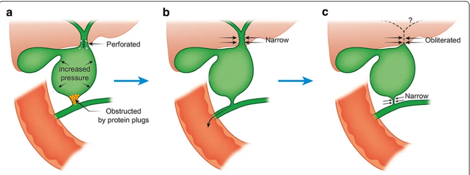
the etiology of development of obliterative cholangiopa-thy. The proposed etiologies of acquired biliary atresia possibly include infection [3] and inflammatory re-sponses [4], but rarely include genetic aberrations [5] and developmental malformations [6]. The onset of ob-literation of the biliary tree also remains unknown, as in most other types of biliary atresia. The most accepted view is that infants with biliary atresia once had intact biliary trees during fetal life, following which the biliary tree began to obliterate as a secondary phenomenon [7], a rational concept that is yet to be proved by clinical ob-servation of the disease. A recent report has described the pathology of biliary atresia could begin in utero as well as perinatally given the elevation of direct bilirubin soon after birth [8]. If the obliterative cholangiopathy consistent with biliary atresia seen in this case at 120 days of age began in the newborn period it certainly progressed very slowly. The reason for the biliary perfor-ation not occurring until 81 days of life is unclear. The present case with type III acquired biliary atresia demon-strates the process of postnatal obliteration of the biliary tree, as shown in the chronological schema of the dis-ease process (Fig. 4). The pathological process possibly occurs in the following sequence. First, biliary perfor-ation occurs due to increased intraluminal pressure in the biliary cyst due to possible obstruction by protein plugs at the distal end of the common bile duct. Second, the resultant decrease in intraluminal pressure in the cyst secondary to adequate drainage of the common bile duct via the indwelling catheter and by spontaneous re-moval of the protein plugs, with subsequent restoration of the patency of the distal end of the common bile duct,
Fig. 3Histopathologic findings of liver (Hematoxylin eosin × 200). Liver biopsy shows a few inflammatory cells infiltrated in the periportal area presenting slight fibrosis, but no significant ductular proliferation noted
Fig. 4Schema of the disease process together with possible etiologies. Biliary perforation secondary to increased luminal pressure in the biliary
cyst due to possible obstruction by protein plugs in the distal end of the common bile duct at 81 days old (Panela). The resultant decrease in
intraluminal pressure, together with adequate drainage of the common bile duct and spontaneous removal of the protein plugs contributes to
healing the site of perforation (Panelb). On the hepatic side, the perihilar bile ducts and common hepatic duct become progressively narrower,
marking the beginning of development of biliary atresia. Finally, the perihilar bile ducts become completely obliterated and the distal side of the
facilitates healing of the site of perforation. On the hepatic side, the perihilar bile ducts and the common hepatic duct become progressively narrower at the be-ginning of the development of the biliary atresia. Finally, the perihilar bile ducts become completely obliterated, and the distal end of the common bile duct becomes narrow, as was seen during the second laparotomy in our patient.
Our case represents cystic biliary atresia, consisting of an extrahepatic biliary cyst with biliary atresia, in which development of the cyst was the primary event rather than secondary to increased luminal pressure due to bil-iary obstruction. The bilbil-iary cyst formed before the hilar biliary obliteration on the distal side seen in the present case. From a clinical standpoint, Davenport [9] divided biliary atresia into four broad groups: syndromic biliary atresia, cystic biliary atresia, cytomegalovirus-associated biliary atresia, and isolated biliary atresia. Among these subgroups, cases with cystic biliary atresia, an uncom-mon and clinically distinct variant of biliary atresia, have been previously reported by many authors. One of the previous reports [10] mentioned that 21 (72%) of the 29 cases with cystic biliary atresia in their clinical series were found to be type III biliary atresia. According to the national registry of the Japanese Biliary Atresia Soci-ety, 131 (32%) of 408 cases with cystic biliary atresia were classified as type III atresia (1989-2015; http://jbas. net/registration/). Among those 131 cases, 31 cases (24%) had yellow or slightly yellow bile, providing evi-dence of the communication between the cyst lumen and intrahepatic biliary tree. The registry provides no further detailed clinical information, such as the process of development of cystic biliary atresia. One previous re-port described the following as characteristics of the cys-tic biliary atresia: diagnosed in the prenatal period, presenting with jaundice from birth, and presence of closed cyst formation in the biliary tree, as confirmed by cholangiography [11]. However, previous reports failed to show the etiology of type III cystic biliary atresia, and lacked chronological evidence of development of the dis-ease [11–13]. It is accepted that type III cystic biliary atresia has a different pathogenesis as compared to atre-sia without biliary cyst formation. The evidence that the cyst develops distal to obstruction of the hilar biliary tree cannot be explained only by an increase of intraluminal pressure, as in our case with type III cystic biliary atresia. In the present case, biliary cyst formation clearly preceded the hilar biliary obliteration, as seen in chronological cholangiograms. The etiological difference between biliary cysts associated with acquired biliary atresia and congenital choledochal cyst perforation, therefore, remains uncertain.
Our case presents perforation of a biliary cyst as the initial manifestation of acquired biliary atresia. The role
of biliary perforation in the process of obliteration of the biliary tree in the present case with type III biliary atre-sia remains unknown. Biliary perforation is a rare condi-tion of various etiologies, such as pancreaticobiliary maljunction with and/or without a choledochal cyst, de-velopmental weakness of the biliary wall, trauma, viral infection, stenosis of the biliary tract, and distal obstruc-tion of the extrahepatic bile duct [14,15]. Among these proposed etiologies, pancreaticobiliary maljunction has been focused on as the most probable etiological hy-pothesis [16, 17]. In the present case, pancreaticobiliary maljunction was found during the second operation. Bil-iary perforation with this congenital anomaly is associ-ated with an abrupt increase in intramural pressure of the biliary tree secondary to impaction of protein plugs [16]. We suspect that protein plugs or inspissated bile were the possible cause of obstruction in the present case, which may have resulted in distal biliary obstruc-tion that resolved following adequate biliary drainage [18]. Our patient’s mother had a history of surgery for pancreaticobiliary maljunction. However, it still remains unknown whether to etiologically associate this history with the onset of biliary atresia in our patient. The com-bination of a mother with pancreaticobiliary maljunction and daughter with biliary atresia has not been previously reported. Further, the site of biliary perforation in the present patient with type III biliary atresia remains un-known, although the site of perforation may have been closely associated with the obliterative process of hilar biliary obstruction. The most common site of biliary perforation is reportedly the anterior wall of the com-mon bile duct, adjacent to its junction with the cystic duct, due to possible embryogenic mural weakness of the common bile duct at its junction with the cystic duct [19, 20]. This mural weakness is vulnerable to ischemia and distal obstruction, resulting in perforation. Some investigators have suggested that extravasated bile in ad-jacent periductal tissues could lead to protracted inflam-mation and fibrosis, causing secondary obliteration of the hilar biliary tract [7].
symptoms, and age at presentation of primary symptoms. Their case was the first case of type III biliary atresia ever reported. The outcome was fair, without jaundice and with normal liver function tests postoperatively.
Ours is the first case demonstrating the pathophysio-logical process of acquired type III biliary atresia in a pa-tient with a perforated biliary cyst, providing a better understanding of the development of type III biliary atresia as an acquired disease.
Abbreviations
BE:Base excess; CK: Creatine kinase; CRP: C-reactive protein; CT: Computed
tomography; HCO3: Bicarbonates; LDH: Lactate dehydrogenase;
MRCP: Magnetic resonance cholangiopancreatography
Availability of data and materials
The authors declare that the data supporting the findings of this study are available within the article.
Authors’contributions
TK, KeO, and KaO collected the clinical details and photographs of this case. TK performed the literature review, and designed and drafted the manuscript. TK and KaO interpreted the radiological images and selected the photographs. KeO, HK, and TF verified the diagnosis and other scientific facts. All authors read and approved the final manuscript.
Ethics approval and consent to participate
Not applicable. Written consent for the standard medical care given to their child as described in this case study was obtained from the guardian.
Consent for publication
Written informed consent was obtained from the patient’s legal guardian for
publication of this case report and accompanying images. A copy of the written consent is available for review by the Editor of the journal.
Competing interests
The authors declare that they have no competing interests.
Publisher’s Note
Springer Nature remains neutral with regard to jurisdictional claims in published maps and institutional affiliations.
Received: 1 July 2017 Accepted: 30 April 2018
References
1. Hartley JL, Davenport M, Kelly DA. Biliary atresia. Lancet. 2009;374(9702):
1704–13.
2. Davenport M, Saxena R, Howard E. Acquired biliary atresia. J Pediatr Surg.
1996;31(12):1721–3.
3. Morecki R, Glaser JH, Cho S, Balistreri WF, Horwitz MS. Biliary atresia and
reovirus type 3 infection. N Engl J Med. 1982;307(8):481–4.
4. Narayanaswamy B, Gonde C, Tredger JM, Hussain M, Vergani D, Davenport
M. Serial circulating markers of inflammation in biliary atresia–evolution of
the post-operative inflammatory process. Hepatology. 2007;46(1):180–7.
5. Shimadera S, Iwai N, Deguchi E, Kimura O, Fumino S, Yokoyama T. The inv
mouse as an experimental model of biliary atresia. J Pediatr Surg. 2007;42(9):
1555–60.
6. Davenport M, Savage M, Mowat AP, Howard ER. Biliary atresia splenic
malformation syndrome: an etiologic and prognostic subgroup. Surgery.
1993;113(6):662–8.
7. Sokol RJ, Mack C, Narkewicz MR, Karrer FM. Pathogenesis and outcome
of biliary atresia: current concepts. J Pediatr Gastroenterol Nutr. 2003;
37(1):4–21.
8. Harpavat S, Finegold MJ, Karpen SJ. Patients with biliary atresia have
elevated direct/conjugated bilirubin levels shortly after birth. Pediatrics.
2011;128(6):e1428–33.
9. Davenport M. Biliary atresia: clinical aspects. Semin Pediatr Surg. 2012;21(3):
175–84.
10. Caponcelli E, Knisely AS, Davenport M. Cystic biliary atresia: an etiologic and
prognostic subgroup. J Pediatr Surg. 2008;43(9):1619–24.
11. De Matos V, Erlichman J, Russo PA, Haber BA. Does“cystic”biliary atresia
represent a distinct clinical and etiological subgroup? A series of three
cases. Pediatr Dev Pathol. 2005;8(6):725–31.
12. Redkar R, Davenport M, Howard ER. Antenatal diagnosis of congenital anomalies
of the biliary tract. J Pediatr Surg. 1998;33(5):700–4.
13. Muise AM, Turner D, Wine E, Kim P, Marcon M, Ling SC. Biliary atresia with
choledochal cyst: implications for classification. Clin Gastroenterol Hepatol.
2006;4(11):1411–4.
14. Hasegawa T, Udatsu Y, Kamiyama M, Kimura T, Sasaki T, Okada A, Mushiake
S. Does pancreatico-biliary maljunction play a role in spontaneous perforation
of the bile duct in children? Pediatr Surg Int. 2000;16(8):550–3.
15. Evans K, Marsden N, Desai A. Spontaneous perforation of the bile duct in
infancy and childhood: a systematic review. J Pediatr Gastroenterol Nutr.
2010;50(6):677–81.
16. Ando H, Ito T, Watanabe Y, Seo T, Kaneko K, Nagaya M. Spontaneous perforation
of choledochal cyst. J Am Coll Surg. 1995;181(2):125–8.
17. Yano H, Matsumoto H. Choledochal cyst following operation for idiopathic
perforation of the biliary tract in childhood. Jpn J Surg. 1983;13(5):441–5.
18. Davenport M, Heaton ND, Howard ER. Spontaneous perforation of the bile
duct in infants. Br J Surg. 1991;78(9):1068–70.
19. Lilly JR, Weintraub WH, Altman RP. Spontaneous perforation of the
extrahepatic bile ducts and bile peritonitis in infancy. Surgery. 1974;
75(5):664–73.
20. Xanthakos SA, Yazigi NA, Ryckman FC, Arkovitz MS. Spontaneous perforation
of the bile duct in infancy: a rare but important cause of irritability and
abdominal distension. J Pediatr Gastroenterol Nutr. 2003;36(2):287–91.
21. Tan CE, Driver M, Howard ER, Moscoso GJ. Extrahepatic biliary atresia: a
first-trimester event? Clues from light microscopy and immunohistochemistry.