Special issue • 2020 • vol.2 • 29-35
CT FEATURES OF PANCOLITIS:
ANALYSIS OF 4 CASES
Romano P.
1, Di Grezia G.
1, Vallebona L.
2, Giordano M.
2, Musto L.A.
1, Gatta G.
21Radiology Department - G Criscuoli Hospital, Sant’Angelo dei Lombardi, Avellino IT 2Radiology Department - University of Campania “Luigi Vanvitelli” Naples, IT
Email: [email protected]
Abstract
INTRODUCTION: Between the infective pancolitis, Clostridium Difficile infections are more common in hospitalized patients. Most of the cases can be resolved with medical treatment but in some patients is necessary to perform a surgical intervention.
METHODS AND MATERIALS: In the period between January and September 2018 we diagnosed four different cases of pancolitis due to Clostridium Difficile infection.
We considered positive for pancolitis an abdominal CT exam with the presence of at least two different signs between increased intestinal wall thickness, target sign, pericolic fat stranding, bowel or peritoneal pneumatosis
RESULTS: All patients have been hospitalized for different reasons, in all of them the CT diagnoses a pancolitis that is regressed in three of four patients with medical treatment and got worse in one case concluding in exitus for the impossibility to undergo a surgical treatment
CONCLUSIONS: Abdomen CT with intravenous contrast media is very useful for the detection and follow-up of a colonic involvement in patients with Clostridium Difficile infection. colonic involvement in patients with Clostridium Difficile infection.
Introduction
Pancolitis is a diffuse inflammation of the bowel that can be differentiate in infective and non-infective. Between the infective pancolitis, Clostridium Difficile infections are more common in hospitalized patients.
Clostridium Difficile is a gram-positive bacterium known as the most common cause of antibiotic-associated diarrhea and colitis; symptoms are very variable going from a mild diarrhea to a pancolitis that can progress until exitus [1].
Most of cases can be resolved with medical treatment, generally metronidazole or vancomycin, but in patients with symptomatology could be necessary to perform a total abdominal colectomy and in rare cases an end ileostomy [2,3].
Aim of the study is to analyze all the features in abdominal CT with intravenous contrast medium that can be useful to diagnose pancolitis in patients with a story of Clostridium Difficile infection.
Methods
In the period between January and September 2018 we diagnosed four different cases of pancolitis due to Clostridium Difficile infection thanks to abdominal CT exams with intravenous contrast medium in patients admitted in the Rehabilitation Section of our Hospital.
To perform a diagnosis of pancolitis we measured: Intestinal wall thickness (normal values: 3 mm for the colon and 4 mm for the rectum). We found a segmentary or total wall thickening with a low attenuation associated with a hyperemic enhancing mucosa stretched over edematous and thickened submucosal fold (accordion sign) .
the "target sign" that consist in the presence of layers of different enhancement in the intestinal wall the pericolic fat stranding, sometimes correlated to free peritoneal or pleural fluid Bowel or peritoneal pneumatosis (Tab.1) [4,5].
We considered positive for pancolitis an abdominal CT exam with the presence of at least two different signs of the previous.
Results
Three of four patients have an age between 70 and 85 years (mean 75.5 years) and were hospitalized in the Rehabilitation section for different reasons (Tab.2).
In all patients the diagnosis of Clostridium Difficile infection required the analysis of a stool sample, but an abdomen CT exam with intravenous contrast medium revealed signs of a pancolitis.
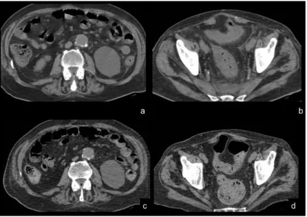
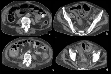
CG had an initial regression in seven days (Fig.1) but the pancolitis represented after two month with an extension and exitus in two days (Fig.2) probably because was necessary a surgical procedure but the high anesthesiologic risk, due the aortic dissection, of the patient didn't allow it.
FM and SG had a total regression of the pancolitis in a few days.
PGN had a clinical improvement in sixteen days but for the resolution seven month were necessary. In the end we had a total regression of the pathology in three case on four and only one went to progression and exitus and only for the impossibility to perform a mandatory surgery. The mortality was 0% at 30 days from diagnosis and 25% at two months, the clinical improvement rate at 30 days from diagnosis was 3/4.
Discussion
Clostridium Difficile's infection are common complication in patients under antibiotic treatment, some of them can progress until pancolitis, most of the cases can be healed with medical treatment but sometimes can be necessary to perform a surgical intervention [6,7].
Gash et al. say that one third of the patients with Clostridium Difficile pancolitis is under 65 years old and the male-female ratio is 3.25-1, also it seems that men are more easily candidates to surgical intervention due to fewer comorbities [2, 8].
When there is a suspicion of Clostridium Difficile infection an abdominal CT exam with intravenous contrast medium it is mandatory to detect signs of pancolitis and to define a severity scale [7]; this way is possible to get an early diagnosis that is crucial to define the patient management and plan, when possible, a surgical procedure or a medical treatment.
The Contrast medium injection rates and acquisition protocols can be very variable depending on the institution [9 -11] we prefer a triphasic exam with an arterial, a portal and a delayed phase for the study of our patients [12, 13].
are consequences of tissue necrosis so they are late signs [14,15], but we always get an early diagnosis. Patients that are hospitalized in rehabilitation section have often an high anesthesiologic risk that preclude the possibility of a surgical intervention; in these patients, when the medical treatment results ineffective is very difficult to find a solution to cure them.
Conclusions
Clostridium Difficile's pancolitis is a severe complication in hospitalized patients and can cause exitus in some cases, especially if clinical conditions are unstable (e.g. patients of rehabilitation section). Abdomen CT with intravenous contrast media is very useful for the detection and follow-up of a colonic involvement in patients with Clostridium Difficile infection.
References
1.
R.M. Dallal, B. G. Harbrecht, A.J. Boujoukas, C,A. Sirio, L.M. Farkas, K.K. Lee, R.L. Simmons; Fulminant Clostridium difficile: An underappreciated and increasing cause of death and complications; Annals of surgery; 2002; vol. 235, No 3, 363-3722.
K. Gash, E. Brown, A. Pullyblank; Emergency subtotal colectomy for fulminant Clostridium difficile colitis – is a surgical solution considered for all patients? Colorectal surgery; 2010; 92; 56-60].3.
Paola Crivelli, Marcello Carboni, Rino Aldo Montella, Antonio Matteo Amadu, Stefano Profili, Maurizio Conti, Giovanni Battista Meloni. Gastroduodenal stenting: is still useful in the treatment of malignant obstruction? La radiologia medica 122 (8), 564-5674.
J. Lim, A.W. Phillips, W.L. Thompson; An unexpected CT finding in a patient with abdominal pain; BMJ Case Reports; 2013; doc. 10.11365.
L. Palau-Davila, R. Lara-Medrano, A.A. Negreros-Osuna, M. Salinas-Chapa, E. Garza-Gonzalez, E.M. Gutierrez-Delgado, A. Camacho-Ortiz; Efficacy of computed tomography for the prediction of colectomy and mortality in patients with Clostrium difficile infection; Annals of medicine and surgery; 2016;12; 101-1056.
D.M. Tang, N.H. Urrunaga, H. De Groot, E.C. vonRosenvinge, G. Xie, L.Y. Ghazi:
Pseudomembranous colitis: not always caused by Clostridium difficile; Case report in Medicine; Vol. 2014; Article ID 812704
7.
A. Kalakonda, S. Garg, S. Tandon, R. Vinajak, S. Dutta; A rare case of infectious colitis; Gastroenterology Reports; 2016; 4; 328-3308.
A. Reginelli, G. Di Grezia, G. Gatta, F. Iacobellis,C. Rossi, M. Giganti, F. Coppolino, L. Brunese; Role of conventional Radiology and Mri defecography of pelvic floor hernias; BMC surgery; 2013; 13; S53
9.
A. Reginelli, R. Capasso, V. Ciccone, M.R. Croce, G. Di Grezia, M. Carbone, N. Maggialetti, A. Barile, P. Fonio, M. Scialpi, L. Brunese; Usefulness of triphasic CT aortc angiography in acute and surveillance: Our esperience in assessment of acute aortic dissection and endoleak; International journal of surgery; 2016; 33; S76-S8410.
V. Valentini, G.L. Buquicchio, M. Galluzzo, S. Ianniello, G. Di Grezia, R. Ambrosio, M. Trinci, V. Miele; Intussusception in adults: The role of MDCT in the indentification of the site and cause of obstruction; Gastroenterology research and pratice; 2016; article ID 562371811.
C. Rossi, A. Reginelli, M. D'Amora, G. Di Grezia,Y. Mandato, A. D'Andrea, L. Brunese, R. Grassi, A. Rotondo; Safety profile and protol prevention of adverse reactions to uroangiographic contrast mediain diagnostic imaging; Journal of biological regulators and homeostatic agents; 2014; vol. 28; 155-165
12.
Faggian A, Alabiso ME, Serra N, Pizza NL,Iasiello F, Tecame M, Somma F, Rossi C, Di Grezia G, Feragalli B, Iacomino A, Grassi R
“Entero-colpo-cysto-defecography vs supine
entero-MRI: which one is the best tool in the differentiation of enterocele, elytrocele and
edrocele?” Journal Biological Regulators and
Homeostatic agents 2013 Jul-Sep; 27(3):861-8
13.
Reginelli A, Iacobellis F, Del Vecchio L, MonacoL, Berritto D, Di Grezia G, Genovese EA, Giganti M, Cappabianca S “VFMSS findings in elderly
dysphagic patients: our experience” BMC
Surgery 2013; 138 (Suppl2): 554
pneumatosis, colon ischemia, cold intussusception, gallstone ileus, and foreign bodies: Our esperience and literature review of incidental gastrointestinal MDCT findings; Biomed research international; 2017; article ID 5716835
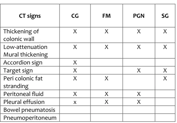
Table 1. Shows abdominal CT signs for each patient.
CT signs CG FM PGN SG
Thickening of colonic wall
X X X X
Low-attenuation Mural thickening
X X X X
Accordion sign X
Target sign X X X
Peri colonic fat stranding
X X X
Peritoneal fluid X X X
Pleural effusion x X X
Bowel pneumatosis Pneumoperitoneum
Table 2. Shows details of patients and the reason of the hospitalization in
Rehabilitation section of our Hospital
Patient name Sex Age Anamnesis
CG Male 70 Rehabilitation post-stenting for thoracic aortic dissection FM Male 77 Rehabilitation post cerebral ischemia with hemorrhage
Figure 1. CG, male, 7o yo
Figure 2. CG, male, 7o yo