R E S E A R C H A R T I C L E
Open Access
Normal sulfation levels regulate spinal cord
neural precursor cell proliferation and
differentiation
Michael Karus
1,4, Samira Samtleben
2, Claudia Busse
3, Teresa Tsai
2, Irmgard D Dietzel
3, Andreas Faissner
1and Stefan Wiese
2*Abstract
Background:Sulfated glycosaminoglycan chains are known for their regulatory functions during neural development and regeneration. However, it is still unknown whether the sulfate residues alone influence, for example, neural precursor cell behavior or whether they act in concert with the sugar backbone. Here, we provide evidence that the unique 473HD-epitope, a representative chondroitin sulfate, is expressed by spinal cord neural precursor cellsin vivo andin vitro, suggesting a potential function of sulfated glycosaminoglycans for spinal cord development.
Results:Thus, we applied the widely used sulfation inhibitor sodium chlorate to analyze the importance of normal sulfation levels for spinal cord neural precursor cell biologyin vitro. Addition of sodium chlorate to spinal cord neural precursor cell cultures affected cell cycle progression accompanied by changed extracellular signal-regulated kinase 1 or 2 activation levels. This resulted in a higher percentage of neurons already under proliferative conditions. In contrast, the relative number of glial cells was largely unaffected. Strikingly, both morphological and electrophysiological characterization of neural precursor cell-derived neurons demonstrated an attenuated neuronal maturation in the presence of sodium chlorate, including a disturbed neuronal polarization.
Conclusions:In summary, our data suggest that sulfation is an important regulator of both neural precursor cell proliferation and maturation of the neural precursor cell progeny in the developing mouse spinal cord.
Keywords:Chondroitin sulfate proteoglycans, Extracellular matrix, Neuronal differentiation, Sulfation
Background
Complex carbohydrate moieties attached either to secreted extracellular matrix molecules or to membrane bound cell adhesion molecules are important players for central nervous system (CNS) development, because they can bind a multitude of cytokines or growth factors [1-3]. Among these carbohydrate structures, the chondroitin sulfate (CS) glycosaminoglycans (GAGs) have been proved to be particularly important for neural precursor cell (NPC) biology, because interference with CS-GAG biol-ogy using the bacterial enzyme chondroitinase ABC (ChABC) strongly affects NPC proliferation and differenti-ation [4-7]. CS-GAGs are highly sulfated, and the sulfdifferenti-ation pattern results from the specific expression pattern of
various CS-sulfotransferases during CNS development and in adulthood [8]. Sulfated CS-GAGs also play a role in pathophysiological situations. Up-regulation of CS pro-teoglycans (PGs) bearing complex CS-GAGs after CNS injury is associated with a specific sulfation pattern on CS-GAGs, mediating potentially inhibitory properties of PGs on axonal regeneration [9,10]. These inhibitory prop-erties can be overcome using ChABC in combination with defined motor tasks [11,12]. However, different sulfation patterns appear to differentially affect axonal regeneration after CNS lesion [13].
Besides CS-GAGs, the heparan sulfate (HS)-GAGs con-stitute a second major class of sulfated glycans in the CNS. HS-GAGs are known for their strong impact on fibroblast growth factor (FGF) and Sonic hedgehog signal-ing [14]. In addition, some other glycan structures can be sulfated as well. Among these, the complex LewisX glycan, * Correspondence:[email protected]
2Group for Molecular Cell Biology, Ruhr-University Bochum, Bochum, Germany
Full list of author information is available at the end of the article
which is expressed by NPCs during development and in adulthood [15-17], and the human natural killer 1 antigen are the most prominent ones [18].
The transfer of sulfate on the glycosaminoglycan back-bone of CS-GAGs and HS-GAGs is catalyzed by CS- or HS-specific sulfotransferases located in the Golgi apparatus [19,20]. 3′-phosphoadenosine 5′-phosphosulfate (PAPS) serves as the sulfate donor. The enzymatic generation of PAPS can be inhibited using sodium chlorate (NaClO3)
[21], resulting in hypo-sulfated GAG chains. Therefore, NaClO3is often used to pharmacologically interfere with
GAG biology. Here, we used NaClO3to further elucidate
the role of sulfation for differentiation and proliferation of spinal cord NPCs cultivated as free floating neurospheres. Neurospheres treated with NaClO3showed a reduction in
growth accompanied by changes in neuronal differentiation and maturation. In contrast, differentiation towards glial lineages seemed to be largely unaffected. Thus, we propose that the proliferation of spinal cord NPCs and the matur-ation of spinal cord NPC-derived neurons depend on a spe-cific sulfation pattern during development.
Methods Animals
All experiments were performed according to inter-national rules using timed-pregnant wild-type NRMI mice kept under standard housing conditions. The age of the embryos was determined following the Theiler Stages, and the day of the vaginal plug was considered as E0.5.
Neurosphere culture
Cultivation of mouse spinal cord NPCs was carried out as previously described [22]. Briefly, the spinal cords of E13.5 to E18.5 mouse embryos were dissected using small for-ceps. Afterwards, the isolated spinal cords were enzymati-cally dissociated with 30 U/mL Papain (Worthington, New Jersey, USA), resulting in a single cell suspension. After centrifugation for 5 min at 200 g the cell sediment was resuspended in neurosphere medium containing DMEM and nutrient mixture F12 (in a ratio of 1:1; both Gibco, Karlsruhe, Germany), 2 % (v/v) B27 (Invitrogen, Karlsruhe, Germany), 1 % (v/v) L-glutamine (Invitrogen) and 1 % (v/v) penicillin/streptomycin (Invitrogen). Under proliferative conditions, either 1,250 cells/mL (clonal density) or 100,000 cells/mL (high density culture) were plated, and either 10 ng/mL (clonal density) or 20 ng/mL (high density culture) EGF and FGF2 (both PreproTech, Rocky Hill, USA) were added. FGF2 containing cultures were additionally supplemented with 0.25 U/mL (clonal density) or 0.5 U/mL (high density culture) heparin (Sigma-Aldrich, Munich, Germany). After one week at 37° C and 5 % (v/v) CO2, neurospheres had formed. For clonal
analyses, the cells were kept for one week without any agi-tation, in order to avoid neurosphere fusion events. As
recently described, 30 mM NaClO3or the respective
solv-ent were added for the solv-entire cultivation period [23]. For differentiation analyses, primary neurospheres were dissociated using trypsin/ethylenediaminetetraacetic acid (EDTA; Invitrogen) for 4 min at 37°C, and single cells were plated at 5,000 cells/well onto poly-DL-ornithine /laminin coated four-well dishes (Greiner, Frickenhausen, Germany) for another 4 days in the presence of 1 % (v/v) FCS and either 30 mM NaClO3or the respective solvent.
Afterwards, the cells were either subjected to immuno-cytochemical or electrophysiological analyses.
Immunological reagents
In the following the primary antibodies and the respective dilutions used in this study are listed. The monoclonal antibodies were: O4 (1:50; mouse IgM) [24], anti-Nestin (1:500; mouse IgG; clone rat-401; Chemicon, Hofheim, Germany), anti-α-Tubulin (1:10,000; mouse IgG; clone DM1a; Sigma-Aldrich), anti-βIII-Tubulin (1:500; mouse IgG; clone SDL3D10; Sigma-Aldrich) and anti-DSD-1-epitope (1:300 (immunofluorescence), 1:100 (west-ern blot); rat IgM; clone 473) [25]. The polyclonal anti-bodies were: anti-GFAP (1:300; rabbit; Dako, Hamburg, Germany), anti-EGFR (1:500; rabbit; Santa Cruz, Hamburg, Germany), anti- receptor protein tyrosine phosphatase (RPTP)-β/ζ(1:300 (immunofluorescence), 1:1000 (western blot); rabbit; batch Kaf13/5) [25], anti-BLBP (1:300; rabbit; Chemicon), anti-GLAST (1:1000; guinea-pig; Chemicon), anti-pH3 (1:300; rabbit; Chemicon), anti-Erk1/2 (1:1000; rabbit; Santa Cruz), anti-pErk1/2 (1:1000; rabbit; Cell Sig-naling Technologies, Beverly, USA), anti-Akt (1:1000; rabbit; Cell Signaling Technology), anti-pAkt (1:1000; rabbit; Cell Signaling Technologies), anti-MAP2 (1:300: rabbit; Chemicon) and anti-Tau (1:300; mouse IgG) [26].
Immunohistochemistry
Pregnant mice were killed by cervical dislocation, and the embryos were immediately removed. The embryos’ trunks were washed once with PBS and then fixed in 4 % (w/v) paraformaldehyde (PFA) at 4°C. Depending on the age of the embryo, the fixation time varied between 30 min (E9.5) and 20 h (E18.5). After fixation the embryos were transferred to 20 % (w/v) sucrose for cryo-protection. Finally, the tissue was embedded in Tissue Tec Freezing Medium (Jung, Nussloch, Germany) and cut into 16 μm thin sections on a cryostat CM3050S (Leica, Solms, Germany). The sections were mounted on superfrost slides (Thermo Scientific, Schwerte, Germany) and stored at−20°C until further use.
sucrose was added for cryoprotection for 4 h at 4°C. Fi-nally, the neurospheres were embedded using Tissue Tec Freezing Medium and cut into 16 μm thin cryosec-tions on a cryostat CM3050S. The cryoseccryosec-tions were rehydrated and blocked for 1 h at room temperature with PBT1 (PBS + 1 % (w/v) BSA + 0.1 % (v/v) Triton X-100) and 1.7 % (w/v) NaCl-PBS (PBS + 0.9 % (w/v) NaCl) in a ratio of 1:1 plus 10 % (v/v) normal goat serum, fol-lowed by incubation with the primary antibodies diluted in PBT1 + 5 % (v/v) normal goat serum overnight at 4°C. The next day, the sections were washed three times with PBS and subsequently incubated with species-specific antibodies coupled with either Cy2 (1:250) or Cy3 (1:500) (Dianova, Hamburg, Germany) diluted in PBS/A (PBS + 0.1 % (w/v) BSA) for 3 h at room temperature. Hoechst 33528 (Sigma) was included (diluted 1:105 in PBS), to additionally label the nuclei. The sections were washed three times with PBS and finally mounted with ImmuMount (Invitrogen).
Immunocytochemistry
After removal of the culture medium, adherent cells were briefly washed twice with PBS/A. In case of membrane bound or extracellular epitopes (O4, 473HD), the incuba-tion with the primary antibody diluted in PBS/A was dir-ectly carried out for 30 min at room temperature. Then, the cells were washed again three times with PBS/A and fixed with 4 % (w/v) PFA for 10 min at room temperature. To detect intracellular epitopes, the fixation was performed prior to the incubation with the primary antibodies diluted in PBT1. After incubation with the primary antibody, the cells were washed three times with PBT1 and the incuba-tion with either Cy3- or Cy2- or HRP (1:500)-coupled species-specific secondary antibodies (Dianova) diluted in PBS/A was carried out at room temperature for 30 min. Hoechst 33528 (1:105) was additionally added to visualize the nuclei. Finally, the cells were washed twice with PBS and mounted in PBS and glycerine (2:1). For BrdU incorp-oration analysis, 1μM BrdU was added to the neurospheres for 1 h. Then, the neurospheres were dissociated, and single cells were plated on a poly-DL-ornithine substrate for two hours. Finally, the BrdU-immunocytochemistry was carried out using the BrdU-labeling and detection Kit I (Roche, Mannheim, Germany) according to the manufacturer’s instructions. The diaminobenzidin staining was carried out by incubating the cells with freshly prepared diaminobenzi-din (Sigma-Aldrich) diluted in double distilled water for 10 min after incubation with the HRP-coupled secondary antibody. Finally, the cells were washed twice with double distilled water and mounted in PBS and glycerine (2:1).
Western blot
Neurospheres were homogenized and solubilized by mechanical agitation in 4°C cold cell lysis buffer (50 mM
Tris–HCl pH 7.4; 150 mM NaCl; 5 mM EDTA; 5 mM ethyleneglycotetraacetic acid; 1 % (v/v) Triton X100; 0.1 % (v/v) Na-desoxycholate; 0.1 % (v/v) SDS) and incu-bated for further 30 min on ice. The lysate was then cleared by centrifugation (16,000 g) at 4°C. The protein concentration was determined using a protein quantifi-cation kit (Pierce, Rockford, USA) according to the man-ufacturer’s instructions. 10 μg protein was separated on a 7 % (v/v) SDS-gel and transferred to a PVDF mem-brane (Roth, Karlsruhe, Germany). After transfer the membrane was blocked with 5 % (w/v) skim milk pow-der in Tris-buffered-Saline (TBS) for one hour at room temperature. The primary antibodies were diluted in 5 % (w/v) skim milk powder in TBS + 0.05 % (v/v) Tween20 (TBST) and incubated at 4°C over night. Subsequently, the membrane was washed three times with TBST for 10 min and the incubation with the HRP-coupled sec-ondary antibodies (1:5000) diluted in 5 % (w/v) skim milk powder in TBST was carried out at room temperature for one hour. Finally, the membrane was washed again three times with TBS, and the signal was detected using enhanced chemiluminescence reagent (Pierce, Rockfort, USA).
Electrical recordings
Whole cell patch-clamp recordings of spinal cord NPC-derived neurons were performed using borosilicate glass pipettes of a mean resistance of 2–6 MΩ. The glass pip-ettes were filled with a solution, mimicking the intracellu-lar ion concentrations (100 mM K-gluconate, 0.1 mM CaCl2, 1.1 mM EDTA, 5 mM MgCl2, 5 mM NaCl, 10 mM
HEPES, 3 mM MgCl2-ATP, pH 7.35, 235 mOsm). For the
experiments, the cell culture medium was removed and the cells were washed twice with the bath solution (110 mM NaCl, 5.4 mM KCl, 1.8 mM CaCl2, 0.8 mM
MgCl2, 10 mM HEPES, 10 mM glucose, pH 7.35,
29 control cells and 31 NaClO3 treated cells were
ana-lyzed. Each cell with a leak current of more than 20 pA was excluded from the final data analysis, resulting in 12 control cells and 16 NaClO3treated cells.
Documentation and data analysis
Pictures were taken at an Axioplan2 microscope with the AxioCam HRc camera using the AxioVision 4.4 and 4.5 software (Zeiss, Jena, Germany). For quantitative analyses of immunocytochemical antigen detections, a minimum of 200 Bisbenzimid-positive nuclei were counted in at least three independent experiments per antibody and culture condition.
To quantify the neurosphere formation from primary spinal cord cells, the number of all neurospheres in the whole culture flask was counted after one week. In order to prevent the formation of non-clonal neurospheres, the culture flasks were not taken out of the incubator during the cultivation period. The diameters of single neuro-spheres as well as the neuronal morphology were analyzed using ImageJ v1.41. Unless otherwise stated, the data are expressed as mean ± SD. Statistical significance was assessed using the paired and unpaired two-tailed Student’s ttest and theP-values are given as *P≤0.05, **P≤0.01, and ***P≤0.001.
Results
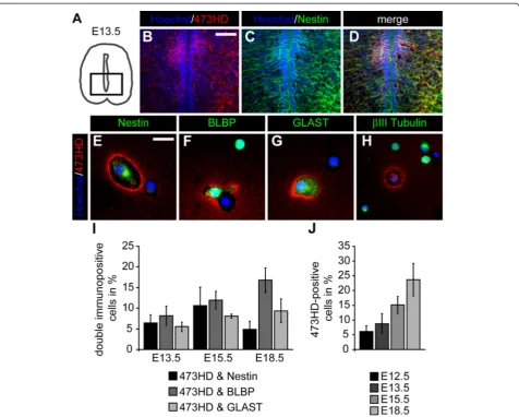
The sulfation-dependent 473HD-epitope is expressed by neural precursor cells in the embryonic mouse spinal cord Since systematic data concerning the expression of GAG chains throughout spinal cord development are missing, we started with an expression analysis of the 473HD-epitope, a representative CS-GAG present on the chondroitin sulfate proteoglycan phosphacan/RPTPβ/ζ [25,27]. Immunohisto-chemical detection of the 473HD-epitope on frontal spinal cord sections between E9.5 and E18.5 show its up-regulation towards the end of neurogenesis at E12.5 (Add-itional file 1: Figure S1A, B). The expression was particularly high in the ventral spinal cord between E13.5 and E15.5 (Additional file 1: Figure S1C, D). Towards the end of em-bryogenesis at E18.5, the 473HD-epitope could be detected within the whole spinal cord except for the central canal re-gion (Additional file 1: Figure S1E). Further immunohisto-chemical analyses on frontal E13.5 spinal cord sections (Figure 1A) revealed that the 473HD-epitope was expressed by Nestin-positive NPCs in vivo (Figure 1B-D). Note that most of the ventricular zone lacks immunoreactivity for the 473HD-epitope except for a distinct region within the ven-tral spinal cord. To investigate the cellular source, we disso-ciated the spinal cord from various embryonic ages and plated single cells in low density for two hours on a poly-DL-ornithine substrate. After that, we immunocytochemi-cally characterized the cells using various cell type specific markers. We observed that many 473HD-positive cells
co-expressed the NPC markers Nestin, BLBP and GLAST (Figure 1E-G). In contrast, we never observed 473HD immunoreactivity on βIII-Tubulin-positive young neurons (Figure 1H). We further quantified the relative number of 473HD-positive cells expressing the NPC markers Nestin, BLBP and GLAST at E13.5, E15.5 and E18.5. Our findings are summarized in Table 1. The percentage of 473HD-positive cells co-expressing one of the mentioned markers was about 5 % for each marker at E13.5 but increased within the next two days to around 10 % (Figure 1I and Table 1). Towards the end of embryogenesis at E18.5, the percentage of Nestin- and-473HD-positive cells decreased again, while the BLBP-and-473HD populations increased and the GLAST-and-473HD populations did not change (Figure 1I and Table 1). Finally, we determined the overall 473HD-positive cell population throughout development and found a general increase in the relative amount of 473HD-positive cells between E12.5 and E18.5, consistent with our immuno-histochemical analyses (E12.5: 6.2 ± 1.9 % (n = 4); E13.5: 9.0 ± 3.3 % (n = 10); E15.5: 15.2 ± 2.7 % (n = 10); E18.5: 23.2 ± 5.3 % (n = 8); Figure 1J).
Sodium chlorate efficiently reduces the level of the sulfation-dependent 473HD-epitope in spinal cord neural precursor cell cultures
Several studies dealing with GAG biology were based on the usage of NaClO3, in order to interfere with the
sulfa-tion levels of the GAG chains. In this study we applied NaClO3and asked whether alterations in sulfation levels
might regulate proliferation, survival and differentiation of spinal cord NPCs grown as free floating neurospheres. We cultured primary neurospheres from E13.5 spinal cord cells and analyzed the expression of the sulfation-dependent 473HD-epitope and its carrier protein RPTPβ/
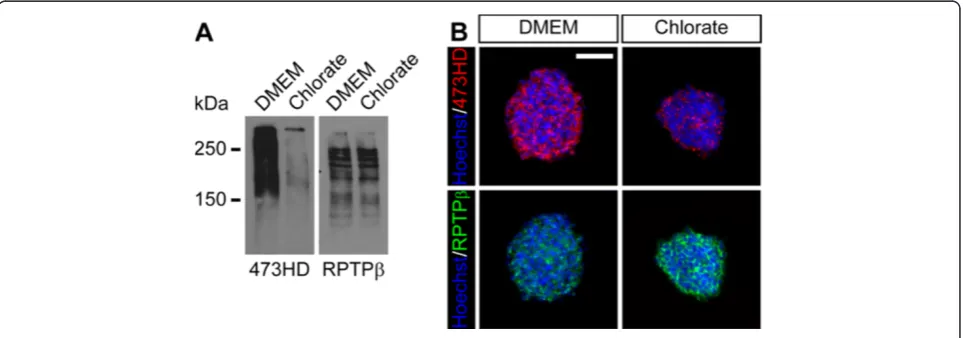
ζ after one week. Western blot analyses of neurosphere detergent extracts revealed that neurospheres expressed high levels of the 473HD-epitope under standard culture conditions. The addition of NaClO3strongly reduced the
473HD levels in comparison to the solvent control (Figure 2A). However, the expression levels of its carrier protein itself appeared not to be affected (Figure 2A). In an independent experiment, neurosphere cryosections were labeled for the 473HD-epitope as well as RPTPβ/ζ. Consistent with the western blot analysis, the 473HD-immunoreactivity was strongly reduced when sulfation levels were decreased using NaClO3 (Figure 2B). The
RPTPβ/ζ immunoreactivity, however, seemed to be enhanced, potentially reflecting an enhanced accessibility of the epitope rather than an increased expression level (Figure 2B), since the western blot analysis did not reveal changes in the RPTPβ/ζexpression. Yet, these data dem-onstrate the suitability of NaClO3to suppress normal
Sodium chlorate affects spinal cord neural precursor cell growth
We recently reported on a decreased neurosphere formation capacity from primary cortical cells upon chlorate treatment [8]. Thus, we started to analyze the influence of NaClO3on
NPC proliferation by assaying the clonal neurosphere for-mation from E13.5 primary spinal cord cells. In order to in-vestigate whether NaClO3 might just affect NPC
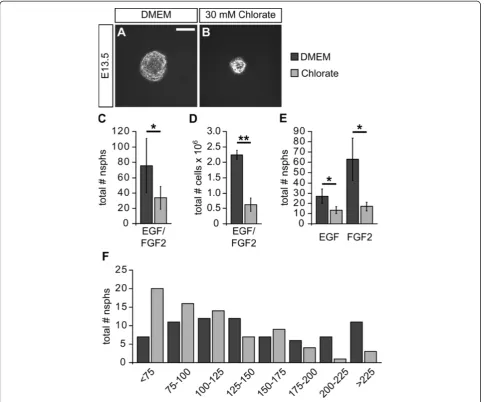
subpopulations, we grew neurospheres under different growth factor conditions. Figure 3A,B shows representative
pictures of neurospheres grown in the presence of EGF and FGF2 under control or NaClO3conditions. Typical
neuro-spheres of 100μm to 200μm diameter formed within one week under control conditions. In contrast, NaClO3-treated
neurospheres were smaller. Moreover, the number of pri-mary neurospheres was significantly reduced upon NaClO3
treatment (without: 75.6 ± 35.2; NaClO3: 33.8 ± 15.1; n = 5;
proliferation rate, was determined after one week. In the presence of NaClO3we observed strongly reduced total cell
numbers in the culture flasks (without: 2,240,000 ± 149,000; NaClO3: 622,000 ± 219,000; n = 4;P= 0.001; Figure 3D).
Next, we analyzed the influence of NaClO3on EGF- or
FGF2-dependent neurosphere formation. Again, the quanti-fication revealed a general decrease in neurosphere forma-tion capacity independently of the growth factor added (EGF only: 27.0 ± 6.9; EGF and NaClO3: 13.3 ± 3.5; n = 3;
P= 0.034; FGF2 only: 63.0 ± 20.7; FGF2 and NaClO3:
17.0 ± 4.4; n = 3;P= 0.044; Figure 3E).
Finally, we further analyzed the effect of NaClO3on the size
of neurospheres grown in the presence of EGF and FGF2 for one week. For that purpose, photomicrographs of single neu-rospheres were used to measure the diameter of respective colonies. Data from four independent experiments revealed that the addition of NaClO3resulted in a shift towards
smal-ler neurospheres at the expense of larger neurospheres (Figure 3F). However, since there were still some large neuro-spheres in the presence of NaClO3, NaClO3itself might only
affect the proliferation rate of a subpopulation of NPCs.
Sodium chlorate leads to G2-phase retention of spinal cord neural precursor cells
In order to further analyze the influence of NaClO3on NPC
proliferation, we examined the cellular composition of neu-rospheres grown for one week in the presence of EGF and FGF2. The percentage of Nestin-positive cells was not altered (without: 82.2 ± 11.7%; NaClO3: 85.6 ± 15.5%; n = 6;
Figure 4A,D). In contrast, the relative number of β III-Tubulin-positive neurons was significantly increased upon NaClO3treatment (without: 1.3 ± 0.5%; NaClO3: 3.2 ± 1.0%;
n = 5; P= 0.014; Figure 4B,E). Finally, we did not observe changes in the percentage of Caspase3-positive cells under-going apoptotic cell death (without: 1.03 ± 0.36%; NaClO3:
1.08 ± 0.44%; n = 4; Figure 4C,F).
Next, we added 1μM BrdU for 1 h after one day and after one week to investigate the overall proliferation rate. Interestingly, the relative number of BrdU-positive cells was not changed for either time point investigated (with-out 1div: 6.3 ± 0.5%; NaClO31div: 7.1 ± 1.1% (n = 3);
with-out 7div: 25.4 ± 8.4%; NaClO3 7div: 33.5 ± 2.2%; (n = 5);
Figure 4G,I), indicating that the S-phase entry was not affected by NaClO3. We next analyzed both the
percent-age of pH3-positive cells after one day and their pH3 staining pattern, to assay for the G2- and M-phase (Figure 4H-H”) [28]. The relative amount of pH3-positive cells was not affected by NaClO3 (without: 3.6 ± 0.6%;
NaClO3: 4.3 ± 1.0%; n = 3; Figure 4J). However,
signifi-cantly more cells remained in the G2-phase after NaClO3
addition, while only a minority of pH3-positive cells pro-ceeded into the M-phase (without G2: 66.0 ± 1.6%; NaClO3 G2: 82.6 ± 1.7%; (n = 4; P= 0.0002); without M:
34.0 ± 1.6%; NaClO3 M: 17.4 ± 1.7%; (n = 4; P= 0.0002);
Figure 4J). These data indicate that NPCs grown in the presence of NaClO3display a G2-phase arrest.
Table 1 Immunocytochemical characterization of 473HD-positive spinal cord cells in the embryonic spinal cord
Marker E12.5 E13.5 E15.5 E18.5
473HD 6.2 ± 1.9% (n = 4)
9.0 ± 3.3% (n = 10)
15.2 ± 2.7% (n = 10)
23.2 ± 5.3% (n = 8)
Nestin/473HD ND 6.4 ± 2.0% (n = 6)
10.6 ± 4.5% (n = 3)
4.9 ± 1.9% (n = 4)
BLBP/473HD ND 8.7 ± 2.4% (n = 5)
12.7 ± 2.4% (n = 5)
17.3 ± 2.6% (n = 4)
GLAST/473HD ND 6.8 ± 2.8% (n = 4)
9.0 ± 2.1% (n = 5)
9.7 ± 2.4% (n = 4)
The data are depicted as mean ± SD. ND: not determined.
Figure 2Sodium chlorate efficiently reduces the expression level of the sulfation-dependent 473HD-epitope. (A)Western blot analysis of neurosphere detergent extracts, showing a reduced 473HD expression level upon NaClO3treatment. The expression level of different RPTPβ/ζisoforms,
known 473HD carrier proteins, did not appear to be changed in the presence of NaClO3.(B)Immunohistochemical detection of the 473HD-epitope
and RPTPβ/ζisoforms on neurosphere cryosections confirmed the reduced 473HD expression levels in NaClO3-treated cultures. Here, the
Sodium chlorate affects Erk-signaling
To gain first insights into potential molecular mechanisms mediating the cell biological influence of NaClO3on spinal
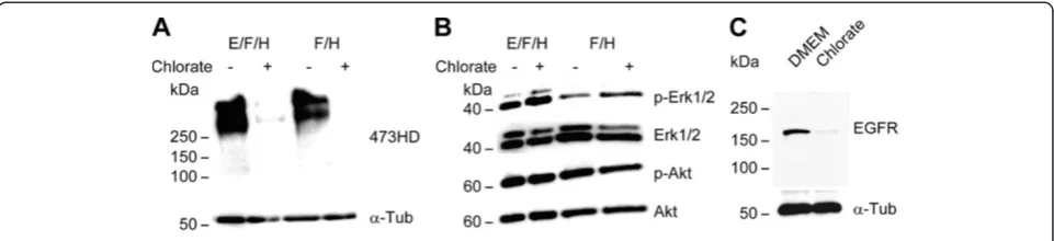
cord NPCs, we analyzed the activation levels of Erk1/2 and Akt, two effector molecules downstream of tyrosine kinase receptors. For that purpose, we used neurospheres from high density cultures grown in the presence of either EGF and FGF2 or FGF2 alone. Both conditions were additionally supplemented with heparin. The addition of NaClO3
resulted in a reduced expression of the 473HD epitope (Figure 5A), confirming the functionality of NaClO3in that
experiment. While the level of phosphorylated Akt (pAkt)
was not changed, we observed increased levels of phos-phorylated Erk1/2 (Figure 5B). Moreover, the overall expres-sion level of the EGFR in the neurospheres was reduced upon NaClO3treatment (Figure 5C). This was in line with
the increased percentage of neurons in the presence of NaClO3.
Sodium chlorate affects neuron numbers under differentiating conditions
Since CS-GAG chains regulate the differentiation of cor-tical NPCs [4-6], we next analyzed the impact of NaClO3
on spinal cord NPC differentiation upon growth factor Figure 3Sodium chlorate affects clonal neurosphere formation from primary spinal cord cells. (A, B)Neurospheres were grown under clonal conditions in the presence of EGF and FGF2. The addition of NaClO3resulted in smaller neurospheres in comparison to the solvent control.(C)
Moreover, the quantification of neurospheres revealed a significantly reduced number of neurospheres in the presence of NaClO3(n = 5;P<0.05).(D)
Consistent with this observation, the total cell number in high density cultures was strongly reduced upon NaClO3treatment after one week (n = 4;P
<0.01).(E)Analysis of EGF- and FGF2-dependent neurosphere formation showed that, under both growth factor conditions, the addition of NaClO3
resulted in a significantly smaller number of neurospheres (n = 3;P<0.05 for both conditions).(F)Using neurosphere size, based on the neurosphere diameter, in the presence or absence of NaClO3,the neurospheres were sorted into different categories. The size distribution histogram shows that
withdrawal. For this purpose, neurospheres grown as high density cultures were dissociated and single cells were ini-tially plated on a poly-DL-ornithine/laminin substrate for four days in the presence of 1 % FCS. The differentiation was immunocytochemically investigated using the cell type
specific markers Nestin, GFAP, O4 and βIII-Tubulin. The general differentiation level was low after four days, since the majority of cells were still Nestin-positive (Additional file 2: Figure S2A). However, several GFAP-positive astro-cytes as well as βIII-Tubulin-positive neurons and O4-Figure 4Sodium chlorate induces G2 arrest of spinal cord neural precursor cells. (A-F)Neurospheres grown as high density culture in the presence of EGF and FGF2 for one week were dissociated, and single cells were plated for 2 h on a poly-DL-ornithine substrate for further analysis. The percentage of Nestin-positive cells was not altered upon NaClO3addition (n = 6). In contrast, the relative number ofβ
III-Tubulin-positive neurons was significantly enhanced (n = 5;P<0.05). NaClO3did not affect the proportion of Caspase3-positive cells (n = 4).(G, I)BrdU
incorporation analysis was performed after one day and after seven days. NaClO3did not affect the BrdU incorporation rate for either time point
investigated (1div: n = 4; 7div: n = 5).(H, J)Moreover, the relative number of pH3-positive cells was also not changed in response to NaClO3.
positive immature oligodendrocytes were also present under both conditions (Additional file 2: Figure S2A, B). Again, counting for the different cell types revealed that the treatment with NaClO3resulted in a higher percentage of
βIII-Tubulin-positive neurons (without: 5.5 ± 1.6%; NaClO3:
8.8 ± 3.4%; n = 7; P= 0.041) (Additional file 2: Figure S2D). In contrast, neither astroglial nor oligodendroglial tiation were significantly affected after four days of differen-tiation (GFAP without: 3.3 ± 2.6%; GFAP with NaClO3:
5.5 ± 4.2%; n =7; O4 without: 1.9 ± 0.6%; O4 with NaClO3:
1.6 ± 1.2%; n = 7) (Additional file 2: Figure S2C, E).
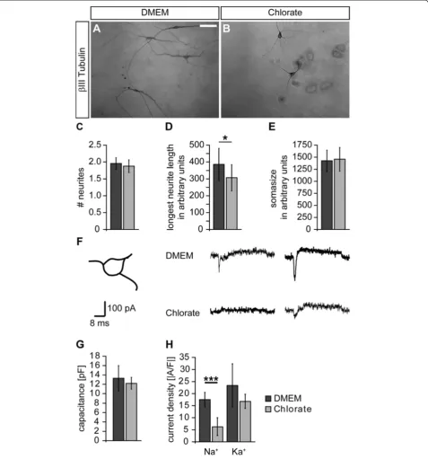
Sodium chlorate affects morphological and functional maturation of neural precursor cell-derived neurons The observation that neuronal differentiation was enhanced in the presence of the sulfation inhibitor NaClO3
prompted us to further examine neuronal maturation with regard to morphology. To address that issue, we plated dis-sociated primary neurosphere cells under differentiating conditions and analyzed the neuronal morphology after four days in terms of neurite number, length of the longest neurite and basal soma size. Figure 6A,B shows representa-tive photomicrographs of neurons grown in the absence and presence of NaClO3. The mean number of neurites
was not affected by NaClO3(without: 1.95 ± 0.17; NaClO3:
1.88 ± 0.19; n = 6; Figure 6C). In contrast, the mean length of the longest neurite was significantly reduced upon NaClO3 treatment (without: 385.5 ± 94.6; NaClO3:
306.8 ± 77.8; n = 6;P= 0.044; Figure 6D). The average soma size was also not changed (without: 1,426 ± 218; NaClO3:
1,454 ± 253; n = 6; Figure 6E).
Next, we analyzed if NaClO3 also affects
electrophysio-logical properties of NPC-derived neurons. Thus, we per-formed whole cell patch-clamp recordings of NPC-derived neurons. To take potential neuronal subpopulations into consideration, we specifically analyzed neurons with a tri-angular cell soma (Figure 6F). Upon supra-threshold depolarization, the neurons usually responded with an
inward current (sodium current component) followed by a prolonged outward current (potassium current component). Following culture in NaClO3, peak sodium currents were
strongly decreased in comparison to cells cultured under control conditions (Figure 6F). To ensure that these differ-ences were not due to differdiffer-ences in cell size, we first deter-mined the cell capacitance and did not observe any changes in response to NaClO3 treatment (without: 13.3 ± 2.7 pF;
NaClO3± 12.2 ± 1.3 pF; n = 5 preparations; Figure 6G).
Then, we measured both the sodium inward and potassium outward current and normalized both components to the cell capacitance, yielding current densities. We found that the sodium current density was significantly reduced in the presence of NaClO3(without: -17.5 ± 2.9 A/F; NaClO3:
-6.3 ± 3.6 A/F; n = 5 preparations; P <0.001; Figure 6H). In contrast, the potassium current density was not significantly changed (without: 23.4 ± 8.9 A/F; NaClO3: 16.8 ± 3.0 A/F;
n = 5 preparations; Figure 6H).
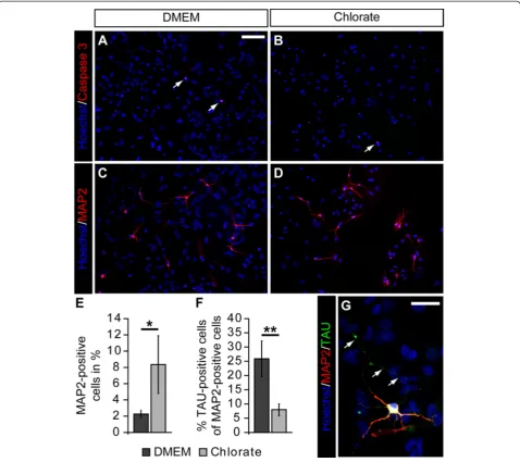
To further analyze the influence of NaClO3 on
neur-onal maturation, we extended the differentiation period for another three days. After that, we first determined the relative number of Caspase3-positive cells to investi-gate whether potential differences might be due to changes in the rate of apoptotic cell death. However, we did not notice any differences in the percentage of Caspase3-positive cells (Figure 7A,B). Next, we counted the number of MAP2-positive cells and found a signifi-cant increase in the percentage of MAP2-positive cells upon NaClO3 treatment (without: 2.3 ± 0.5%; NaClO3:
8.4 ± 3.6% (n = 4; P= 0.015)). Finally, we analyzed the polarization of the neurons in our differentiation assay by staining for MAP2 (dendritic compartment) in com-bination with Tau (axonal compartment). While under control conditions, a substantial number of MAP2-positive cells also had a Tau-MAP2-positive axon; the relative number of clearly polarized neurons in the presence of NaClO3 was significantly reduced (without: 25.9 ± 6.3%;
NaClO3 8.0 ± 2.1%; (n = 4;P= 0.002)). We also observed
Figure 5Sodium chlorate increases Erk1/2 activation levels in primary neurospheres. (A)Treatment of primary neurospheres grown either in the presence of EGF and FGF2 or FGF2 alone with NaClO3reduced the expression level of the sulfation-dependent 473HD epitope.(B)In
contrast, the phosphorylation of Erk1/2 (pErk1/2) was enhanced in response to NaClO3. However, NaClO3did not affect the phosphorylation of
Figure 6Sodium chlorate affects morphological and functional maturation of neural precursor cell-derived neurons. (A, B)In order to analyze the neuronal morphology, theβIII-Tubulin immunoreactivity was visualized using diaminobenzidine. In the control situation the neurons often displayed a multipolar morphology with long neurites. Upon NaClO3treatment the neurites appeared to be shorter.(C-E)To assess the
morphological maturation, the neurite number, the length of the longest neurite and the soma size were measured. The latter two are given in arbitrary units. While the neurite number and the soma size were not altered in the presence of NaClO3(n = 6), the mean length of the longest
neurite was significantly reduced (n = 6;P<0.05).(F)Whole cell patch-clamp recordings of individual NPC-derived neurons were performed under voltage clamp conditions. To take neuronal subpopulations into account, only neurons exhibiting a triangular soma shape were analyzed. Upon depolarization, the neurons responded with a brief sodium inward current followed by a delayed potassium outward current. The inward component was strongly reduced in the presence of NaClO3.(G)The determination of the cell capacitance revealed no changes in the overall
cell size upon NaClO3treatment (n = 5 preparations).(H)Analysis of the two current components expressed as current density clearly showed a
reduced sodium current density in the presence of NaClO3(n = 5 preparations;P<0.001). In contrast, the potassium current density was not
a similar phenomenon in two independent experiments after a ten-day differentiation period (data not shown).
Discussion
GAG chains on proteins of the extracellular matrix or on membrane bound cell adhesion molecules are com-plex carbohydrates that participate in many biological processes during development and in adulthood, in part via their highly specific sulfation patterns. Sulfation of GAGs is performed by sulfotransferases that have been shown to be particularly relevant in developmental
processes [8,29-34]. In this study, we initially investi-gated the expression of the 473HD-epitope, a unique CS-motif, during embryonic spinal cord development and found that it is expressed at late embryonic ages. Its pronounced expression within the NPC compartment, moreover, suggests a functional role of sulfated GAG chains in the regulation of spinal cord NPC biology. Along these lines it has been shown that highly sulfated complex GAG structures are expressed by cortical NPCs [35-37] and regulate proliferation and differentiation of embryonic and adult cortical NPCs [4-7,38]. Thus, we Figure 7Sodium chlorate attenuates neuronal polarization of neural precursor cell-derived neurons. (A-D, G)Photomicrographs of
differentiated NPCs stained for cleaved Caspase3, MAP2, and Tau after seven days of differentiation.(A-D)NaClO3treatment did not result in an increased
apoptotic cell death after seven days. However, in line with an increased percentage ofβIII-Tubulin-positive neurons after four days, the relative number of MAP2-positive cells was enhanced in response to NaClO3.(E)The quantification of MAP2-positive neurons revealed a significant increase in the relative amount of
neurons in NaClO3treated cultures (n = 4;P= 0.015).(F, G)Interestingly, the percentage of Tau-positive cells among MAP2-positive neurons was strongly reduced
analyzed the role of changes in sulfation on the prolif-eration and differentiation potential of mouse embryonic spinal cord NPC using NaClO3 as a potent inhibitor of
eukaryotic sulfation reactions. Low concentrations of NaClO3(2 mM to 5 mM) preferentially inhibit sulfation
of CS-GAGs, whereas higher concentrations (15 mM to 30 mM) are required to inhibit sulfation of HS-GAGs as well [39]. We used a concentration of 30 mM, to assure an almost complete sulfation blockage of both CS-GAGs and HS-GAGs. To monitor the effectiveness of NaClO3,
we analyzed the expression level of the 473HD-epitope, and observed an almost complete reduction of the epi-tope after NaClO3treatment. This observation is in line
with a former study demonstrating that the 473HD-epitope depends on a distinct sulfation pattern [40]. Some residual 473HD immunoreactivity in the western blot as well as in the immunohistochemical analyses is likely due to the fact that NaClO3might not affect every
neurosphere cell as soon as the neurospheres reach a certain size. However, since the total amount of the car-rier protein RPTPβ/ζ does not change, our data prove the functionality of NaClO3 to specifically affect
sulfa-tion events.
We next analyzed clonal neurosphere formation cap-acity from E13.5 spinal cord NPCs. Treatment of neuro-sphere cultures with 30 mM NaClO3 resulted in
significantly fewer and smaller neurospheres. This effect was independent from the growth factor used to propa-gate the neurospheres (EGF, FGF2 or both; Figure 4 C-E). Looking at the diameter of the spheres, it became obvious that there are more small-diameter neurospheres and fewer large-diameter neurospheres when the cultures were treated with NaClO3 (Figure 4A,B,F). This
phenomenon has been observed previously for the em-bryonic telencephalic neurospheres [8]. The reasons for the altered neurosphere growth could be that NPCs within the neurosphere divide more slowly in the pres-ence of NaClO3;NPCs die more frequently; or NPCs have
a higher differentiation capacity, and therefore NaClO3
might affect stem or precursor cell maintenance. Our BrdU incorporation analysis did not reveal any changes in the S-phase of the cell cycle. However, while the relative number of pH3-positive cells present in the G2-phase of the cell cycle was significantly enhanced, the percentage of M-phase cells was significantly decreased, indicating a potential G2-phase arrest in the presence of NaClO3.
Al-though a G2-arrest is a common phenomenon preceding apoptotic cell death, especially in cancer cells [41], we did not observe an increased percentage of Caspase3-positive cells, Thus, it is unlikely that NaClO3affected cell death
rates in our culture system. Besides the difference with re-gard to the G2/M-phase of the cell cycle, we also docu-mented a significant increase of βIII-Tubulin-positive young neurons in response to NaClO3 treatment.
Furthermore, the overall expression level of EGFR in the neurosphere cultures was reduced. This is also in line with an increased percentage of βIII-Tubulin-positive neurons, since the acquisition of the EGFR is generally considered a hallmark for the developmental switch from neurogenic NPCs towards gliogenic NPCs [42]. However, based on our data, we conclude that NaClO3 primarily
affects spinal cord NPC proliferation by changing the cell cycle kinetics. This results in a higher number of neurons already under proliferative conditions. However, it remains unsolved whether NaClO3truly promotes
neuro-genic differentiation. It might be that the enhanced per-centage of neurons simply reflects the reduced proliferation capabilities of the surrounding cells.
Regarding potential molecular mechanisms, we ini-tially reasoned that the suppression of sulfation might compromise growth factor signaling, leading to changes in the activation levels of downstream effector mole-cules such as Erk1/2 and Akt. Surprisingly, we observed an increased Erk1/2 activation in the presence of NaClO3. Yet an increased Erk1/2 activation has already
been reported previously in the context of a G2 arrest in different cancer cell lines [43,44]. It is unlikely that the increased Erk1/2 activation depends on increased receptor activation, because the lack of sulfation should result in a reduced growth factor signaling strength. Along these lines, we recently showed that the enzym-atic degradation of CS-GAGs compromises FGF signal-ing in cortical neurosphere cultures, resultsignal-ing in a reduced MAP kinase activation [5]. Here, we think that NaClO3might reduce the activation levels of
phospha-tases through a yet unknown mechanism. In this con-text, it is noteworthy that RPTPβ/ζ bears sulfated CS-GAGs [25] and regulates Erk1/2 activation levels in human keratinocytes [45]. Therefore, the sulfation sta-tus of RPTPβ/ζmight have a critical influence on its ac-tivation level and as a consequence also on the Erk1/2 activation levels.
Our laboratory has recently shown that degradation of CS-GAGs from telencephalic NPCs using ChABC results in a pronounced glial differentiation at the ex-pense of neuronal cell types [6]. With regard to spinal cord NPCs, we observed a higher proportion of neuronal cells already under proliferative conditions. Under differ-entiating conditions we documented an enhanced rela-tive number ofβIII-Tubulin-positive cells after four days and MAP2-positive cells after one week following NaClO3treatment. These data are in line with previous
To further elucidate the phenotype of theβ III-Tubulin-positive cells, we took a closer look at the morphology of these cells and valued the morphology as a level of matur-ation. The spinal cord NPC-derived neurons exhibited no changes with respect to soma size and total primary neur-ite number upon NaClO3treatment. But they significantly
differed in the length of their longest neurite. This was surprising, since CSPGs and HSPGs are primarily known for their inhibitory influence on neurite outgrowth, par-ticularly under pathological conditions [10,47]. Moreover, CS-GAGs have recently been shown to influence the morphology of NPC-derived neurons in terms of neurite number, length and branching [4,48]. Yet there is growing evidence that specific CS-GAGs also promote neurite out-growth depending on both the mode of their presentation (that is, homogenous substrate or alternating substrate) and on the neuronal cell type [13,25,49]. Thus, we believe that our data hint towards an attenuated maturation of spinal cord NPC-derived neurons in the presence of NaClO3 in vitro. In line with this hypothesis, we also
documented a strongly reduced neuronal polarity after seven and ten days in culture. To check whether this delayed morphological maturation is also relevant at a functional level, we analyzed the differentiated neurons grown from NaClO3-treated dissociated cultures in
com-parison with the control group using whole cell patch-clamp recordings. We determined that the overall cell capacitance of the cells was not changed. This is in line with the fact that the soma size was not changed either. However, we observed a significantly reduced sodium current density, while the potassium current density remained unaltered. Lower sodium currents suggest a delay in functional neuronal maturation, because sodium currents usually increase upon differentiation of neuronal precursors during embryogenesis [50]. Furthermore, neur-ite outgrowth and sodium channel- dependent excitability have previously been shown to be tightly coupled during functional neuronal maturation [51]. Interestingly, the addition of FGF2 to rat postnatal hippocampal neurons significantly enhances the sodium current density, demon-strating a growth factor signaling-dependent regulation [52]. This might be mediated by highly sulfated GAG chains. In this context, it is noteworthy that we recently demonstrated changes in spontaneous synaptic activity of primary hippocampal neurons upon ChABC-mediated CS-GAG degradation [53].
Conclusions
Based on our data, we believe that normal sulfation levels of CSPGs and HSPGs during spinal cord histogenesis pro-mote cellular maturation in general and neuronal matur-ation in particular. Since both CSPGs and HSPGs also show a pronounced expression under pathological condi-tions, our data particularly highlight the need for a better
understanding of the impact of specific PGs and their sul-fation patterns for neural development and regeneration.
Additional files
Additional file 1:Figure S1.CS-GAGs are expressed during early gliogenesis of mouse spinal cord development. (A-E) Photomicrographs of frontal spinal cord sections stained against the 473HD epitope. At the beginning of neurogenesis (E9.5) the 473HD epitope was mainly expressed in the meninges and the surrounding parenchyma. Towards the end of neurogenesis (E12.5) the 473HD epitope appeared at the ventral central canal. One day later the immunoreactivity has expanded into the prospective ventral white matter and to a less extent into the dorsal spinal cord. Note that both the floor plate and the roof plate lacked any immunoreactivity. At E15.5 the immunoreactivity was even stronger in the ventral spinal cord and still low in the dorsal part. Another three days later the 473HD epitope was evenly distributed throughout the whole spinal cord except for the region around the central canal. To visualize the nuclei, the cells were counterstained with Hoechst 33528. Scale bar: 50μm (A, B), 100μm (C-D).
Additional file 2:Figure S2.Sodium chlorate promotes neuronal differentiation of spinal cord NPCs. (A, B) After one week in the presence of EGF and FGF2 neurospheres were dissociated, and the differentiation capacity was immunocytochemically analyzed using cell type specific markers. The overall differentiation was low, since about 75% of all cells still expressed the NPC marker Nestin after that time period. (C) The quantification of GFAP-positive astrocytes showed no differences between the NaClO3and control conditions (n = 7). (D) However, the
number ofβIII-Tubulin-positive neurons was significantly increased in the presence of NaClO3(n = 7;P<0.05). (E) The amount of O4-positive
immature oligodendrocytes was not changed (n = 7). Hoechst 33528 was added, to visualize the cell nuclei. Scale bar: 50μm.
Abbreviations
AKT: Protein kinase B (also PKB); BLBP: Brain lipid binding protein; BrdU: 5-bromo2’-deoxy-uridine; CNS: Central nervous system; CSPG: Chondroitin sulfate proteoglycan; Div: Day(s) in vitro; E0.5: E9.5, E12.5, E18.5, Embryonic day 0.5, 9.5, 12.5, 18.5; EGF: Epidermal growth factor; EGFR: Epidermal growth factor receptor; ERK: Extracellular signal-regulated protein kinase;
FGF: Fibroblast growth factor; FGFR: Fibroblast growth factor receptor; GAG: Glycosaminoglycan; GFAP: Glial fibrillary acidic protein; GLAST: Glutamate Aspartate transporter; H: Hour(s); HRP: Horse-reddish peroxidase; MAP2: Microtubule-associated protein 2; mM: Milli Molar; NaClO3: Sodium Chlorate; NPC: Neural precursor cell; PBS: Phosphate-buffered saline; PG: Proteoglycan; pH3: Phosphor-Histone 3; RPTP: Receptor protein tyrosine phosphatase.
Competing interest
The authors declare that they have no competing interests.
Acknowledgments
MK was supported through the PhD program of the International Graduate School of Neuroscience (IGSN), the Research School at the Ruhr-University supported by the DFG (GSC 98/1), and the Wilhelm and Günther Esser Foundation. We gratefully acknowledge grant support to AF from the Ruhr-University (President’s special programme call 2008).
Author details
1Department of Cell Morphology and Molecular Neurobiology,
Ruhr-University Bochum, Bochum, Germany.2Group for Molecular Cell Biology, Ruhr-University Bochum, Bochum, Germany.3Department of
Neurobiochemistry, Ruhr-University Bochum, Bochum, Germany.4International Graduate School of Neuroscience, Ruhr-University Bochum, Bochum, Germany.
Authors’contributions
designed the electrophysiological experiments. AF designed part of the immunohistochemical experiments. SW conceived the study and helped drafting the manuscript. All authors read and approved the final manuscript.
Received: 21 November 2011 Accepted: 6 March 2012 Published: 8 June 2012
References
1. Kleene R, Schachner M:Glycans and neural cell interactions.Nat Rev
Neurosci2004,5:195–208.
2. Yanagisawa M, Yu RK:The expression and functions of glycoconjugates in neural stem cells.Glycobiology2007,17:57R–74R.
3. Yu RK, Yanagisawa M:Glycosignaling in neural stem cells: involvement of glycoconjugates in signal transduction modulating the neural stem cell fate.J Neurochem2007,103(Suppl 1):39–46.
4. Gu WL, Fu SL, Wang YX, Li Y, Lu HZ, Xu XM, Lu PH:Chondroitin sulfate proteoglycans regulate the growth, differentiation and migration of multipotent neural precursor cells through the integrin signaling pathway.BMC Neurosci2009,10:128.
5. Sirko S, von Holst A, Weber A, Wizenmann A, Theocharidis U, Gotz M, Faissner A:Chondroitin sulfates are required for fibroblast growth factor-2-dependent proliferation and maintenance in neural stem cells and for epidermal growth factor-dependent migration of their progeny.Stem
Cells2010,28:775–787.
6. Sirko S, von Holst A, Wizenmann A, Gotz M, Faissner A:Chondroitin sulfate glycosaminoglycans control proliferation, radial glia cell differentiation and neurogenesis in neural stem/progenitor cells.Development2007,
134:2727–2738.
7. Tham M, Ramasamy S, Gan HT, Ramachandran A, Poonepalli A, Yu YH, Ahmed S:CSPG is a secreted factor that stimulates neural stem cell survival possibly by enhanced EGFR signaling.PLoS One2010,5:e15341. 8. Akita K, von Holst A, Furukawa Y, Mikami T, Sugahara K, Faissner A:
Expression of multiple chondroitin/dermatan sulfotransferases in the neurogenic regions of the embryonic and adult central nervous system implies that complex chondroitin sulfates have a role in neural stem cell maintenance.Stem Cells2008,26:798–809.
9. Properzi F, Carulli D, Asher RA, Muir E, Camargo LM, van Kuppevelt TH, ten Dam GB, Furukawa Y, Mikami T, Sugahara K, Toida T, Geller HM, Fawcett JW:
Chondroitin 6-sulphate synthesis is up-regulated in injured CNS, induced by injury-related cytokines and enhanced in axon-growth inhibitory glia.
Eur J Neurosci2005,21:378–390.
10. Silver J, Miller JH:Regeneration beyond the glial scar.Nat Rev Neurosci 2004,5:146–156.
11. Garcia-Alias G, Barkhuysen S, Buckle M, Fawcett JW:Chondroitinase ABC treatment opens a window of opportunity for task-specific rehabilitation.
Nat Neurosci2009,12:1145–1151.
12. Wang D, Ichiyama RM, Zhao R, Andrews MR, Fawcett JW:Chondroitinase combined with rehabilitation promotes recovery of forelimb function in rats with chronic spinal cord injury.J Neurosci2011,31:9332–9344. 13. Lin R, Rosahl TW, Whiting PJ, Fawcett JW, Kwok JC:6-sulphated
chondroitins have a positive influence on axonal regeneration.PLoS One 2011,6:e21499.
14. Eswarakumar VP, Lax I, Schlessinger J:Cellular signaling by fibroblast growth factor receptors.Cytokine Growth Factor Rev2005,16:139–149. 15. Capela A, Temple S:LeX/ssea-1 is expressed by adult mouse CNS stem
cells, identifying them as nonependymal.Neuron2002,35:865–875. 16. Capela A, Temple S:LeX is expressed by principle progenitor cells in the
embryonic nervous system, is secreted into their environment and binds Wnt-1.Dev Biol2006,291:300–313.
17. Hennen E, Czopka T, Faissner A:Structurally distinct LewisX glycans distinguish subpopulations of neural stem/progenitor cells.J Biol Chem 2011,286:16321–16331.
18. Liedtke S, Geyer H, Wuhrer M, Geyer R, Frank G, Gerardy-Schahn R, Zahringer U, Schachner M:Characterization of N-glycans from mouse brain neural cell adhesion molecule.Glycobiology2001,11:373–384. 19. Habuchi O:Diversity and functions of glycosaminoglycan
sulfotransferases.Biochim Biophys Acta2000,1474:115–127. 20. Kusche-Gullberg M, Kjellen L:Sulfotransferases in glycosaminoglycan
biosynthesis.Curr Opin Struct Biol2003,13:605–611.
21. Jones KS, Petrow-Sadowski C, Bertolette DC, Huang Y, Ruscetti FW:Heparan sulfate proteoglycans mediate attachment and entry of human T-cell
leukemia virus type 1 virions into CD4+ T cells.J Virol2005,
79:12692–12702.
22. Karus M, Denecke B, Ffrench-Constant C, Wiese S, Faissner A:The extracellular matrix molecule tenascin C modulates expression levels and territories of key patterning genes during spinal cord astrocyte specification.Development2011,138:5321–5331.
23. Sirko S, Akita K, Von Holst A, Faissner A:Structural and functional analysis of chondroitin sulfate proteoglycans in the neural stem cell niche.
Methods Enzymol2010,479:37–71.
24. Sommer I, Schachner M:Monoclonal antibodies (O1 to O4) to
oligodendrocyte cell surfaces: an immunocytological study in the central nervous system.Dev Biol1981,83:311–327.
25. Faissner A, Clement A, Lochter A, Streit A, Mandl C, Schachner M:Isolation of a neural chondroitin sulfate proteoglycan with neurite outgrowth promoting properties.J Cell Biol1994,126:783–799.
26. Kempf M, Clement A, Faissner A, Lee G, Brandt R:Tau binds to the distal axon early in development of polarity in a microtubule- and microfilament-dependent manner.J Neurosci1996,16:5583–5592. 27. Garwood J, Schnadelbach O, Clement A, Schutte K, Bach A, Faissner A:
DSD-1-proteoglycan is the mouse homolog of phosphacan and displays opposing effects on neurite outgrowth dependent on neuronal lineage.
J Neurosci1999,19:3888–3899.
28. Bossard C, Jarry A, Colombeix C, Bach-Ngohou K, Moreau A, Loussouarn D, Mosnier JF, Laboisse CL:Phosphohistone H3 labelling for histoprognostic grading of breast adenocarcinomas and computer-assisted
determination of mitotic index.J Clin Pathol2006,59:706–710. 29. Conway CD, Howe KM, Nettleton NK, Price DJ, Mason JO, Pratt T:Heparan
sulfate sugar modifications mediate the functions of slits and other factors needed for mouse forebrain commissure development.J Neurosci 2011,31:1955–1970.
30. Ishii M, Maeda N:Oversulfated chondroitin sulfate plays critical roles in the neuronal migration in the cerebral cortex.J Biol Chem2008,
283:32610–32620.
31. Ishii M, Maeda N:Spatiotemporal expression of chondroitin sulfate sulfotransferases in the postnatal developing mouse cerebellum.
Glycobiology2008,18:602–614.
32. Ito Z, Sakamoto K, Imagama S, Matsuyama Y, Zhang H, Hirano K, Ando K, Yamashita T, Ishiguro N, Kadomatsu K:N-acetylglucosamine 6-O-sulfotransferase-1-deficient mice show better functional recovery after spinal cord injury.J Neurosci2010,30:5937–5947.
33. Properzi F, Lin R, Kwok J, Naidu M, van Kuppevelt TH, Ten Dam GB, Camargo LM, Raha-Chowdhury R, Furukawa Y, Mikami T, Sugahara K, Fawcett JW:Heparan sulphate proteoglycans in glia and in the normal and injured CNS: expression of sulphotransferases and changes in sulphation.Eur J Neurosci2008,27:593–604.
34. Wilson VA, Gallagher JT, Merry CL:Heparan sulfate 2-O-sulfotransferase (Hs2st) and mouse development.Glycoconj J2002,19:347–354. 35. Ford-Perriss M, Turner K, Guimond S, Apedaile A, Haubeck HD, Turnbull J,
Murphy M:Localisation of specific heparan sulfate proteoglycans during the proliferative phase of brain development.Dev Dyn2003,227:170–184. 36. Kabos P, Matundan H, Zandian M, Bertolotto C, Robinson ML, Davy BE, Yu
JS, Krueger RC Jr:Neural precursors express multiple chondroitin sulfate proteoglycans, including the lectican family.Biochem Biophys Res
Commun2004,318:955–963.
37. von Holst A, Sirko S, Faissner A:The unique 473HD-Chondroitinsulfate epitope is expressed by radial glia and involved in neural precursor cell proliferation.J Neurosci2006,26:4082–4094.
38. Kerever A, Schnack J, Vellinga D, Ichikawa N, Moon C, Arikawa-Hirasawa E, Efird JT, Mercier F:Novel extracellular matrix structures in the neural stem cell niche capture the neurogenic factor fibroblast growth factor 2 from the extracellular milieu.Stem Cells2007,25:2146–2157.
39. Fjeldstad K, Pedersen ME, Vuong TT, Kolset SO, Nordstrand LM, Prydz K:
Sulfation in the Golgi lumen of Madin-Darby canine kidney cells is inhibited by brefeldin A and depends on a factor present in the cytoplasm and on Golgi membranes.J Biol Chem2002,277:36272–36279. 40. Clement AM, Nadanaka S, Masayama K, Mandl C, Sugahara K, Faissner A:
The DSD-1 carbohydrate epitope depends on sulfation, correlates with chondroitin sulfate D motifs, and is sufficient to promote neurite outgrowth.J Biol Chem1998,273:28444–28453.
42. Lillien L, Raphael H:BMP and FGF regulate the development of EGF-responsive neural progenitor cells.Development2000,127:4993–5005. 43. Guo Y, Zhang X, Meng J, Wang ZY:An anticancer agent icaritin induces
sustained activation of the extracellular signal-regulated kinase (ERK) pathway and inhibits growth of breast cancer cells.Eur J Pharmacol2011,
658:114–122.
44. Wei F, Xie Y, Tao L, Tang D:Both ERK1 and ERK2 kinases promote G2/M arrest in etoposide-treated MCF7 cells by facilitating ATM activation.Cell
Signal2010,22:1783–1789.
45. Xu Y, Xia W, Baker D, Zhou J, Cha HC, Voorhees JJ, Fisher GJ:Receptor-type protein tyrosine phosphatase beta (RPTP-beta) directly
dephosphorylates and regulates hepatocyte growth factor receptor (HGFR/Met) function.J Biol Chem2011,286:15980–15988.
46. Sasaki N, Hirano T, Ichimiya T, Wakao M, Hirano K, Kinoshita-Toyoda A, Toyoda H, Suda Y, Nishihara S:The 3′-phosphoadenosine
5′-phosphosulfate transporters, PAPST1 and 2, contribute to the maintenance and differentiation of mouse embryonic stem cells.PLoS One2009,4:e8262.
47. Busch SA, Silver J:The role of extracellular matrix in CNS regeneration.
Curr Opin Neurobiol2007,17:120–127.
48. See J, Bonner J, Neuhuber B, Fischer I:Neurite outgrowth of neural progenitors in presence of inhibitory proteoglycans.J Neurotrauma2010,
27:951–957.
49. Klausmeyer A, Conrad R, Faissner A, Wiese S:Influence of glial-derived matrix molecules, especially chondroitin sulfates, on neurite growth and survival of cultured mouse embryonic motoneurons.J Neurosci Res2010,
89:127–141.
50. Dietzel ID:Voltage-gated ion currents in embryogenesis.Perspect Dev
Neurobiol1995,2:293–308.
51. Brackenbury WJ, Calhoun JD, Chen C, Miyazaki H, Nukina N, Oyama F, Ranscht B, Isom LL:Functional reciprocity between Na + channel Nav1.6 and beta1 subunits in the coordinated regulation of excitability and neurite outgrowth.Proc Natl Acad Sci U S A2010,107:2283–2288. 52. Niederkinkhaus V, Marx R, Hoffmann G, Dietzel ID:Thyroid hormone
(T3)-induced up-regulation of voltage-activated sodium current in cultured postnatal hippocampal neurons requires secretion of soluble factors from glial cells.Mol Endocrinol2009,23:1494–1504.
53. Pyka M, Wetzel C, Aguado A, Geissler M, Hatt H, Faissner A:Chondroitin sulfate proteoglycans regulate astrocyte-dependent synaptogenesis and modulate synaptic activity in primary embryonic hippocampal neurons.
Eur J Neurosci2011,33:2187–2202.
doi:10.1186/1749-8104-7-20
Cite this article as:Karuset al.:Normal sulfation levels regulate spinal cord neural precursor cell proliferation and differentiation.Neural Development20127:20.
Submit your next manuscript to BioMed Central and take full advantage of:
• Convenient online submission
• Thorough peer review
• No space constraints or color figure charges
• Immediate publication on acceptance
• Inclusion in PubMed, CAS, Scopus and Google Scholar
• Research which is freely available for redistribution