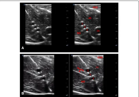
Ultrasound-guided regional anesthesia simulation: use of meat glue in inexpensive and realistic nerve block models
Full text
Figure


Related documents
First step engages sending the employed bee to locate out the Association rules to assess their values then the Association rules were chosen by the onlooker based on the data
The second column indicates whether or not the changepoint is statistically significant for the changepoint type given in the first column; they are “Yes” or “?” in the
this data and data from building information systems applies a simple machine learning
Likewise, non-signifi- cant regression coefficients were obtained when the exchangeable sodium content of only the sur- face layer in the soil was tested against
processes. which conveys best the sense of the word "ideology" as originally envisaged by Marx. A s a result of these struggles, a schism has c o m e to exist between
provide information on contaminant levels in the various fish species in the Detroit River and provide 81.. guidance on the safest consumption
Although spirit- levelled height differences are usually quite precise, the derived orthometric heights for a region or nation are supposed to be the result of an
Western blot analysis for pTau ( Figure 8G and Figure S8 ) revealed significant elevations (p<0.04) in sonicated cortex but not in the hippocampus from Group C rats,
