INTRODUCTION
Malignant gliomas (MGs), including glioblastomas (GBMs) and anaplastic as-trocytomas (AAs), are the most common primary brain tumors (1). The prognosis for patients diagnosed with MG remains poor, with a median survival time of up to 3 years (2,3). Current conventional treatment protocols include maximally safe surgical resection followed by frac-tioned radiation therapy of the tumor and surrounding brain parenchyma and systemic chemotherapy with alkylating compounds. The efficacy of alkylating compounds, however, such as ni-trosoureas or temozolamide, is fairly lim-ited by the epigenetic inactivation of the DNA repair enzyme methylguanine methyltransferase (MGMT). Other DNA repair pathways, such as the DNA mis-match repair and the base excision repair pathways, have also been proposed as significant mechanisms of resistance to alkylating agents. Defects in these
path-ways can cause errors in DNA base pair-ing, which arise during DNA replication, and consequent chemoresistance to alky-lating agents (4).
In this review, developments in molec-ularly targeted therapies for MGs are critically evaluated, and advances in the molecular and genetic pathogenesis of these lethal brain malignancies are also discussed.
MOLECULAR PATHOGENESIS OF GLIOMAS
The biological features of MGs consist of high resistance to apoptosis and florid necrosis (5). Briefly, common molecular, genetic, and epigenetic alterations in pri-mary GBMs include amplification of the epidermal growth factor receptor (EGFR), deletion or mutation of homozy-gous cycldependent kinase (CDK) in-hibitor p16INK4A(CDKN2A), and alter-ations in tumor suppressor phosphatase and tensin homolog (PTEN) on
chromo-some 10 (6). Primary and secondary GBMs share similar characteristics, and few molecular and genetic alterations make them distinguishable from one an-other. For instance, human double-minute 2 (HDM2) and p53 mutations are often observed to be amplified in second-ary GBMs (7).
In regard to AAs, inactivating muta-tions of tumor-suppressor gene TP53and elevated expression of platelet-derived growth factor (PDGF) ligands and recep-tors are commonly observed in grade III AAs (8). Loss of heterozygosity in chro-mosome 10qhas also been detected in primary high-grade AAs, and the inacti-vation of PTEN is observed in approxi-mately 40% of AAs that have lost chro-mosome 10q(9).
Mutations in p16 are also involved, be-cause hypermethylation in the promotor region of p16 has been detected in sev-eral cases of MGs, thus silencing p16 ex-pression and possibly contributing to tumor genesis (10). Additionally, Bcl2-like 12 (Bcl2L12) interacts with and neu-tralizes caspase-7; and increased Bcl2L12 expression inhibits apoptosis (11). The astrocyte elevated gene-1 (AEG-1) has also been implicated in the pathogenetic hallmark of MGs (12). AEG-1is overex-pressed in the majority of human MG samples, and cooperates with the Ha-ras
Andreas A Argyriou and Haralabos P Kalofonos
Department of Medicine—Division of Clinical Oncology, University Hospital, University of Patras Medical School, Rion-Patras, Greece
This review critically evaluates current knowledge of molecularly targeted therapies of malignant gliomas. Various molecularly targeted single-agent therapies, including targeted therapies of growth and survival, have been evaluated in clinical trials but have failed to demonstrate a significant survival benefit compared with standard treatment regimens. The efficacy of multi-targeted kinase inhibitors or combinations of single-multi-targeted kinase inhibitors is a promising strategy, but requires additional clini-cal evaluation before definitive conclusions can be made. Important areas for further research include the assessment of serum or tissue biomarkers, the elucidation of prognostic molecular markers, and the determination of whether the mechanism of ac-tion of a drug is appropriate to the genetic alteraac-tions observed within individual tumors.
© 2009 The Feinstein Institute for Medical Research, www.feinsteininstitute.org Online address: http://www.molmed.org
doi: 10.2119/molmed.2008.00123
Address correspondence and reprint requests to Haralabos P Kalofonos, Department of Medicine—Division of Oncology, University Hospital, University of Patras Medical School, Rion-Patras 26504, Greece. Phone: + 30 2610 999535; Fax: + 30 2610 994645; E-mail: kalofon@ med.upatras.gr.
family of retrovirus-associated DNA se-quences (RAS) to promote cellular trans-formation and subsequently to augment invasion and growth of transformed cells (8,9). Furthermore, oncogenic Ha-ras in-duces AEG-1 expression by modulating the phosphatidylinositol 3-kinase (PI3K)-Akt signaling pathway, thus contributing to the growth of MGs (13).
MOLECULARLY TARGETED THERAPY
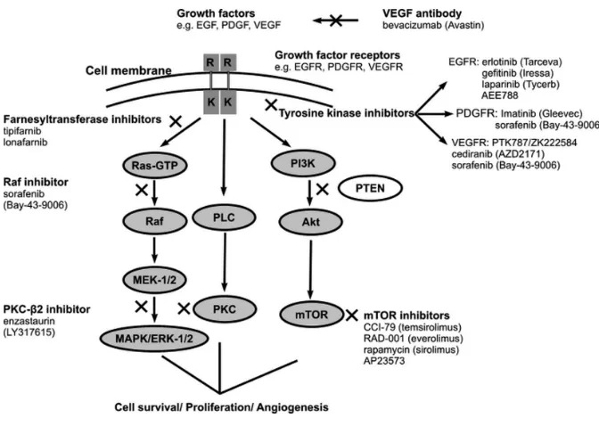
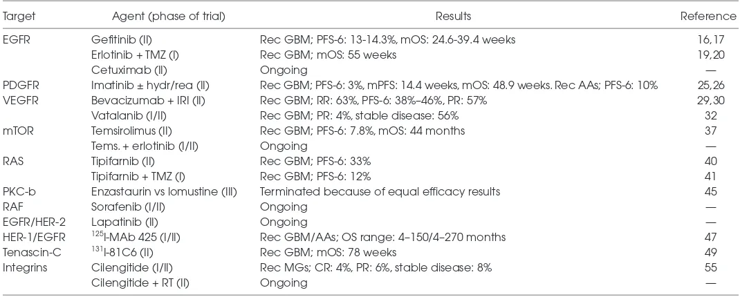
Elevated expression or mutation of re-ceptors and intracellular downstream ef-fectors has been observed in MGs (14). These pathways are regulated by several growth factors linked to tyrosine kinase, such as the EGFR, insulin-like growth factor receptor (IGFR), PDGF receptor (PDGFR), and vascular EGF receptor (VEGFR). Specific targeting of these sig-naling pathways that lead to uncon-trolled cellular proliferation and cell mi-gration and invasion could provide new molecularly targeted treatment options for MGs. The growth factor signaling pathways and their inhibition in MGs are shown in Figure 1 (14), and Table 1 sum-marizes the major clinical trials of molec-ularly targeted therapies in MGs.
EGFRs
The EGFR belongs to the ErbB family of tyrosine kinase receptors. The pres-ence of EGFRvIII, a specific variant of the EGFR lacking exons 2 to 7, is an in-dependent predictor of poor survival in patients with primary MGs (15). Treat-ment options that target EGFRs include gefitinib (ZD-1839) and erlotinib (OSI-774). However, efficacy of these agents is modest.
Gefitinib was evaluated in an open-label, single-center phase II clinical trial in patients (n = 57) with the first recur-rence of a GBM (16). Each patient ini-tially received 500 mg of gefitinib (orally, once a day), and the dose was escalated up to 1000 mg in patients receiving enzyme-inducing antiepileptic drugs or dexamethasone. Quantification of gefi-tinib antiglioma efficacy was assessed by 6-month progression-free survival (PFS-6) and brain magnetic resonance
imaging (MRI) quantification of tumor response (radiographic response). The study population had a PFS-6 of 13% (7 of 53 patients) and a median overall survival (OS) time from treatment initia-tion of 39.4 weeks, but no radiographic response was observed (16). In a multi-center, open-label, single-arm phase II clinical trial of gefitinib in patients (n = 28) with recurrent MG after surgery plus radiotherapy and first-line chemother-apy, overall median time to progression (TTP)was 8.4 weeks, PFS-6 was 14.3%, and median OS was 24.6 weeks (17). None of the patients presented objective radiographic response (17), and it was concluded that gefitinib demonstrated limited efficacy against GBM compared with the standard Stupp regimen
(radio-therapy plus temozolamide; median PFS 6.9 months; PFS-6 53.9%, and median OS 58 weeks) (18).
In several phase I/II clinical trials, OS rates for erlotinib and gefitinib treatment were similar, but erlotinib was more ef-fective than gefitinib treatment in terms of objective radiographic responses (19,20). A multicenter, open-label phase I clinical trial evaluated the efficacy of er-lotinib plus radiation therapy in patients (n = 19) with GBM. With a median fol-low-up of 52 weeks, progression was as-sessed in 16 patients and 13 deaths oc-curred. Median TTP was 26 weeks and median OS was 55 weeks (19). Addition-ally, in an open-label phase I dose-escalation clinical trial, patients (n = 83) with stable or progressive malignant
mary gliomas received erlotinib alone or combined with temozolomide (20). Of the patients assessed (n = 57), eight tients demonstrated a PR and six pa-tients demonstrated a median PFS of greater than 6 months, which included four patients with a PR (20). Erlotinib treatment was equally as effective as the standard Stupp regimen (median OS 58 weeks; median PFS 6.9 months; 7 [11.3%] CRs and 17 [27.4%] PRs) (18). The favor-able tolerability profile and evidence of antitumor activity suggest that addi-tional evaluation of erlotinib treatment is warranted. However, it should also be noted that responders to drugs targeting EGFR usually have intact PTEN and EGFRvIII and no phospho-Akt (21,22).
In an in vitrostudy, administration of cetuximab, a human-murine chimeric anti-EGFR mAb, increased apoptosis in EGFR-amplified GBM cells (23). Cetux-imab treatment alone or in combination with radiation therapy or chemotherapy was also assessed in vivoin female athymic nude mice 4 to 6 weeks old (23). Treated mice received cetuximab (0.5 mg, intraperitoneal injection twice weekly)
for 5 wk, and the control group received an IgG-1 isotype-matched antibody (0.5 mg, intraperitoneal injection twice weekly) for the same period. Treatment with cetuximab significantly increased median OS compared with the control treatment, with median survival for ce-tuximab-treated mice (65 days) increased by more than 200% compared with IgG-treated mice (24 days) (23).
PDGFR
PDGFRs regulate angiogenesis and are overexpressed in approximately 75% of MGs (24). Administration of imatinib (STI-571), a PDGFR inhibitor, either as monotherapy or in combination with hy-droxyurea or radiotherapy, has been as-sociated with modest activity in open-label, phase II clinical trials in patients with MG (25,26).
An open-label phase II trial of imatinib monotherapy (400 mg, once a day) in pa-tients (n = 55) with anaplastic glioma or GBM demonstrated minimal efficacy (25). Radiographic response was <6% for both types of brain tumors; in patients with GBM (n = 34) two patients (6%)
demonstrated partial responses (PRs) and six patients (18%) demonstrated sta-ble disease, but there were no complete responses (CRs) (25). One patient with GBM was removed from the study be-cause of toxicity. Among the patients (n = 21) with anaplastic glioma, there were no CRs or PRs, and five patients had stable disease (25). The PFS-6 was 10% (2 of 21) in patients with anaplastic glioma, and PFS-6 was just 3% (1 of 33) in patients with GBM (25). In an open-label, single-arm phase II clinical trial, patients with recurrent GBM (n = 33) received imatinib mesylate (500 mg, twice a day) plus hy-droxyurea (orally) on a continuous daily schedule (26). At a median follow-up of 58 weeks, 27% of patients (9 of 33) were progression free at 6 months, with a me-dian PFS of 14.4 weeks (26). Radi-ographic responses were observed in 3 patients (9%), 14 (42%) achieved stable disease, and the median OS rates were 48.9 weeks (26). In all cases, the re-sponses observed in these clinical trials in patients with recurrent GBM were in-ferior to those observed with the stan-dard Stupp regimen (PFS-6, 53.9%;
me-Table 1.Major clinical trials (completed and/or are ongoing) and their main efficacy results with each drug category.a
Target Agent (phase of trial) Results Reference
EGFR Gefitinib (II) Rec GBM; PFS-6: 13-14.3%, mOS: 24.6-39.4 weeks 16,17
Erlotinib + TMZ (I) Rec GBM; mOS: 55 weeks 19,20
Cetuximab (II) Ongoing —
PDGFR Imatinib ± hydr/rea (II) Rec GBM; PFS-6: 3%, mPFS: 14.4 weeks, mOS: 48.9 weeks. Rec AAs; PFS-6: 10% 25,26
VEGFR Bevacizumab + IRI (II) Rec GBM; RR: 63%, PFS-6: 38%–46%, PR: 57% 29,30
Vatalanib (I/II) Rec GBM; PR: 4%, stable disease: 56% 32
mTOR Temsirolimus (II) Rec GBM; PFS-6: 7.8%, mOS: 44 months 37
Tems. + erlotinib (I/II) Ongoing —
RAS Tipifarnib (II) Rec GBM; PFS-6: 33% 40
Tipifarnib + TMZ (I) Rec GBM; PFS-6: 12% 41
PKC-b Enzastaurin vs lomustine (III) Terminated because of equal efficacy results 45
RAF Sorafenib (I/II) Ongoing —
EGFR/HER-2 Lapatinib (II) Ongoing —
HER-1/EGFR 125I-MAb 425 (I/II) Rec GBM/AAs; OS range: 4–150/4–270 months 47
Tenascin-C 131I-81C6 (II) Rec GBM; mOS: 78 weeks 49
Integrins Cilengitide (I/II) Rec MGs; CR: 4%, PR: 6%, stable disease: 8% 55
Cilengitide + RT (II) Ongoing —
a
dian, PFS 6.9 months; overall response rate 38.7%, including 7 [11.3%] CRs and 17 [27.4%] PRs) (18,27).
VEGFR
VEGFRs are overexpressed in MGs (14); therefore, inhibiting their function may block angiogenesis and limit peri-tumoral edema. A phase II clinical trial in patients (n = 16) with recurrent GBM used a series of MRI protocols to assess the efficacy of cediranib (AZD-2171; AstraZeneca, Wilmington, DE, USA), a rapid-onset, reversible, orally adminis-tered VEGFR tyrosine kinase inhibitor, as indicated by relative vessel size and per-meability, tumor contrast enhancement, and edema-associated parameters (28). Relative cerebral blood volume of larger vessels (gradient echo images) and smaller vessels (spin echo images), as well as cerebral blood flow, were calcu-lated by use of a standard deconvolution technique. Permeability was measured by using dynamic contrast-enhanced MRI techniques. In addition, correlations between temporal changes in these pa-rameters and molecular markers in blood (angiogenic cytokines) and cellular bio-markers of vascular response were as-sessed. Cediranib treatment normalized tumor blood vessels in patients with re-current GBM and alleviated edema (28). Moreover, relative tumor vessel size sig-nificantly decreased as early as 1 day after the onset of AZD-2171 treatment (P< 0.05), and remained decreased at day 28. At day 56, the relative vessel size reversed (day 56 versus day 28; P< 0.05) toward abnormal values (28).
An open-label phase II trial in patients (n = 23) with MG assessed the efficacy of bevacizumab (10 mg/kg administered every 21 days), a humanized mAb anti-body against VEGF, and irinotecan (CPT-11) (29). Patients administered enzyme-inducing antiepileptic drugs (EIAEDs) received a 340-mg/m2dose of irinotecan, whereas patients not taking EIAEDs re-ceived 125 mg/m2. A response rate of 63%, a median PFS time of 23 weeks and a PFS-6 of 38%, were observed (29). The synergistic beneficial effect was
con-firmed by the same researchers in a sec-ond phase II clinical trial in patients with recurrent GBM (n = 35). Two cohorts of patients were included. The initial cohort of patients (n = 23) received bevacizumab (10 mg/kg) plus irinotecan every 2 wk. After this regimen was deemed safe and effective, the irinotecan schedule was changed to a regimen of 4 doses in 6 weeks. The second cohort of patients (n = 12) received bevacizumab (15 mg/kg) every 21 days and irinotecan on days 1, 8, 22, and 29. Each cycle was 6 wk long. Patients in the second cohort (n = 35) demonstrated a PFS-6 of 46% and a 6-month OS of 77%, and PRs were ob-served in 57% of patients (30). Overall, regimens consisting of bevacizumab plus irinotecan demonstrated similar survival and progression rates compared with the standard Stupp regimen (18). Of note, however, is that trials assessing the effi-cacy of bevacizumab plus irinotecan (29,30) enrolled a relatively small num-ber of patients and therefore were not powered adequately to provide more sig-nificant results.
Vatalanib (PTK-787; ZK-222584; Novar-tis AG, Basel, Switzerland), an oral con-trolled-release PDGF/VEGF-receptor ty-rosine kinase angiogenesis inhibitor, was assessed in preclinical models for effi-cacy against VEGF-dependent glioma vascularization and growth. Vatalanib significantly limited VEGF-mediated glioma growth, thereby providing a promising new treatment option for MGs (31). Vatalanib was evaluated in patients with recurrent MGs in a open-label, non-randomized phase I/II clinical trial as a monotherapy (32) as well as in a simi-larly designed phase I/II clinical trial with temozolamide or lomustine (33). Preliminary results showed that vata-lanib monotherapy (1200 or 1500 mg/d; n = 47) led to 2 patients (4%) with PRs, 31 patients (56%) with stable disease, and 14 patients (25%) with disease progres-sion (32), compared with the standard Stupp regimen (OR rate of 38.7%, includ-ing 11.3% CRs and 27.4% PRs) (18,26). However, final results from these trials are awaited to potentially support the
significance of using vatalanib in patients with recurrent MSs, because disappoint-ing efficacy data were obtained in other clinical trials in patients with primary and recurrent MGs (5,14,34).
Mammalian Target of Rapamycin Signaling
RAS
MGs often show increased RAS activ-ity (cell growth and differentiation) be-cause of mutation or amplification of up-stream growth factor receptors (39). Farnesyltransferases are part of the RAS signal transduction pathway, and farne-syltransferase inhibitors, including tipi-farnib (R-115777; Johnson and Johnson Pharmaceutical Research and Develop-ment, Brunswick, NJ, USA), and lona-farnib (Sch-66336), have been assessed and shown to have modest survival ben-efits in phase I and II clinical trials in pa-tients with recurrent MGs (40,41). For ex-ample, in an open-label, nonrandomized, phase II clinical trial to determine the ef-ficacy and safety of tipifarnib in patients (n = 67) with recurrent GBM, eight pa-tients (11.9%) had a PFS of >6 months (40). In addition, a PFS-6 of 33% was ob-served in a nonrandomized phase I clini-cal trial of temozolamide and lonafarnib in patients (n = 15) with recurrent GBM (41). However, PFS rates following ad-ministration of tipifarnib or temozo-lamide plus lonafarnib were inferior to those observed after administration of the standard Stupp regimen (18).
Protein Kinase C
Enzastaurin (LY-317615; Eli Lilly, Indi-anapolis, IN, USA), a selective inhibitor of activated protein kinase C (PKC)β suppressed tumor cell proliferation (42). In addition, enzastaurin treatment sup-pressed the phosphorylation of glycogen synthase kinase 3β(GSK3β), a serine/ threonine PK, in GBM xenograft tumor tissues. Enzastaurin also limited the growth of human GBM xenografts (43). These effects are supported by data from a preclinical study that investigated the effects of enzastaurin and radiotherapy in vitro, and in vivocompared with either treatment alone (44). This study demon-strated that combining cerebral irradia-tion with enzastaurin decreased the fol-lowing parameters: tumor volume, irradiation-induced tumor satellite for-mation, upregulation of VEGF expres-sion in vitroand in vivo, and enhanced microvessel density in vivo(44).
How-ever, in an open-label, multicenter phase III clinical trial that compared enzastau-rin with lomustine treatment in patients (n = 266) with recurrent GBM, treatment effects were modest (45). Median PFS, OS, and PFS-6 rates were not signifi-cantly different between treatment arms, and therefore enzastaurin was not supe-rior to lomustine in patients with recur-rent GBM (45).
Ligand–Toxin Conjugates
The Her1/EGFR-expressing tumors are specifically targeted by radioisotopes or toxic compounds conjugated to Her1/EGFR-specific antibodies or lig-ands, including 125iodine (I)-MAb 425, TP-38, and DAB389-EGF (46). Regional administration of radiolabeled mAbs tar-geting tumor-specific antigens expressed by MGs has demonstrated encouraging antitumor activity and acceptable toxic-ity in clinical trials (34).
The 125I-MAb 425, an IgG2a antibody that binds to a protein determinant on the external domain of human EGFRs, is internalized upon binding to EGFRs and downregulates EGFR expression without stimulating receptor tyrosine kinase ac-tivity (47). In an open-label, nonrandom-ized phase I/II clinical trial, adjuvant ad-ministration of 125I-MAb 425 (50 μCi, intravenous, once a week) in patients (n = 180) with MGs significantly in-creased median survival compared with controls receiving radiotherapy alone (47). The actuarial OS range for GBM and AA patients was 4 to 150 and 4 to 270 months, respectively (47). A similar study investigated the putative benefits of teleradiotherapy and 125I-MAb 425 radioimmunotherapy administered after neurosurgery in high-grade gliomas compared with teleradiotherapy alone (48). A median OS of 14 months for both treatment groups was observed, with no improvement in disease-free survival or OS in either treatment group after neuro-surgery (48). Therefore, radiotherapy and radioimmunotherapy with anti-EGFR 125
I-MAb 425 was not beneficial com-pared with radiotherapy alone for the adjuvant treatment of high-grade
gliomas following neurosurgery (48). Therefore, compared with the standard Stupp regimen (OS range for GBM was 13.2 to 16.8 months) (18), 125I-MAb 425 greatly increased the OS range. In addi-tion, mAb-806 (Life Science Pharmaceuti-cals) and 3C10 mAb are mAbs directed against EGFR-vIII with conjugated toxins or radioisotopes and may represent other targeted treatment options for MGs (34).
The administration of the mAb against tenascin-C, an extracellular matrix glyco-protein ubiquitously expressed by malig-nant gliomas, has also been evaluated in clinical trials (49,50). In a nonrandom-ized, phase II, dose-response clinical trial in patients (n = 33) with primary MGs, 131
sur-vival (P< 0.002) (50). Therefore, addi-tional clinical trials are warranted to de-termine the effectiveness of 131I-81C6 for the treatment of MGs.
TP-38 is a recombinant chimeric pro-tein composed of transforming growth factor αcombined with a mutated form of Pseudomonasexotoxin (51). Binding specificity of TP-38 for cells in the brain was demonstrated by the ability of non-radiolabeled TP-38 to block the binding of 125I-EGF to EGFR-expressing
non–small cell lung cancer cell lines (51). TP-38 has also demonstrated toxicity to human glioma cell lines (51). However, in a pilot phase I clinical trial, TP-38 was associated with an inferior clinical re-sponse (52), compared with the Stupp regimen (18). Efficacy results of this pilot study (52) showed that after TP-38 admin-istration, the median OS of patients (n = 20) with recurrent malignant brain tumors was 23 weeks. Median OS for patients with residual disease at the time of TP-38 therapy was 18.7 wk, whereas for those without radiographic evidence of residual disease median survival was 32.9 weeks. Overall, 2 of 15 patients (14%) with resid-ual disease at the time of therapy demon-strated radiographic responses. One pa-tient (7%) had CR and another (7%) had a PR with >50% tumor shrinkage 34 weeks after TP-38 therapy (52).
However, interpretation of data from trials, such as those described above, is challenging because of methodological problems, mainly consisting of the small sample sizes enrolled and the open-label study design (48–50,52).
Integrins
Integrins are cell surface receptors that play important roles in tumor invasion (53). Cilengitide has demonstrated some efficacy against MGs in both a preclinical study (54) and in a nonrandomized phase I clinical trial of cilengitide (2400 mg/m2) for treating 51 patients with MGs, includ-ing 37 with GBMs (55). Among the evalu-able patients, 2 patients (4%) demon-strated CRs, 3 patients demondemon-strated PRs (6%), and 4 (8%) demonstrated stable dis-ease. Considering these preliminary
re-sults, cilengitide appears to be a promis-ing treatment agent against MGs, and therefore the final results of this study are awaited before definite conclusions can be drawn. To our knowledge, a larger randomized phase II trial of cilengitide is currently ongoing in patients with newly diagnosed GBMs concurrent with radia-tion therapy.
Histone Deacetylase Inhibitors
Epigenetic changes to the genome through DNA methylation and covalent modification of the histones that form the nucleosome are key to maintenance of the differentiated state of the cell. Thus, inhibition of deacetylation, which is controlled by histone deacetylases, may lead to chromatin remodeling, up-regulation of key tumor repressor genes, differentiation, or apoptosis. Histone deacetylase inhibitors, by altering func-tional epigenetic modifications, are addi-tional potential anticancer agents for the treatment of MGs.
Structurally diverse histone deacety-lase inhibitors, including vorinostat and romidepsin (FK-228; Gloucester Pharma-ceuticals, Cambridge, MA, USA) have demonstrated their ability to inhibit pro-liferation and induce differentiation and/or apoptosis of tumor cells in cul-ture and in animal models (56), suggest-ing that treatment with vorinostat may enhance radiation-induced cytotoxicity in MGs (57).
MONITORING RESPONSE TO
MOLECULARLY TARGETED THERAPIES
A critical issue that remains to be fully explored is the identification of the opti-mal method to evaluate the response and biologic activity of molecularly targeted therapies in gliomas (58). Because antian-giogenic therapy is acknowledged to be the most promising treatment approach against MGs, research has been focused in the development of objective methods to evaluate its efficacy.
Pharmacodynamic Surrogate Markers
Measurements of serum levels of VEGF and other angiogenic cytokines
have been considered to provide useful information toward the monitoring of re-sponse of molecularly targeted therapies in solid tumors (58). This method is not applied in MGs, however, because the levels of VEGF are not increased in pa-tients with MGs (59). Levels of VEGF and basic FGF in cerebrospinal fluid have been associated with brain tumor vascularity and patient survival (60). Therefore measurement of VEGF and basic FGF in cerebrospinal fluid may be a suitable method to clinically monitor the response to antiangiogenic therapy. In addition, the levels of circulating en-dothelial progenitor cells (CEPs) in the peripheral blood correlate with anti-angiogenic drug activity (61), and there-fore CEPs have been measured to clini-cally monitor response to antiangiogenic therapy (58). Results were conflicting, however, and therefore the significance of CEPs as pharmacodynamic markers to monitor antiangiogenic drug activity in MGs remains to be conclusively demonstrated.
Radiological Functional Techniques
Apart from the standard MRI imaging techniques, novel molecular imaging techniques—such as arterial spin label-ing, perfusion MRI (62), 1H-magnetic res-onance spectroscopy (63), and blood oxy-genation level–dependent imaging (64)— have recently been shown to provide quantitative measurements of brain tumor perfusion. Single-photon emission computed tomography and positron emission tomography imaging have been recently applied in the clinical setting and may be sensitive quantitative tech-niques to objectively assess the efficacy of antiangiogenic therapy in MGs, pro-viding some useful information (65). Re-cently, coupling of antibodies against
mainly degraded by the preliminary or experimental results and the limited availability in the general setting. In any case, this issue of great importance war-rants further study.
CONCLUSIONS
Various single-agent therapies, such as gefitinib and imatinib, that target growth and survival pathways have failed to demonstrate a significant survival bene-fit. Therefore, more effective therapies may be those that target multiple signal-ing pathways simultaneously by multi-targeted kinase inhibitors or combina-tions of kinase inhibitors that target single kinases. Additional clinical trials are required to elucidate whether multi-targeting strategies will improve survival rates in patients with MGs.
Pharmacokinetic evaluation of drugs is important to assess therapeutic drug lev-els and identify potential drug interac-tions. Important areas for additional pharmacodynamic research include the assessment of serum or tissue biomark-ers, the elucidation of prognostic molecu-lar markers, and the use of biomarkers to determine if the mechanism of drug ac-tion is appropriate to genetic alteraac-tions within individual tumors. Moreover, bio-logical endpoints, such as measures of target inhibition, should be included in the design of clinical trials that evaluate standard or novel targeted therapies against MGs.
DISCLOSURE
We declare that the authors have no competing interests as defined by Molec-ular Medicine, or other interests that might be perceived to influence the re-sults and discussion reported in this paper. No funding source had a role in the preparation of this paper or in the decision to submit it for publication.
REFERENCES
1. Central Brain Tumor Registry of the United States (CBTRUS). (2006) Statistical report: primary brain tumors in the United States, 1998–2002. Chicago: CBTRUS. (Available from: http://www.cbtrus. org/reports//2004-2005/2005report.pdf). 2. Kleihues P, Cavenee WK (2000) World Health
Or-ganization classification of tumors. Pathology and genetics: Tumours of the nervous system. 2nd edition. Albany: WHO Publications Centre, USA. 314 pp.
3. Stupp R, Reni M, Gatta G, Mazza E, Vecht C (2007) Anaplastic astrocytoma in adults. Crit. Rev. Oncol.Hematol 63:72–80.
4. Sarkaria JN, et al. (2008) Mechanisms of chemore-sistance to alkylating agents in malignant glioma. Clin Cancer Res.14:2900–8.
5. Omuro AM, Faivre S, Raymond E. (2007) Lessons learned in the development of targeted therapy for malignant gliomas. Mol. Cancer Ther. 6:1909–19. 6. Ohgaki H. (2005) Genetic pathways to
glioblas-tomas. Neuropathology25:1–7.
7. Houillier C, et al. (2006) Prognostic impact of mo-lecular markers in a series of 220 primary glioblastomas. Cancer106:2218–23. 8. Merlo A. (2003) Genes and pathways driving
glioblastomas in humans and murine disease models. Neurosurg. Rev.26:145–58.
9. Ohgaki H, Kleihues P. (2007) Genetic pathways to primary and secondary glioblastoma. Am. J. Pathol.170:1445–53.
10. Wolter M, et al. (2001) Oligodendroglial tumors frequently demonstrate hypermethylation of the CDKN2A (MTS1, p16INK4a), p14ARF, and CDKN2B (MTS2, p15INK4b) tumor suppressor genes. J. Neuropathol. Exp. Neurol.60:1170–80. 11. Stegh AH, et al. (2007) Bcl2L12 inhibits
post-mitochondrial apoptosis signaling in glioblas-toma. Genes. Dev.21:98–111.
12. Emdad L, et al. (2007) Astrocyte elevated gene-1: Recent insights into a novel gene involved in tumor progression, metastasis and neurodegen-eration. Pharmacol. Ther.114:155–70.
13. Kang DC, et al. (2005) Cloning and characteriza-tion of HIV-1-inducible astrocyte elevated gene-1, AEG-1. Gene 353:8–15.
14. Reardon DA, Wen PY. (2006) Therapeutic ad-vances in the treatment of glioblastoma: rationale and potential role of targeted agents. Oncologist
11:152–64.
15. Pelloski CE, et al.(2007) Epidermal growth factor receptor variant III status defines clinically dis-tinct subtypes of glioblastoma. J. Clin. Oncol.
25:2288–94.
16. Rich JN, et al. (2004) Phase II trial of gefitinib in recurrent glioblastoma. J. Clin. Oncol.22:133–42. 17. Franceschi E, et al.(2007) Gefitinib in patients
with progressive high-grade gliomas: A multi-centre phase II study by Gruppo Italiano Cooper-ativo di Neuro-Oncologia (GICNO). Br. J. Cancer
96:1047–51.
18. Stupp R, et al. (2005) Radiotherapy plus concomi-tant and adjuvant temozolomide for glioblas-toma. N. Engl. J. Med.352:987–96.
19. Krishnan S, et al. (2006) Phase I trial of erlotinib with radiation therapy in patients with glioblas-toma multiforme: Results of North Central Can-cer Treatment Group protocol N0177. Int. J. Rad. Oncol. Biol. Phys. 65:1192–9.
20. Prados MD, et al. (2006) Phase 1 study of
er-lotinib HCl alone and combined with temozolo-mide in patients with stable or recurrent malig-nant glioma. Neuro. Oncol. 8:67–78.
21. Mellinghoff IK, et al.(2005) Molecular determi-nants of the response of glioblastomas to EGFR kinase inhibitors. N. Engl. J. Med.353:2012–24. 22. Haas-Kogan DA, et al. (2005) Epidermal growth
factor receptor, protein kinase B/Akt, and glioma response to erlotinib. J. Natl. Cancer Inst.97:880–7. 23. Eller JL, et al. (2005) Anti-epidermal growth factor
receptor monoclonal antibody cetuximab aug-ments radiation effects in glioblastoma multiforme in vitro and in vivo. Neurosurgery56:155–62. 24. Ranza E, Facoetti A, Morbini P, Benericetti E,
Nano R. (2007) Exogenous platelet-derived growth factor (PDGF) induces human astrocy-toma cell line proliferation. Anticancer Res.
27:2161–6.
25. Wen PY, et al.(2006) Phase I/II study of imatinib mesylate for recurrent malignant gliomas: North American Brain Tumor Consortium Study 99–08.
Clin. Cancer Res.12:4899–907.
26. Reardon DA, et al.(2005) Phase II study of ima-tinib mesylate plus hydroxyurea in adults with recurrent glioblastoma multiforme. J. Clin. Oncol.
23:9359–68.
27. Yaman E, et al. (2008) Temozolomide in newly di-agnosed malignant gliomas: administered con-comitantly with radiotherapy, and thereafter as consolidation treatment. Onkologie31:309–13. 28. Batchelor TT, et al.(2007) AZD2171, a pan-VEGF
receptor tyrosine kinase inhibitor, normalizes tumor vasculature and alleviates edema in glioblastoma patients. Cancer Cell11:83–95. 29. Vredenburgh JJ, et al.(2007) Phase II trial of
beva-cizumab and irinotecan in recurrent malignant glioma. Clin. Cancer Res.13:1253–9.
30. Vredenburgh JJ, et al.(2007) Bevacizumab plus irinotecan in recurrent glioblastoma multiforme.
J. Clin. Oncol. 25:4722–9.
31. Goldbrunner RH, et al. (2004) PTK787/ZK222584, an inhibitor of vascular endothelial growth factor receptor tyrosine kinases, decreases glioma growth and vascularization. Neurosurgery55:426–32. 32. Conrad C, et al.(2004) A phase I/II trial of
single-agent PTK787/ZK222584, a novel oral angiogen-esis inhibitor, in patients with recurrent GBM.
Proc. Am. Soc. Clin. Oncol. Abstract 1512. 33. Reardon D, Friedman H, Yung WKA. (2004) A
phase I/II trial of PTK787/ZK222584 (PTK/ZK), a novel oral angiogenesis inhibitor, in combina-tion with either temozolomide or lomustine for patients with recurrent glioblastoma multiforme (GBM). Proc. Am. Soc. Clin. Oncol.23:110. 34. Argyriou AA, Antonacopoulou A, Iconomou G,
Kalofonos HP. (2008) Treatment options for ma-lignant gliomas, emphasizing towards new mole-cularly targeted therapies. Crit. Rev. Oncol. Hema-tol.2008, Jul 3 [Epub ahead of print].
35. Hay N, Sonenberg N. (2004) Upstream and downstream of mTOR. Genes. Dev.18:1926–45. 36. Goudar RK, et al.(2005) Combination therapy of
vascular endothelial growth factor receptor 2 (AEE788) and the mammalian target of rapamycin (RAD001) offers improved glioblastoma tumor growth inhibition. Mol. Cancer Ther. 4:101–12. 37. Galanis E, et al.(2005) Phase II trial of
tem-sirolimus (CCI-779) in recurrent glioblastoma multiforme: a North Central Cancer Treatment Group Study. J. Clin. Oncol.23:5294–304. 38. Chang SM, et al.(2005) Phase II study of CCI-779
in patients with recurrent glioblastoma multi-forme. Invest. New Drugs23:357–61.
39. Adjei AA. (2001) Blocking oncogenic Ras signal-ing for cancer therapy. J. Natl. Cancer Inst.
93:1062–74.
40. Cloughesy TF, et al.(2006) Phase II trial of tipi-farnib in patients with recurrent malignant glioma either receiving or not receiving enzyme-inducing antiepileptic drugs: a North American Brain Tumor Consortium Study. J. Clin. Oncol.
24:3651–6.
41. Gilbert MR, et al.(2006) A phase I study of temo-zolamide (TMZ) and the farnesyltransferase in-hibitor (FTI), lonafarnib (Sarasar, SCH66336) in recurrent glioblastoma. Proc. Am. Soc. Clin. Oncol. 24:1556.
42. da Rocha AB, Mans DR, Regner A, Schwarts-mann G: (2002) Targeting protein kinase C: new therapeutic opportunities against high-grade ma-lignant gliomas? Oncologist7:17–33.
43. Graff JR, et al.(2005) The protein kinase Cβ-selective inhibitor, enzastaurin (LY317615.HCl), suppresses signaling through the AKT pathway, induces apo-ptosis, and suppresses growth of human colon cancer and glioblastoma xenografts. Cancer Res.
65:7462–9.
44. Tabatabai G, et al. (2007) Synergistic antiglioma activity of radiotherapy and enzastaurin. Ann. Neurol. 61:153–61.
45. Fine HA, et al.(2008) Enzastaurin (ENZ) versus lomustine (CCNU) in the treatment of recurrent, intracranial glioblastoma multiforme (GBM): a phase III study. J. Clin. Oncol. 26: Abstract 2005. 46. Halatsch ME, Schmidt U, Behnke-Mursch J,
Unterberg A, Witz CR. (2006) Epidermal growth factor receptor inhibition for the treatment of glioblastoma multiforme and other malignant brain tumours. Cancer Treat. Rev.32:74–89. 47. Quang TS, Brady LW. (2004)
Radioimmunother-apy as a novel treatment regimen: 125I-labeled
monoclonal antibody 425 in the treatment of high-grade brain gliomas. Int. J. Radiat. Oncol. Biol. Phys.58:972–5.
48. Wygoda Z, et al.(2006) Use of monoclonal anti-EGFR antibody in the radioimmunotherapy of malignant gliomas in the context of EGFR ex-pression in grade III and IV tumors. Hybridoma (Larchmt)25:125–32.
49. Akabani G, et al.(2005) Dosimetry and radio-graphic analysis of 131I-labeled anti-tenascin 81C6 murine monoclonal antibody in newly di-agnosed patients with malignant gliomas: a phase II study. J. Nucl. Med.46:1042–51. 50. McLendon RE, et al. (2007) Tumor resection
cav-ity administered iodine-131-labeled antitenascin
81C6 radioimmunotherapy in patients with ma-lignant glioma: neuropathology aspects. Nucl. Med. Biol.34:405–13.
51. Pastan I, Chaudhary V, FitzGerald DJ. (1992) Re-combinant toxins as novel therapeutic agents.
Ann. Rev. Biochem. 61:331–54.
52. Sampson JH, et al.(2003) Progress report of a Phase I study of the intracerebral microinfusion of a recombinant chimeric protein composed of transforming growth factor (TGF)-βand a mu-tated form of the Pseudomonas exotoxin termed PE-38 (TP-38) for the treatment of malignant brain tumors. J. Neurooncol. 65:27–35.
53. D’Abaco GM, Kaye AH. (2007) Integrins: molec-ular determinants of glioma invasion. J. Clin. Neurosci. 14:1041–8.
54. MacDonald TJ, et al. (2001) Preferential suscepti-bility of brain tumors to the antiangiogenic ef-fects of an avintegrin antagonist. Neurosurgery
48:151–7.
55. Nabors LB, et al. (2004) A phase I trial of EMD 121974 for treatment of patients with recurrent malignant gliomas. Neuro Oncol 6:379. 56. Vigushin DM, Coombes RC. (2002) Histone
deacetylase inhibitors in cancer treatment. Anti-cancer Drugs 13:1–13.
57. Chinnaiyan P, Vallabhaneni G, Armstrong E, Huang SM, Harari PM. (2005) Modulation of ra-diation response by histone deacetylase inhibi-tion. Int. J. Radiat. Oncol. Biol. Phys.62:223–9. 58. Shaked Y, et al. (2005) Cellular and molecular
surrogate markers to monitor targeted and non-targeted antiangiogenic drug activity and deter-mine optimal biologic dose. Curr. Cancer Drug Targets5:551–9.
59. Takano S, et al. (1996) Concentration of vascular endothelial growth factor in the serum and tumor tissue of brain tumor patients. Cancer Res.
56:2185–90.
60. Peles E, et al. (2004) Angiogenic factors in the cerebrospinal fluid of patients with astrocytic brain tumors. Neurosurgery 55:562–7; discussion 567–8.
61. Kerbel R, Folkman J. (2002) Clinical translation of angiogenesis inhibitors. Nat. Rev. Cancer2:727–39. 62. Cha S, et al. (2003) Dynamic, contrast-enhanced
perfusion MRI in mouse gliomas: correlation with histopathology. Magn. Reson. Med. 49:848–55. 63. Kaminogo M, et al. (2001) Diagnostic potential of
short echo time MR spectroscopy of gliomas with single-voxel and point-resolved spatially lo-calised proton spectroscopy of brain. Neuroradiol-ogy43:353–63.
64. Ogawa S, Lee TM, Kay AR, Tank DW. (1990) Brain magnetic resonance imaging with contrast dependent on blood oxygenation. Proc. Natl. Acad. Sci.USA 87:9868–72.
65. Jouanneau E. (2008) Angiogenesis and gliomas: current issues and development of surrogate markers. Neurosurgery 62:31–50; discussion 50–2.
66. Anderson SA, et al. (2005) Noninvasive MR
im-aging of magnetically labeled stem cells to di-rectly identify neovasculature in a glioma model.
Blood105:420–5.

