S H O R T R E P O R T
Open Access
Developmental attenuation of
N-methyl-D-aspartate receptor subunit
expression by microRNAs
Caroline Corbel
1,2, Israel Hernandez
1, Bian Wu
1and Kenneth S. Kosik
1*Abstract
Background:N-methyl-D-aspartate receptors (NMDARs) are a subtype of ionotropic glutamate receptors and are expressed throughout the central nervous system (CNS). Their activity is required for excitatory synaptic transmission, the developmental refinement of neural circuits and for the expression of many forms of synaptic plasticity. NMDARs are obligate heterotetramers and the expression of their constituent subunits is developmentally and anatomically regulated. In rodent cortex and hippocampus, the GluN2B subunit is expressed at high levels early in development and decreases to plateau levels later while expression of the GluN2A subunit has a concomitant increase. Regulation of GluN2A and GluN2B expressions are incompletely understood. Here, we showed the influence of miRNAs in this process.
Findings:Two miRNAs, miR-19a and miR-539 can influence the levels of NMDARs subunits, as they target the mRNAs encoding GluN2A and GluN2B respectively. MiR-539 also modified the expression of the transcription factor REST, a known regulator of NMDAR subunit expression.
Conclusions:miR-19a and miR-539, in collaboration with REST, serve to set the levels of GluN2A and GluN2B precisely during development. These miRNAs offer an entry point for interventions that affect plasticity and a novel approach to treat neurodegenerative diseases.
Keywords:microRNA (miRNA), Synaptic plasticity, Glutamate receptor, Developmental switch, NMDA receptor, miR-19a, miR-539
Findings
N-methyl-D-aspartate receptors (NMDARs) are a sub-type of ionotropic glutamate receptors and are widely expressed throughout the nervous system [1]. They are required for induction and expression of many forms of synaptic plasticity [2] and are implicated in the develop-mental refinement of neural circuits [3]. NMDARs are obligate heterotetramers, requiring two GluN1 subunits and two other subunit types (GluN2A-D or GluN3A-B). In the cortex and hippocampus, GluN2B expression is initially high in neurons, but decreases during develop-ment as the expression of GluN2A increases [4]. These shifting expression patterns are thought to affect the
threshold for and the magnitude of long-term potenti-ation (LTP) [5, 6]. The experimental eliminpotenti-ation of GluN2B in the adult increased the number of functional synapses and the absence of GluN2A increased the strength of unitary connections [7, 8]. GluN2B subunit-containing NMDA receptors promote plasticity-induced spine growth [9] and hippocampal-dependent learning [10]. NMDARs mediate the synaptotoxic effects of β-amyloid oligomers on LTP [11–13] and excitotoxicity [14]. Although GluN2A and GluN2B subunits are clearly involved in many developmental and pathological pro-cesses, the molecular factors controlling subunit expres-sion are incompletely understood. The only control element so far identified is the transcriptional repressor REST, which maintains the expression of GluN2B [15].
MicroRNAs (miRNAs) regulate gene expression post-transcriptionally and typically undergo large profile shifts in expression when cells change identity during * Correspondence:[email protected]
1Neuroscience Research Institute, Department of Molecular, Cellular and Developmental Biology, University of California, Santa Barbara, CA 93106, USA
Full list of author information is available at the end of the article
development or oncogenesis [16, 17]. However, as the fate of a neuron narrows during development the role of miRNAs is less clear. A recent study implicated miR-124 in the distribution and regulation of another glutamate re-ceptor subunit, GluA2 [18]; however, the involvement of miRNAs in the expression of NMDAR subunits is hereto-fore unreported. Here, we demonstrate that miR-19a and miR-539 expression patterns are complementary to those of GluN2A and GluN2B.
Results
MicroRNA control over developmental switching of NMDAR subunits
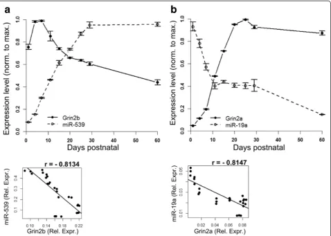
The expression pattern of Grin2a and Grin2b (mRNAs for GluN2A and GluN2B, respectively) in the rat hippo-campus was determined by qPCR (Fig. 1a,b). Grin2b mRNA expression was maximal between P1 and P7 after which expression decreased gradually and was sustained at approximately half of peak levels (Fig. 1a) consistent with previous reports [4]. Conversely, Grin2a mRNA
expression was very low at P1, increased after P7 and plateaued at P20 (Fig. 1b). TargetScan (www.targetsca-n.org) [19] version 6.2 and miRDB (www.mirdb.org) [20] were used to find candidate miRNAs that target NMDAR mRNAs. miRDB had one target for Grin2b mRNA (miR-539) and no targets for Grin2a. TargetScan had 12 targets for Grin2a and 68 targets for Grin2b mRNA, one of which was miR-539. We further narrowed the candidate list by choosing miRNAs that were selective for single NMDAR subunits and chose the candidates pre-senting the best seed matches and context scores (site-type, 3′-supplementary pairing, local AU content, position con-tribution as detailed in [21]) among this subset. This ana-lysis produced three candidates for Grin2a (miR-19a,−351,
and −137) and three candidates for Grin2b (miR-539,
−3541, −296). We measured the expression levels of
these candidate miRNAs in rat hippocampus by qPCR over the same developmental interval as Grin2a and Grin2b mRNAs. Only miR-19a and miR-539 showed an expression pattern that was inversely correlated
with its predicted NMDAR subunit target (correlation coefficients: r = -0.8147 for Grin2a/miR-19a and r = -0.8134 for Grin2b/miR-539) (Fig. 1a,b). MiR-19a levels fell steeply shortly after birth and then flattened out by P11. In contrast, miR-539 showed a gradual increase beginning after birth and through the first month of post-natal life. Other candidate miRNAs (miR-351, miR-137, miR-3541 and miR-296) showed uncorrelated patterns of expression or did not vary with time (not shown).
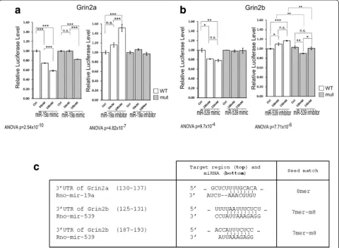
To demonstrate that miR-19a and miR-539 target mRNAs encoding GluN2A and GluN2B respectively we performed luciferase assays (Fig. 2a,b). HEK293 cells co-transfected with a miR-19a mimic showed decreased luciferase activity of Luc-Grin2a 3′UTR by 41.6 %
(±1.9 % of control, p < 0.001), whereas the mimic had much less effect on Luc-Grin2a-mut 3′UTR (−17.5 ± 0.5 %, p < 0.001). This finding is consistent with miR-19a binding to the 3′ UTR of Grin2a mRNA as a putative target. Co-transfection with the miR-19a locked nucleic acid (LNA) inhibitor increased the luciferase activity of Luc-Grin2a 3′UTR (51.7 ± 9.6 %, p < 0.001) with no sig-nificant effect on Luc-Grin2a-mut 3′UTR, confirming the target relationship (Fig. 2a). Similarly, co-transfection with the miR-539 mimic decreased the luciferase activity from Luc-Grin2b 3′UTR by 21.9 % (± 3.4 %, p < 0.01) but not from Luc-Grin2b-mut 3′UTR (1 %, p > 0.05) (Fig. 2b). Co-transfection with the miR-539 inhibitor increased the luciferase activity from Luc-Grin2b 3′UTR by 16.8 % (± 1.1 %, p < 0.001), whereas no significant change
was observed for the Luc-Grin2b-mut 3′UTR between control and the maximal concentration (150nM) (p > 0.05) (Fig. 2b), confirming that miR-539 targets Grin2b mRNA. The concentration of miR-539 mimic required to attenuate luciferase activity in these experiments was higher than the concentration of miR-19a mimics, consist-ent with the presence of two miRNA response elemconsist-ents (MREs) for miR-539 in Grin2b mRNA (Fig. 2c).
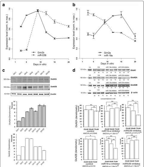
Cultured hippocampal neurons are a well characterized experimental system and are more easily manipulated than the intact hippocampus. To confirm that the devel-opmental profile of NMDAR subunit expression from cul-tured neurons compared favorably with our results from acutely isolated hippocampal tissue, we extracted RNA from hippocampal cultures between DIV3 and DIV20. The time-dependent changes in the expression of Grin2a
and Grin2b mRNA paralleled the changes observed in tis-sue (Fig. 3a,b). Western blots confirmed that GluN2A and GluN2B protein levels followed the mRNA expression pattern (Fig. 3c). MiR-19a and miR-539 approximated the expected inverse pattern of the targeted subunit: miR-19a expression gradually declined after DIV9, and miR-539 ex-pression increased sharply. The initial increase in GluN2B protein (from DIV1 to DIV5) likely resulted from regrowth of dendrites following cell plating [22] and reduced the strength of the correlations.
To examine the role of these miRNAs in determining NMDAR subunit expression patterns, we used LNA miRNA inhibition. Based on the above observations, DIV8 was selected for the treatment. It was not possible to follow the expressions of miRNAs with this technique due to the recognized problem that the antisense
inhibitor can directly inhibit the qPCR reaction [23], an issue also recognized when performing Northern blots [24]. Grin2a and Grin2b mRNA levels were not notably changed following LNA treatment (not shown). The ab-sence of a miRNA effect on a validated target mRNA is frequently observed and is probably due to translational inhibition in the absence of mRNA target degradation. Therefore, the protein level was used to detect miRNA efficacy. We next examined whether 539 and miR-19a affect NMDAR subunit protein expression. We assessed GluN2A and GluN2B protein expression on cultured rat neurons following LNA treatment at DIV8 (Fig. 3d). In neurons treated with the miR-19a inhibitor, protein expression of GluN2A increased (21.3 ± 0.6 % of control, p < 0.01) and in neurons treated with the miR-539 inhibitor, protein expression of GluN2B increased (12.5 ± 0.1 % for GluN2B, p < 0.01). Interestingly, each of the miR inhibitors also induced a reciprocal effect in the regulated subunit that it does not directly target. The miR-19a inhibitor was associated with a decrease in GluN2B decreased (−29.5 ± 0.71 %, p < 0.01) and miR-539 inhibitor was associated with a decrease in GluN2A (−40 ± 0.52 % of control, p < 0.01). These findings sup-port the roles of miR-19a and miR-539 on their targets and suggest additional controls that maintain reciprocal levels of the mature and immature subunits. To examine whether other NMDA receptor subunits were affected by LNA treatment, we used an antibody for GluN1 but detected no difference in protein levels following LNA treatment (Fig. 3d).
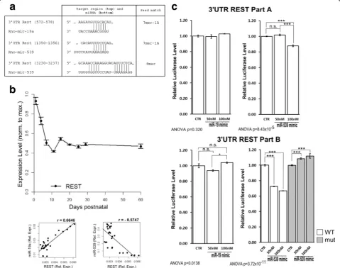
The GluN2 subunit switching network is linked to REST
The repressor element 1 silencing transcription factor (REST) silences genes by epigenetic remodeling [15]. Grin2b is a REST target and knockdown of RESTin vivo prevents the developmental decline in GluN2B, but not GluN2A. Interestingly, the 3′UTR of REST has putative sites for miR-19a and miR-539 (Fig. 4a). To relate the REST expression pattern to those of the GluN2 sub-units, miR-19a and miR-539 expressions, we tracked REST mRNA expression in vitro by qPCR (Fig. 4b). REST expression dramatically decreased from P1 to P11, then plateaued through adulthood (P > 60). To test whether REST was a target for miR-19a and/or miR-539 we used the luciferase assay. The miR-539 mimic reduced the luciferase signal when fused to the 3′ UTR of REST and especially for the REST PartB (−33.12 ± 0.39 % of the signal compared to the con-trol, p < 0.001). On the other hand, the miR-19a mimic had no effect on the luciferase signal (3.92 ± 1.06 % of the signal compared to the control (REST PartB, 50nM), p < 0.05) (Fig. 4c). Thus, it is likely that REST operates within the gene regulatory network in conjunction with the implicated miRNAs to regulate NMDAR maturation.
Discussion
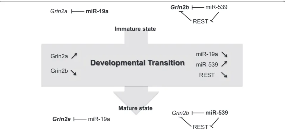
The regulation of changes in NMDAR subunit compos-ition during development suggests a complex graded dy-namical system: as the GluN2 subunit composition changes during maturation, miR-539 goes up and REST
goes down (Fig. 5). Concomitantly, miR-19a decreases as GluN2A reaches its mature level. REST targeting by miR-539 may smoothly implement the transition by dir-ectly or indirdir-ectly coordinating the developmental re-duction of both Grin2b and REST. These transitions require an extensively parameterized network of control elements capable of operating as a closed-loop system from the time of induction until a new equilibrium is reached. miRNAs act in small-scale gene regulatory net-works with defined topologies [25, 26]. These network motifs function in recurrent regulatory circuits, often with transcription factors [27] and, in this case, imple-ment transitions in the NMDAR subunit composition.
Methods
Ethics approval
All animal care procedures were reviewed and approved by the Institutional Animal Care Committee at the University of California, Santa Barbara and found to be in compliance with guidelines on Animal Care.
Hippocampus rat brain extraction and rat primary hippocampal neuron cultures
Hippocampi from postnatal days (from P1 to P > 60) rats (strain Sprague-Dawley from Charles River Laboratories) were dissected. Neurons for culture were dissected from E18 Sprague Dawley rats in 5 mL of dissection media
containing 500 μL of Trypsin 2.5 % and grown in complete growth medium (500 mL Neurobasal media, 10 mL B-27 Supplement, 1.25 mL L-Glutamine and 1 mL P/S).
RNA extraction
Total RNA containing microRNA was extracted from the frozen hippocampi of each animal or cultures by using a mirVana miRNA isolation kit (#AM1560, Life technologies).
Western blot
After neuronal cells lysates obtainment, the following antibodies were used: anti-GluN2A (#AB1555P, Millipore); anti-GluN2B (#05-920, Millipore); anti-β-Actin (#A5441, Sigma) and anti-NMDAR1 (GluN1) (#MAB363, Chemicon International). Western Blots were quantitated using the Image J software.
LNA treatment
At DIV8, neurons were transfected with either comple-mentary sequence based locked nucleic acid inhibitors of miR-19a or miR-539 (miR-19a, #410118-00, Exiqon; miR-539, #MC11336, Exiqon), or a scrambled control (miRCURY LNA Inhibitor Control, #199004-00, Exiqon) using Lipofectamine 2000. The following day, protein lysates were collected.
qPCR measurements
Quantitative PCRs were carried out using specific primers (Table 1) and SYBR Green PCR Master Mix (#4367659, Life Technologies). Grin2a, Grin2b and REST expressions were normalized to GAPDH expression. For miRNAs, 10 nanograms of the total RNA containing microRNA were reverse-transcribed with a TaqMan MicroRNA Reverse Transcriptase kit (#PN 4366596, Applied Biosystems) and analyzed with the TaqMan
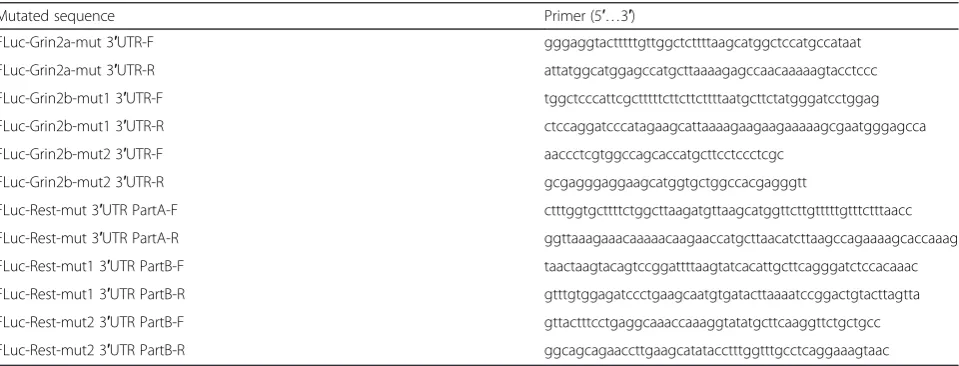
Table 2Primers for mutagenesis
Mutated sequence Primer (5′…3′)
FLuc-Grin2a-mut 3′UTR-F gggaggtactttttgttggctcttttaagcatggctccatgccataat
FLuc-Grin2a-mut 3′UTR-R attatggcatggagccatgcttaaaagagccaacaaaaagtacctccc
FLuc-Grin2b-mut1 3′UTR-F tggctcccattcgctttttcttcttcttttaatgcttctatgggatcctggag
FLuc-Grin2b-mut1 3′UTR-R ctccaggatcccatagaagcattaaaagaagaagaaaaagcgaatgggagcca
FLuc-Grin2b-mut2 3′UTR-F aaccctcgtggccagcaccatgcttcctccctcgc
FLuc-Grin2b-mut2 3′UTR-R gcgagggaggaagcatggtgctggccacgagggtt
FLuc-Rest-mut 3′UTR PartA-F ctttggtgcttttctggcttaagatgttaagcatggttcttgtttttgtttctttaacc
FLuc-Rest-mut 3′UTR PartA-R ggttaaagaaacaaaaacaagaaccatgcttaacatcttaagccagaaaagcaccaaag
FLuc-Rest-mut1 3′UTR PartB-F taactaagtacagtccggattttaagtatcacattgcttcagggatctccacaaac
FLuc-Rest-mut1 3′UTR PartB-R gtttgtggagatccctgaagcaatgtgatacttaaaatccggactgtacttagtta
FLuc-Rest-mut2 3′UTR PartB-F gttactttcctgaggcaaaccaaaggtatatgcttcaaggttctgctgcc
FLuc-Rest-mut2 3′UTR PartB-R ggcagcagaaccttgaagcatatacctttggtttgcctcaggaaagtaac Table 1Sequences of primers for mRNA detection
Gene symbol Primer (5′…3′)
MicroRNA Assay containing specific primers for miR-19a or miR-539 (hsa-miR-19a-3p, #000395 and hsa-miR-539-5p, #001286, Applied Biosystems) and U6 as a control (#001973).
Luciferase assay
The 3′UTR of the rat Grin2a or Grin2b mRNA were generated by PCR and inserted between the SpeI and HindIII restriction sites downstream of the Firefly Luciferase of the plasmid pMIR-REPORT system (#AM5795, Applied Biosystems). Due to the size of the rat 3′UTR of REST, its sequence was inserted be-tween the SacI and PmeI restriction sites in two dif-ferent parts: PartA (1217 bp), which contains the MRE for miR-19a and PartB (2176 bp) which contains the two MREs for miR-539.
The 3-nucleotides mutants of the Luc-3′UTR Grin2a, Luc-3′UTR Grin2b, Luc-3′UTR REST Part A and Part B were prepared using the primers indicated in (Table 2).
HEK293T cells were transfected with phRL-TK, the FLuc-3′UTR Grin2a or FLuc-3′UTR Grin2b or FLuc-3′ UTR REST and the corresponding miRNA mimics (miR-Vana miR-19a mimic, #MC10649, Ambion; miR(miR-Vana miR-539 mimic, #MC11336, Ambion and a miRVana miRNA mimic Negative Control, #4464058, Life Tech-nologies) or miRNA inhibitors (miR-19a, #410118-00, Exiqon; miR-539, #410328-00, Exiqon or miRCURY LNA Inhibitor Control, #199004-00, Exiqon).
Statistical analysis
Data were expressed as mean ± SEM. One-way ANOVA followed by Tukey’s post-hoc comparisons tests were performed in all statistical. Significance levels are *:
p< 0.05; **: p< 0.01; ***: p< 0.001; n.s.: not significant and r: Pearson’s correlation coefficient.
Abbreviations
DIV:Days in vitro; NMDAR: N-Methyl-D-Aspartate Receptor; miRNA: microRNA; MRE: miRNA Response Element; LTP: Long-Term Potentiation; LNA: Locked Nucleic Acid.
Competing interests
The authors declare that they have no competing interests.
Authors’contributions
CC, IH and KSK conceived of the design of the study. CC and IH contributed to the preparation of hippocampi and hippocampal cultures. CC and IH performed RNA extractions and qPCR measurements. CC performed Western Blots, LNA treatments and Luciferase assays. CC, IH and BW performed the statistical analyses. CC, IH and KSK analyzed the data. CC and KSK drafted the manuscript. All authors read and approved the final manuscript.
Acknowledgements
This work was supported by the Larry L. Hillblom Foundation and Rainwater Charitable Foundation. We thank Snigdha Chatterjee and Katharina Günther for preparing hippocampal cultures, Cheng Wu for statistical advice and Kosik lab members, especially Kenneth Tovar, for insightful conversations.
Author details 1
Neuroscience Research Institute, Department of Molecular, Cellular and Developmental Biology, University of California, Santa Barbara, CA 93106, USA.2Present address: EA4250-Laboratoire d’Ingénierie des Matériaux de Bretagne, Equipe Génie des Bioprocédés et Biomolécules, Université de Bretagne Sud, CER Yves Coppens, Vannes 56017, France.
Received: 23 June 2015 Accepted: 2 September 2015
References
1. Paoletti P. Molecular basis of NMDA receptor functional diversity. Eur J Neurosci. 2011;33(8):1351–65.
2. Harney SC, Rowan M, Anwyl R. Long-term depression of NMDA receptor-mediated synaptic transmission is dependent on activation of metabotropic glutamate receptors and is altered to long-term potentiation by low intracellular calcium buffering. J Neurosci. 2006;26(4):1128–32.
3. Scheetz AJ, Constantine-Paton M. Modulation of NMDA receptor function: implications for vertebrate neural development. FASEB J.
1994;8(10):745–52.
4. Monyer H, Burnashev N, Laurie DJ, Sakmann B, Seeburg PH. Developmental and regional expression in the rat brain and functional properties of four NMDA receptors. Neuron. 1994;12(3):529–40.
5. Yashiro K, Philpot BD. Regulation of NMDA receptor subunit expression and its implications for LTD, LTP, and metaplasticity. Neuropharmacology. 2008;55(7):1081–94.
6. Xu Z, Chen RQ, Gu QH, Yan JZ, Wang SH, Liu SY, et al. Metaplastic regulation of long-term potentiation/long-term depression threshold by activity-dependent changes of NR2A/NR2B ratio. J Neurosci.
2009;29(27):8764–73.
7. Hall BJ, Ripley B, Ghosh A. NR2B signaling regulates the development of synaptic AMPA receptor current. J Neurosci. 2007;27(49):13446–56. 8. Gray JA, Shi Y, Usui H, During MJ, Sakimura K, Nicoll RA. Distinct modes of
AMPA receptor suppression at developing synapses by GluN2A and GluN2B: single-cell NMDA receptor subunit deletion in vivo. Neuron.
2011;71(6):1085–101.
9. Lee MC, Yasuda R, Ehlers MD. Metaplasticity at single glutamatergic synapses. Neuron. 2010;66(6):859–70.
10. von Engelhardt J, Doganci B, Jensen V, Hvalby O, Gongrich C, Taylor A, et al. Contribution of hippocampal and extra-hippocampal NR2B-containing NMDA receptors to performance on spatial learning tasks. Neuron. 2008;60(5):846–60.
11. Hu NW, Klyubin I, Anwyl R, Rowan MJ. GluN2B subunit-containing NMDA receptor antagonists prevent Abeta-mediated synaptic plasticity disruption in vivo. Proc Natl Acad Sci U S A. 2009;106(48):20504–9.
12. Rammes G, Hasenjager A, Sroka-Saidi K, Deussing JM, Parsons CG. Therapeutic significance of NR2B-containing NMDA receptors and mGluR5 metabotropic glutamate receptors in mediating the synaptotoxic effects of beta-amyloid oligomers on long-term potentiation (LTP) in murine hippocampal slices. Neuropharmacology. 2011;60(6):982–90.
13. Kessels HW, Nabavi S, Malinow R. Metabotropic NMDA receptor function is required for beta-amyloid-induced synaptic depression. Proc Natl Acad Sci U S A. 2013;110(10):4033–8.
14. Martel MA, Ryan TJ, Bell KF, Fowler JH, McMahon A, Al-Mubarak B, et al. The subtype of GluN2 C-terminal domain determines the response to excitotoxic insults. Neuron. 2012;74(3):543–56.
15. Rodenas-Ruano A, Chavez AE, Cossio MJ, Castillo PE, Zukin RS. REST-dependent epigenetic remodeling promotes the developmental switch in synaptic NMDA receptors. Nat Neurosci. 2012;15(10):1382–90.
16. Kosik KS. MicroRNAs and cellular phenotypy. Cell. 2010;143(1):21–6. 17. Fineberg SK, Kosik KS, Davidson BL. MicroRNAs potentiate neural
development. Neuron. 2009;64(3):303–9.
18. Ho VM, Dallalzadeh LO, Karathanasis N, Keles MF, Vangala S, Grogan T, et al. GluA2 mRNA distribution and regulation by miR-124 in hippocampal neurons. Mol Cell Neurosci. 2014;61:1–12.
19. Lewis BP, Burge CB, Bartel DP. Conserved seed pairing, often flanked by adenosines, indicates that thousands of human genes are microRNA targets. Cell. 2005;120(1):15–20.
21. Garcia DM, Baek D, Shin C, Bell GW, Grimson A, Bartel DP. Weak seed-pairing stability and high target-site abundance decrease the proficiency of lsy-6 and other microRNAs. Nat Struct Mol Biol. 2011;18(10):1139–46. 22. Williams K, Russell SL, Shen YM, Molinoff PB. Developmental switch in the
expression of NMDA receptors occurs in vivo and in vitro. Neuron. 1993;10(2):267–78.
23. Thomson DW, Bracken CP, Szubert JM, Goodall GJ. On measuring miRNAs after transient transfection of mimics or antisense inhibitors. PLoS One. 2013;8(1):e55214.
24. Horwich MD, Zamore PD. Design and delivery of antisense oligonucleotides to block microRNA function in cultured Drosophila and human cells. Nat Protoc. 2008;3(10):1537–49.
25. Milo R, Shen-Orr S, Itzkovitz S, Kashtan N, Chklovskii D, Alon U. Network motifs: simple building blocks of complex networks. Science. 2002;298(5594):824–7.
26. Shen-Orr SS, Milo R, Mangan S, Alon U. Network motifs in the transcriptional regulation network of Escherichia coli. Nat Genet. 2002;31(1):64–8. 27. Martinez NJ, Ow MC, Barrasa MI, Hammell M, Sequerra R, Doucette-Stamm
L, et al. A C. elegans genome-scale microRNA network contains composite feedback motifs with high flux capacity. Genes Dev. 2008;22(18):2535–49.
Submit your next manuscript to BioMed Central and take full advantage of:
• Convenient online submission
• Thorough peer review
• No space constraints or color figure charges
• Immediate publication on acceptance
• Inclusion in PubMed, CAS, Scopus and Google Scholar
• Research which is freely available for redistribution