C A S E R E P O R T
Open Access
Lifesaving surgery for a ruptured invasive
thymoma using the hemi-clamshell
approach: a case report
Nobuyuki Yoshiyasu
*, Fumitsugu Kojima, Yuya Ishikawa and Toru Bando
Abstract
Background:Among anterior mediastinal tumors, a teratoma is known to rupture with growth, but there have been few previous reports about thymoma rupture. We here report a rare case of an invasive thymoma with intrapulmonary and intrathoracic rupture requiring emergency life-saving surgery. To our knowledge, this is the first such case in the literature.
Case presentation:A 56-year-old woman suddenly experienced right precordial pain and hemoptysis. Enhanced computed tomography revealed a large mediastinal tumor pressing against the pulmonary hilar vascularity, with extravasation of blood into the right lung. Tumor rupture into the lungs was suspected. Given the deterioration of her respiratory status and hemodynamics, thymomectomy with removal of the involved tissues was urgently performed using the hemi-clamshell approach and intrapericardial dissection, with veno-arterial extracorporeal membrane oxygenation on standby. She survived, and no recurrence has been noted for 2 years postoperatively. Conclusions:A large thymoma can suddenly rupture into the thorax, similar to the rupture of a teratoma. Additionally, in cases with hemoptysis, an appropriate procedure should be selected to reach both the pulmonary hilum and thorax for complete resection, as hemoptysis might suggest tumor invasion into the lungs.
Keywords:Emergency surgery, Hemi-clamshell approach, Ruptured invasive thymoma
Background
Among anterior mediastinal tumors, a teratoma is known to rupture with growth, but a ruptured thymoma, requiring emergency surgery, has rarely been reported, and the use of the hemi-clamshell approach in such a case has not been described to date. However, this approach does have some advan-tages for patients with hemodynamic instability due to compression by a large tumor or its severe adhesion to the pulmonary hilum [1].
Here, we present a rare case of an invasive thymoma with intrapulmonary and intrathoracic rupture treated with emergency thymomectomy involving the hemi-clamshell approach and intrapericardial dissection. To our know-ledge, this is the first such case in the literature.
Case presentation
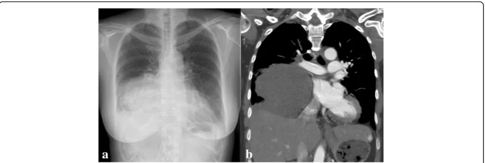
A mass shadow was incidentally detected in the right lower lung field on routine chest radiography and
chest computed tomography (Fig. 1a, b) in a
56-year-old woman with a menopausal disorder.
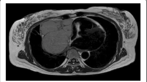
Mag-netic resonance imaging (T2-weighted imaging)
revealed a capsulated mass (10 × 11 cm in diameter) extending from the anterior mediastinum toward the middle mediastinum (Fig. 2). On fluorine-18 fluoro-deoxyglucose (FDG) positron-emission tomography, increased FDG uptake was seen, with a maximum standardized uptake value of 9.6. Surgery was planned for the mediastinal tumor suspected to be a low-grade thymoma. As her menopausal disorder was being treated with estrogen therapy, which is associated with an increased risk of venous thromboembolism and pulmonary thromboembolism, we scheduled surgery 1 month after therapy discontinuation.
* Correspondence:[email protected]
Department of Thoracic Surgery, St. Luke’s International Hospital, 9-1 Akashi-cho, Chuo-ku, Tokyo 104-8560, Japan
However, before the scheduled date, she suddenly complained of acute pain in the right precordial region, which radiated toward the back. The pain exacerbated and hemoptysis occurred when she coughed; thus, she visited the emergency room at our hospital. Her vital signs were stable, except for tachypnea with oxygen desaturation. Auscultation indicated decreased breath sounds in the right lower lung. Her electrocardiogram was normal, without ST elevation. Second chest radiography showed an enlarged tumor with an
irregu-lar shape (Fig. 3a), whereas enhanced computed
tomography revealed a diffuse ground-glass shadow in the right lung, with occlusion of the intermediate bronchus and right inferior pulmonary vein by the tumor (Fig. 3b). Furthermore, there was extravasation of blood into the right lung, as well as pleural effusion. We presumed that her sudden-onset symptoms were caused by intrapulmonary rupture of the mediastinal tumor. Emergency surgery was performed with veno-arterial extracorporeal membrane oxygenation (VA-ECMO) on standby, because of the possibility of deterioration of
the respiratory status and hemodynamics during anesthesia induction. After anesthesia induction on one-lung ventilation, we confirmed that blood pressure and oxygen saturation did not change markedly owing to alteration of body position. We initially performed median sternotomy and found that the large tumor had infiltrated strongly into the right lung and phrenic nerve on the pericardium around the hilar area. Thus, after securing the right main pulmonary artery with an incision into the serous pericardium between the superior vena cava (SVC) and ascending aorta, we con-ducted a hemi-clamshell approach to obtain a better surgical view (Fig. 4). Finally, we performed thymo-mectomy with right pneumonectomy and partial resec-tion of the pericardium and completely excised the thymoma, without requiring VA-ECMO. The right phrenic nerve had to be sacrificed, and a part of the diaphragm was resected with a stapler because of suspected direct invasion. She had an uneventful post-operative recovery, and she was discharged from the hospital 9 days after the surgery.
Pathological examination revealed that the mediastinal tumor was stage III according to the Masaoka–Koga classification and a type B2 invasive thymoma accor-ding to the World Health Organization histological classification. The tumor had infiltrated into the right upper and middle lobes and the pericardium. Vascular breakdown due to tumor compression resulted in pulmonary hemorrhage (Fig. 5). Although the margin was negative, radiation therapy was performed. She sur-vived, and no recurrence has been detected for 2 years postoperatively.
Discussion
An invasive thymoma occurs in the anterior media-stinum and frequently infiltrates into the mediastinal pleura, lungs, pericardium, or blood vessels. The tumor
Fig. 1Chest radiography (a) and chest computed tomography (b) at the first visit show a large mass in the right lower lung field
Fig. 3Second chest radiography (a) shows an enlarged mass with an irregular shape. Enhanced computed tomography (b) after the admission demonstrates occlusion of the right inferior pulmonary vein due to tumor compression
Fig. 4The surgical view in the hemi-clamshell approach with intrapericardial dissection. The right main pulmonary artery was tied using blue rubber bands, while the ascending aorta was covered with gauze. The right lung with the thymoma appeared dark red, indicating a hematoma (asterisk)
may progress in a variety of ways. Cases involving extension into the SVC and right atrium or thymic veins have been reported previously [2, 3]. In the present case, the invasive thymoma mainly developed at the side of the thoracic cavity. The direction of pro-gression may depend on the origin of the tumor, even in the same anterior mediastinum.
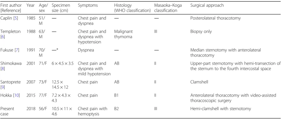
Among anterior mediastinal tumors, mediastinal teratomas are known to infiltrate into the thorax, as these tumors contain some pancreatic tissue, which secretes pancreatic enzymes and causes histolysis and consequent rupture [4]. In contrast, an invasive thy-moma with intrathoracic rupture has rarely been reported [5–10], but has been suggested to be caused by ischemia of the tumor wall owing to rapid growth. To our knowledge, this is the first case of intrapul-monary and intrathoracic rupture of an invasive thym-oma requiring emergency surgery. In this case, marked pulmonary hemorrhage was observed in the middle lobe specimen (Fig. 4). Microscopic observation indi-cated widespread collapse of blood vessels and lung tissues. We assumed that the internal pressure of the tumor had increased as it was sandwiched between the intermediate bronchus and the heart, and it then rup-tured into the lungs, where the pressure was most eas-ily released owing to tumor invasion of the lungs. Six cases of penetration of thymomas into the thorax have been reported to date [5–10]. These patients com-plained of chest pain and/or dyspnea, but none of them had hemoptysis, as in the present case (Table1). When a patient complains of not only chest discom-fort, but also hemoptysis, it should be suspected that the thymoma has infiltrated into the lungs and has progressed to a relatively advanced stage. Therefore, an
appropriate approach should be selected to facilitate access to the pulmonary hilum for complete resection in order to remove the tissues surrounding the lung.
In the present case, we successfully performed complete resection of the tumor by using the hemi-clamshell approach and performing intrapericardial dissection in pneumonectomy. When an intratho-racic operation is required for a mediastinal tumor, the hemi-clamshell approach provides a better oper-ating field than median sternotomy [11]. The hemi-clamshell approach can provide multi-angle views of the pulmonary hilum, posterior–anterior mediasti-num, and diaphragm. Thus, this approach for
lo-bectomy or even pneumonectomy with large
thymomectomy will be useful to control hilar and interlobar anatomic dissection. Furthermore, if the tumor adheres severely to the surrounding tissues at
the pulmonary hilum, intrapericardial dissection
should be performed to control unexpected
hemorrhage. After incision of the fibrous and serous pericardium via median sternotomy, the right main pulmonary artery can be secured on the dorsal side between the SVC and ascending aorta.
In conclusion, a large invasive thymoma can suddenly rupture into the thorax, similar to the rupture of a tera-toma. Additionally, in cases with hemoptysis, an appro-priate procedure, such as the hemi-clamshell approach, should be used to reach both the pulmonary hilum and thorax for complete resection, as hemoptysis might sug-gest tumor invasion into the lungs.
Abbreviations
FDG:Fluorine-18 fluorodeoxyglucose; SVC: Superior vena cava; VA-ECMO: Veno-arterial extracorporeal membrane oxygenation
Table 1Ruptured thymoma case series
First author [Reference] Year Age/ sex Specimen size (cm) Symptoms Histology (WHO classification)
Masaoka–Koga classification
Surgical approach
Caplin [5] 1985 51/
M ―
Chest pain and
dyspnea ― ―
Posterolateral thoracotomy
Templeton [6]
1988 63/
M ―
Chest pain and dyspnea with hypotension
Malignant thymoma
III Biopsy only
Fukuse [7] 1991 70/
M ―
* Dyspnea ― ― Median sternotomy with anterolateral
thoracotomy
Shimokawa [8]
2001 71/F 6 × 4.5 × 3.5 Chest pain and dyspnea with mild hypotension
AB II Upper-part sternotomy with hemi-transection of
the sternum to the fourth intercostal space
Santoprete [9]
2007 73/F 12.5 ×
14.5 × 12
Chest pain AB II Clamshell
Hokka [10] 2015 77/F 7.2 × 4.3 × 4.3
Chest pain B1 II Anterolateral thoracotomy with video-assisted
thoracoscopic surgery
Present case
2018 56/F 10.5 × 11 × 4.6
Chest pain with hemoptysis
B2 III Hemi-clamshell with sternotomy
―No description
Acknowledgements
We would like to thank Editage (http://www.editage.jp) for English language editing.
Author contributions
NY acquired the data and drafted the manuscript. TB performed the preoperative checks and diagnosis. YI, FK, and TB performed the operation. FK and TB participated in the case report design. All authors read and approved the final manuscript.
Funding
No funding was associated with this report.
Availability of data and materials
Data sharing is not applicable to this article.
Ethics approval and consent to participate
Not applicable.
Consent for publication
Informed consent was obtained from the patient for the publication of this case report.
Competing interests
The authors declare that they have no competing interests.
Publisher’s Note
Springer Nature remains neutral with regard to jurisdictional claims in published maps and institutional affiliations.
Received: 4 December 2018 Accepted: 12 February 2019
References
1. Lebreton G, Baste JM, Thumerel M, Delcambre F, Velly JF, Jougon J. The hemiclamshell approach in thoracic surgery: indications and associated morbidity in 50 patients. Interact Cardiovasc Thorac Surg. 2009;9:965–9. 2. Liu CH, Peng YJ, Wang HH, Cheng YL, Chen CW. Spontaneous rupture of a
cystic mediastinal teratoma complicated by superior vena cava syndrome. Ann Thorac Surg. 2014;97:689–91.
3. Terada Y, Ono N, Noguchi T, Kamakari K, Kitano M. A case of thymoma protruding into the superior vena cava through the thymic vein. Ann Thorac Surg. 2004;77:1088–90.
4. Sasaka K, Kurihara Y, Nakajima Y, Seto Y, Endo I, Ishikawa T, et al. Spontaneous rupture: a complication of benign mature teratomas of the mediastinum. AJR Am J Roentgenol. 1998;170:323–8.
5. Caplin JL, Gullan RW, Dymond DS, Bradley SM, Hill IM, Banim SO. Hemothorax due to rupture of a benign thymoma. Jpn Heart J. 1985;26:123–5.
6. Templeton PA, Vainright JR, Rodriguez A, Diaconis JN. Mediastinal tumors presenting as spontaneous hemothorax, simulating aortic dissection. Chest. 1988;93:828–30.
7. Fukuse T, Matsukura T, Nakamura A, Kosaka S, Tamada J. Mediastinal hematoma due to thymoma hemorrhage--a case report. Nihon Kyobu Geka Gakkai Zasshi. 1991;39:930–4 [Article in Japanese].
8. Shimokawa S, Watanabe S, Sakasegawa K, Tani A. Ruptured thymoma causing mediastinal hemorrhage resected via partial sternotomy. Ann Thorac Surg. 2001;71:370–2.
9. Santoprete S, Ragusa M, Urbani M, Puma F. Shock induced by spontaneous rupture of a giant thymoma. Ann Thorac Surg. 2007;83:1526–8.
10. Hokka D, Ogawa H, Tane S, Tanaka Y, Tauchi S, Maniwa Y. Ruptured thymoma causing a hemothorax: a case report. Oncol Lett. 2015;10:1810–2. 11. Lardinois D, Sippel M, Gugger M, Dusmet M, Ris HB. Morbidity and validity