Copyright 0 1990 by the Genetics Society of America
Characterization
of TRP-1 mRNA Levels in Dominant and Recessive
Mutations at the Mouse brown
( b )
Locus
Ian J. Jackson,* Doreen Chambers,* Eugene M. Rinchikt and Dot C. Bennett*
*MRC Human Genetics Unit, Western General Hospital, Edinburgh EH4 2XU, Scotland, ?Biology Division, Oak Ridge National
Laboratory, Oak Ridge, Tennessee 37831-8077, and iDepartment of Anatomy, St. George's Hospital Medical School, London SW17
ORE, England
Manuscript received March 2 2 , 1990 Accepted for publication June 20, 1990
ABSTRACT
T h e mouse brown locus encodes a putative membrane-bound metalloenzyme, tyrosinase-related
protein-1 (TRP-I). We have examined the effect on m R N A expression of the locus of a number of mutant alleles. T h e common null mutant allele, brown, produces wild-type levels of TRP-I mRNA,
which is nonfunctional. Another recessive aliele, cordovan-Hanuell, has an intermediate, dark-brown
phenotype and produces only very low levels of presumably normal TRP-1 mRNA. Two dominant alleles appear to act by killing the melanocyte in which they are expressed. One of them, Light, has normal size and amounts of TRP-1 mRNA. T h e other, White-based brown, produces no detectable
TRP-1 mRNA. I t has a gross DNA rearrangement at the 5' end, and we speculate that this results in
activation of transcription of sequences not usually seen in melanocytes, and that this is toxic to the cell. The relationship between phenotype and molecular structure at the locus is discussed, and we draw some general principles applicable to other developmental genes.
T
HE formation of the mouse coat color is a devel- opmental system particularly amenable to mo- lecular genetic study (SILVERS 1979; JACKSON 1985). Pigmentation is not essential to the viability of the laboratory mouse, and new mutations will therefore generally survive. Furthermore, coat color is a partic- ularly striking characteristic, thus new mutations are readily identified. A large number of mutations at many loci have been found to affect coat color. Six loci in particular [non-agouti ( a ) , brown ( b ) , albino ( c ) ,dilute ( d ) , pink-eyed dilution
( p )
and piebald (s)] have been used for many years in specific-locus mutation experiments, which have produced many new allelesat these loci (RUSSELL 195 1; SEARLE 1974). Some mutations affect only pigment or pigment-related functions, while others have marked developmental effects, usually due to deletions of DNA associated with the locus (RUSSELL 197 1 ; RUSSELL, MONTGOM-
ERY and RAYMER 1982).
T h e cDNA corresponding to the product of the murine chromosome 7 c locus, tyrosinase, has been cloned (KWON et a l . 1987; YAMAMOTO et al. 1987;
MULLER et a l . 1988). We have recently shown that a different cDNA clone, known as pMT4 (SHIBAHARA
et al. 1986), encodes another protein, tyrosinase-re- lated protein-1 (TRP-I), which shares approximately 40% amino acid identity with tyrosinase, and maps to
the b locus on chromosome 4 (JACKSON 1988). An- other tyrosinase-related protein (TRP-2) has been identified (called clone 5A), but not as yet mapped (JACKSON 1988; I. J . JACKSON, unpublished results). Genetics 1 2 6 451-459 (October, 1990)
These three proteins have several common features. All have a hydrophobic residue near to the C termi- nus, which is probably a transmembrane domain (it is known that tyrosinase is a membrane-bound enzyme) [see HEARING and JIMENEZ (1989) for example]. All
three also have striking similarity to tyrosinases of lower eukaryotes (Neurospora crassa) and of prokar- yotes (Streptomyces species) in two particular domains
(LERCH, LONCONI and JORDI 1982; HUBER, HINTER-
MAN and LERCH 1985; MULLER et a l . 1988). One of these regions is also remarkably similar to the copper- binding domain of hemocyanins, particularly in con- servation of the histidine residues (GAYKAMA et a l . 1984; HUBER, HINTERMAN and LERCH 1985). Neu- rospora and Streptomyces tyrosinases are known to contain two copper atoms per molecule (SOLOMON
198 1 ; H U B E R , HINTERMAN and LERCH 1985). In the mouse it would appear that there are at least three membrane-bound, copper-containing enzymes, which are most likely localized on the inner membrane faces of the melanosomes, the site of pigment synthesis within the melanocyte.
452 I . J. Jackson et al.
Mice doubly heterozygous for certain radiation-
induced brown mutations are fully viable but have
complete deletions of t h e T R P - I locus and have a
brown phenotype indistinguishable from the classical
brown (E. M. RINCHIK unpublished results). Brown is
therefore the null phenotype and results in melano-
cytes containing brown eumelanin instead of the wild-
type black. Previously, examination of brown melano-
cytes led to the suggestion that its gene product played
a role in melanosome morphology (RITTENHOUSE
1968). The sequence of TRP-1, however, would lead
us now to propose that it is a membrane-bound me-
talloprotein that has some enzymatic function and
shares a common ancestry with tyrosinase. While null
mutations of tyrosinase (the c locus) cause complete
absence of pigment, this is not the case at t h e b locus.
The function of wild-type T R P - 1 is therefore one that
is not essential for pigment synthesis, but is necessary
for black rather than brown pigment to be formed.
T h e enzymology of melanin synthesis is not well
understood, but there are a number of candidate
enzymes which could be represented by TRP-1 (KOR-
NER a n d PAWALEK 1980, 1982; BARBER et a l . 1984).
In this work we have examined expression of T R P -
1 mRNA from a number of alleles at the b locus. This
locus is unusual among pigmentation genes as it has
mutant alleles both recessive and dominant to wild
type (Black). T h e recessive alleles result in production
of either brown or dark-brown eumelanin. T h e two
dominant alleles, Light ( B t f ) a n d White-based brown (B”),
result in the tips of the hairs being pigmented, but the
bases being much paler (MACDOWELL 1950; HUN-
SICKER 1969). Both are expressed when heterozygous
with wild type (Black) but are more extreme in phe-
notype when homozygous. The phenotype appears to
be d u e t o a “suicide activation” of t h e b locus, resulting
in death, or failure to remain in the hair bulb, late in
the hair cycle, of those melanocytes which are under-
taking pigment synthesis. In Light mice, at each hair
growth cycle, pigment is made for a time, before the
cells begin t o die a n d are incorporated into the hair
shaft. By the end of the growth cycle there are no
melanocytes visible in the hair bulb (QUEVEDO a n d
CHASE 1958; SWEET a n d QUEVEDO 1968; QUEVEDO,
FLEISCHMANN a n d DYCKMAN 1981). New growths of
hair, following molting, have pigmentation restored,
b u t successive rounds of hair growth have less pigment
at the hair tip, possibly reflecting a decreasing pool of
melanocyte precursor cells on which to draw or, less
likely, a more rapid suicide of newly recruited melan-
ocytes (perhaps due to their having a low level of b
locus transcription before recruitment).
Table 1 summarizes the mutants examined in this
study, their origin and their phenotypes. We report
the characterization of expression of the alleles a n d
present the basis for detailed further molecular analy-
sis of the relationship between genotype and pheno-
type at the b locus.
MATERIALS A N D METHODS
Mice and cell lines: C57BL/6J, cordovan-Hanuell and
Light mice are maintained at the Animal Unit of the Western
General Hospital. White-based brown mice are maintained at
the Biology Division of Oak Ridge National Laboratory.
The B16C3 melanoma cells were obtained originally from
J. KREIDER and grown as described previously (BENNETT 1983). Melan-a and melan-b lines were described by BEN-
NETT, COOPER and HART (1 987) and BENNETT et a/. (1989)
and grown as described.
Hybridization probes: Most hybridizations used the 1.6-
kb internal Hind111 fragment of pMT4, recloned into Blues-
cribe (Stratagene) (JACKSON 1988; SHIBAHARA et al. 1986). This fragment detects the diagnostic D- and B-haplotype fragments, in addition to several others. T o examine the 5’
end of TRP-1 an 800-bp EcoRI to PstI fragment from a
genomic clone of the TRP gene was used. The TRP-2 probe
was the 1200-bp EcoRI fragment of clone 5A (JACKSON
1988), subcloned into Bluescribe. Actin mRNA was detected
using the cDNA from clone PAM91 (MINTY et al. 1981)
The fragments were isolated from low gelling temperature a arose, diluted with 3 volumes of water and labeled with
”!P using the method O f FEINBERG and VOGELSTEIN (1983).
DNA methods: DNA was made by homogenization of
organs in STE [ 100 mM NaCI, 50 mM Tris-HCI (pH 8), and
10 mM EDTA], followed by treatment with proteinase K
(100 pg/ml) in 0.5% sodium dodecyl sulfate (SDS) for several
hours. After phenol extraction and chloroform extraction the DNA was precipitated with ethanol and dissolved in 10 mM Tris-HCI (pH 7.5) and 1 mM EDTA.
Restriction digestions were performed using manufactur- ers’ recommended buffers.
DNA was electrophoresed through 0.8% agarose in TAE [40 mM Tris-acetate (pH 7.5) and 1 mM EDTA] and trans- ferred to Hybond-N (Amersham) nylon filters according to manufacturer’s instructions.
Southern blot hybridizations were carried out in 3 X SSC,
10 X Denhardt’s solution supplemented with 100 pg/ml
denatured salmon sperm DNA, 0. I % SDS and 0.1 % sodium
pyrophosphate (JEFFREYS and FLAVELL 1977) [ 1 X SSC is
0.15 M NaCl and 0.015 M sodium citrate (pH 7) and 1 X
Denhardt’s solution is 0.02% bovine serum albumin, 0.02%
Ficoll, 0.02% and polyvinylpyrridoline] or in 0.5 M sodium
phosphate (pH 7.2), 7% SDS (CHURCH and GILBERT 1984)
and washed at 68” down to 0.1 X SSC and 0.1% SDS before
autoradiography.
RNA methods: RNA was made from fresh or frozen
melanoma or melanocyte cell pellets, and from fresh or
frozen dorsal skin of l-4-day-old mice. Cells or skin were
homogenized in 3 M LiCI, 6 M urea (LOVELL-BADGE 1987),
the RNA precipitated overnight, centrifuged and washed in
homogenization buffer. After resuspension in 10 mM Tris-
HCI (pH 7.5) and 0.1% SDS, the RNA was digested with
proteinase K at 100 pg/ml at 65” for several hours, phenol
extracted, chloroform extracted, and ethanol precipitated.
The RNA was electrophoresed through 1 % agarose gels
containing 2.2 M formaldehyde in 40 mM MOPS (pH 7), 5
mM sodium acetate and 1 mM EDTA (LEHRACH et al. 1977)
before blotting in 10 X SSC to nitrocellulose filters.
Northern-blot hybridizations were carried out in 50%
formamide, 4 X SSC, 10 X Denhardt’s solution, 50 mM
sodium phosphate, 100 pg/ml salmon sperm DNA and 0.1%
SDS overnight at 48” and washed down to 0.3 X SSC at
Expression of brown Locus Alleles 453
TABLE 1
Alleles at the brown locus
Allele Symbol Phenotype Origin
Black E Black eumelanin Wild type
brown b Brown eumelanin Mouse fancy
cordovan-Hanuell b'" Dark-brown eumelanin Radiation mutagenesis
Light B ' I Dominant; hair tip more pigmented than base, and Spontaneous
White-based brown B =' Dominant; similar to Light but more extreme, and Radiation mutagenesis is black or dark-brown
pigment is brown
Reverse transcription and the polymerase chain reac-
tion: Double-stranded cDNA was prepared using a kit sup-
plied by Boehringer Mannheim Ltd., according to the man-
ufacturer's instructions, except that oligo(dT) primer was
replaced with 36 pg/ml random hexanucleotides (dNs) (Pharmacia).The oligonucleotide primers used for the po-
lymerase chain reaction (PCR) were: TCCGAATTCAA-
AGGGGTGGATGACCG (bases 293 to 3 12 plus a terminal
EcoRI site) and GACACATAGTAATGCATCC (633 to
61 5) from the tyrosinase cDNA (MULLER et al. 1988);
CAATTAACAGCTGGCATCA (-175 to -157) and
GGAAGGTTTCTCTGCTGA (-97 to -1 14) from the 5'
untranslated region of TRP-1 cDNA; and GCTGCAG-
GAGCCTTCTTTC (714 to 732) and GACGCTGC-
ACTGCTGGTCT (961 to 979) from the middle of TRP-1
cDNA (SHIBAHARA et al. 1986).
T h e reactions were performed essentially as in SAIKI et
al. (1988). Each reaction contained 50 mM KCI, 10 mM Tris
(pH 8.3), 1.5 mM MgC12, 0.01% NP-40, all six oligonucleo- tide primers at 3 pg/ml, all four deoxynucleotide triphos-
phates at 0.1 mM and 2.5 units Taq DNA polymerase
(Amersham) in a volume of 50 pl. Some reaction mixes were
irradiated with UV by placing on a UV Products transillu-
minator for 10 min before addition of enzyme and substrate
(SARKAR and SOMMER 1990). Each cDNA amplification used
the product of reverse transcription of approximately 0.1
pg skin total RNA. T h e reaction was cycled 25 times
through 90 sec at 92", 90 sec at 53" and 120 sec at 72". Thirty percent of the reaction was analyzed by electropho-
resis on gels of 4% Nusieve GTG agarose (FMC Bioprod-
ucts) for ethidium bromide staining, or of 1.5% agaraose
(Sigma) for Southern blotting.
RESULTS AND DISCUSSION
Both TRP-1 and TRP-2 are melanocyte-specific, highly expressed mRNAs. Northern blot analysis on a range of tissues reveals no evidence of expression in cells other than melanocytes and melanoma. In situ hybridization to sections-of neonatal mouse skin shows that TRP-1 is expressed exclusively in the melanocytes of the hair follicles (K. STEEL, D. R. DAVIDSON and I.
J. JACKSON, unpublished results).
SHIBAHARA et al. (1 986) reported two discrete TRP- 1 mRNA species visible on Northern blots. Although the existence of two species is demonstrated by cDNA clones differing at their 3' ends (SHIBAHARA et al. 1986), when blots are washed at high stringency we see only a single species (see Figures 1, 2 and 3, for example), although additional bands can be seen at
lower stringency. T h e high abundance of TRP-1 mRNA in melanocytes permits its detection in neo- natal skin samples, where melanocytes comprise only a small minority of cells. Approximately the same signal is obtained on a Northern blot of 10 pg of 2-
day-old skin total RNA, as from 100 ng of melanoma total RNA. Hybridization of TRP-2 cDNA to North- ern blots reveals a slightly smaller mRNA, which, in this study, we use as a control, not only for the integrity and loading of RNA, but also to confirm the presence of melanocytes in the skin samples examined.
The brown ( b ) mutation: T h e common, recessive brown ( b ) mutation is an old mutation of the mouse fancy. We have shown (JACKSON 1988) that the mu- tation present in a number of different laboratory inbred and outbred stocks is associated with a 4.9-kb TuqI TRP-1 fragment, the D haplotype. All other mice have the B haplotype comprising a 3.7- and 1.2- kb TuqI fragment.
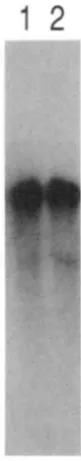
Figure 1 shows autoradiography of a Northern blot, probing total RNA made from melanoma cells and melanocytes grown in vitro. B16 melanoma cells and melan-a cells (BENNETT, COOPER and HART 1987) are both derived from C57BL/6 inbred mice, which are wild type (Black) at the b locus. Melan-b cells (BENNETT et al. 1989) are derived from homozygous brown mice of the outbred Q stock. After washing to remove the first probe, the filter was rehybridized with TRP-2 to check for loading. There is no signticant difference in the abundance or size of TRP-1 mRNA between Black and brown melanocytes, or between melanoma and cultured melanocytes. T h e Q-stock brown muta- tion is associated with the D haplotype (JACKSON 1988), and this result is therefore most likely applica- ble to the common brown mutation seen in all labora- tory mice.
I . Jackson et
(0 ~ (0
zab
Z a b
phenotype (ZDARSKY, FAVOR and JACKSON 1990).
The cordovan-Hamell (b'") mutation: A number of alleles of brown have been described that are inter- mecliate in phenotype between black and brown. They have been variously called dark-brown or cordovan. T h e first intermediate allele to be described, called cordovan ( F ) , arose spontaneously (MILLER and POTAS
19.5.5). We have examined transcription of another intermediate allele, cordovan-Harule11
(6'").
which arose in a y-irradiation mutagenesis experiment in a (C[IH/HeH X 1 OI/H)FI male receivingapproximatelv 600 rad (6 Gy) over a period of about 12 weeks (BATCHELOR, PHILLIPS and SEARLE 1966).We have examined mRNA levels of the 6'" allele in R N A prepared from neonatal skin of litters segregat- ing if" and b. I t is not possible to distinguish mice homozygous for cordovan-Hanuell from those hetero- zygous with brown ( i e . , the mutation is fully dominant over brown). Furthermore it is difficult to distinguish these mice from homozygous brown animals when they are onlv a few days old. However, as the br" mutation arose on mice carrving the B haplotvpe, Southern blots of D N A from these neonates allow distinction to be made between animals homozygous for either B or D haplotypes (br" or b, respectively), or compound heterozygotes (bib'").
We prepared R N A from the skin and DNA from the kidney of each member of a 2-day-old litter derived from parents that were conlpound heterozv- gotes,
bib'".
Analvsis of the D N A bv Southern blot hybridization to TuqI digests determined the genotype1 2 3 4 5 6 7
TRP-1
TR
P-2
I'IGL'RF 2.-Sor111r1.11 l h t l ~ v l ~ ~ i d i m ~ i o ~ ~ t o 1ot:d K S A prepared
from tlorwl skin of e;~cll n ~ c n ~ l ) r r o f ;I 2-d;ly-old litter. l h e filter W ~ I S h!lwidisc4 ;IS I- i ~u r e I ; lirst I O slww I'KP-I (upper panel), srcontllv t o s h n v 1'RI'-2 (lo\ver panrl). Gcnot!pes, tlrternlined I)! Southorn I h t :l1l;ll!sis of Tnql-digested D N A . are: animal 5 . b/h: ;lninl;lls I , 4 . f i ;und 7, h'"/h; anitlmls 2 ; u n d 3. h"'/h'".
of each member of the litter. Figure 2a is an autora- diograph of a Northern blot of the skin RNAs probed
with plvT4. TRP-2 was hybridized subsequently to
confirm the RNAs were undegraded and equally loaded (Figure 2b). Animal 5 is the only b/b animal of the litter. As the mRNA abundance is the same in Black (+/+) or brown ( b / b ) melanocytes (see above), this animal serves as an indicator of wild-type TRP-I mRNA level. Animals 2 and 3 are both homozygous br"/br", and the rest are
bib'".
Comparison of the hybridization signals, within t h e litter and with the control hybridization to TRP-2, shows that the homo- zygous cordovan-Hanuell mice have very low levels of TRP-I mRNA. T h e heterozygotes have about 50%wild-type amount of m R N A , largely due to the brown (6) allele. Hybridization to TRP-I in the
br"/br"
skin is detectable in lanes 2 and 3, but we estimate from scanning the autoradiograph that it is present at ap- proximately 1 % of the abundance of b/b or+/+
mice. As the phenotype of cordovan-Hanuell is not brown, but is somewhat darker, we would expect the melan- ocytes to have reduced, but not absent, TRP-1 activ- ity. I t therefore is likely that the low level of TRP-1 seen represents normal m R N A , and that the pheno- type is due to a greatly reduced level of its (normal) protein product.The effect of dosage of TRP-1 on pigmentation is
Expression of brown Locus Alleles 455
TABLE 2
Dosage of TRP-1 and quantized phenotype
A p p t - m i t ~ ~ a t v l v v d of
( ~ I O I \ ~ V I l r I w i d i / i t l g ( C )
O l ~ ~ v r v v d t t 1 K S A p w w t I 1 v r i active
IIIKV:\ (7;) I'llrnotvpr
I l l n 100 100 Hl;1ck
If111 I O 0 5n Black
Il','/h',, I 1 l h r k 1)ro\\.n
h " / b 5 0 0 . 5 I h r k brown
/ ) / / I I O 0 0 BI-O\CIl
. \ w t t ~ ~ i n g t11;tt hrorc*n T R P - 1 mRS:\ is fully inactive. and cor-
d O ~ Y / 1 7 - ~ ~ O ~ l ' d / T R P - I INKS;\ i s full! itctive.
though complete lack of active protein ( b / b ) leads to a brown phenotype, half normal levels ( B / b ) results in a phenotype indistinguishable from wild type ( R is dominant over b). A reduction to about 1 % wild-type levels of TRP-1 in
br"/br"
mice leads to an clear change in pigmentation from black toward brown, but further reduction to about 0.5% (b'"/b) has no further effect (br" is dominant over 6 ) . Two explanations present themselves. First the effect of TRP-1 on pigment synthesis might show a threshold or quantized effect, meaning that there exist discrete forms of pigment, governed by a particular dose of TRP-1, rather than a continuum of pigment quality between black and brown. Alternatively, such quantization might be per- ceptual; there may be a continuum, but we cannot perceive the difference between more than a limited number of states. A different situation occurs at the albino (c) locus, at which many intermediate alleles [such as chinchilla ( d " ) ] , are recessive to wild type but only semidominant over albino (so, for example, cCh/c'" mice are darker than c'"/c) (SILVERS 1979). T h e
difference between the b and c loci might be due to the function of their products. Tyrosinase, from the c locus regulates the amount of pigment produced while the b-locus product, TRP-I, governs the quality (or possibly stability) of the pigment color.
The molecular basis of the reduced TRP-1 mRNA abundance is currently under investigation. T h e na- ture of the change w i l l be informative as to the regu- lation of m R N A levels in general and in melanocytes
in particular. T h e reduction may be due to decreased transcription or increased degradation of the R N A or its precursor. I t should be noted that we have not been able to detect by restriction fragment analysis any difference between this mutant TRP-1 gene and the wild-type allele, even though the mutation arose following y-irradiation.
The dominant Light (B") mutation: Light ( I l l f ) is the
better studied of the dominant alleles of brown (QUS-
EVEDO and CHASE 1958; SWEET and QUEVEDO 1968;
QUEVEDO, FIXISCHMANN and DYCKMAN 1981). It
arose spontaneously on a C 5 8 background, and South- ern blot examination of D N A from honlozygous Light mice shows no evidence of alteration of gene struc-
1 2
"-
-
~ I G L ' R E J.-Sortllern I d o t hvbridimtion I O total R N A prepared
Irotn t~ors;lI skin of :I :$-tI;t!-oId C.57L3IA/6 tnouse ( B / B . i . ~ . +/+) (I;lne I ) ;Inti ;I R-tI;ly-oItI I1omwygous ~ i g h t mouse (B''/Bf') (I;ltle 2). T h e filter W;IS hyl)ridi7ed to detect T R P - I as described in the test.
ture. We have examined t h e TRP-I mRNA level in the skin of homozygous B" neonates (whose genetic status was confirmed by haplotype analysis) compared with age-matched C57BL/6 (wild-type) animals. At this early stage of the hair cycle we would not vet expect to see a depletion in melanocyte numbers. Figure 3 shows a Northern blot of these RNAs probed with the pMT4 plasmid, which indicates that there is no difference in TRP-1 mRNA size or abundance.
We conclude that the Light mutation most likely exerts its effect through a mutant protein which causes the observed death of the melanocytes. This protein probably has some residual, normal TRP-1 activity, as
t h e pigment produced by homozygous mutant melan- ocytes is dark brown rather than brown, but, in addi- tion the protein must be toxic to the cell. Either it is itself toxic (through mislocalization in the melanocyte, for example), or its enzymatic function has been al-
tered so that a toxic product accumulates in the cell.
The dominant White-based brown (B") mutation:
T h e phenotype of Il'hite- based brown ( B " ' ) mice is very similar to that of Light, although it has not been studied in the same detail. We propose that the prod- uct of the B " ' allele, like that of R'f, causes melanocyte deat.h. but, unlike R", homozygous R''/Ra' mice, or
compound heterozygotes with brown (B"'/b), have brown pigment at the hair tips. Thus the mutation results in a recessive null phenotype (brown) in addi- tion to the dominant (white-base) phenotype. The mutation arose in a spermatogonium of a (101/RI x
CJH/RI)FI male, exposed to 600 rad (6 Gy) of y-
456 I. J . Jackson rf al.
*.
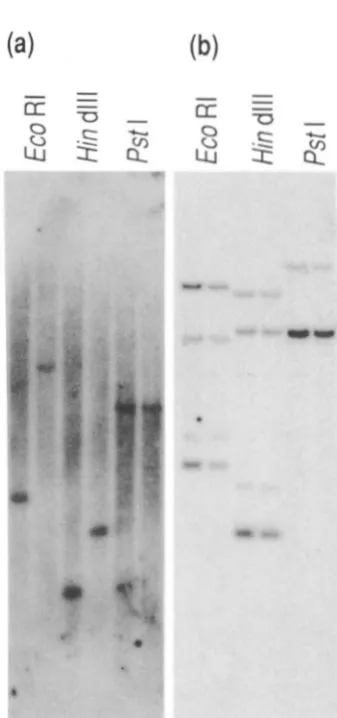
I . ' I G ~ . R ~ : 4.-Southcrn I h t llvbridi/atiot1 t o I)\:\ 1)rcp1rcd 1 1 . o 1 1 1
( 3 1 1/1 I C mice and from Ilorllo/vgous Il'hitr-hosrd hroaln ( N ; ' / R " )
1nicc. 1 1 1 c w 11 I x I i r of I K I ( ks, tligc\trtl wit11 thc restriction C I I / ~ I I I ~ intlic;~tctl a b o v e . t11c lcft-l~and r~.;~c-k i s Il%i/r-ho.wd hrorcjn I)SZI\ and
I O ( . right-hand t ~ ~ c k i s ( 3 1 I / I le I)\:\. I';IIICI ;I \ v m I1vl~ridi/ctl with
the 8OO-l)p /:'roKI t o !'.</I gwomic clone h g m c n t ; t n d p ; I I w l I) is
t h . S I I I I C . liltel. Idlvl)ritli/ctl with rllr I . f i - k b llintllll cl)S;\ lrag-
111('111.
Figure 4 shows this allele is associated w i t h a D N A rearrangement detected by T R P - 1 cDNA and ge- nomic probes. T h e mutation arose i n a hybrid mouse, which has two different chromosomes 4, but we do not kno~v on which chromosome it occurred. We have compared D N A restriction fragments from homozv- gous
R"'
mice w i t h C3H D N A , but we observe essen- tiallv identical fragments M.llen compared with 10 1 DNA (with the exception of variants in an unlinked fragment cross-hybridizing with the 3' end). Probes encompassing most of the coding region of the T R P - 1 gene clo not detect any'clifferences between the B " ' allele and the C3H wild-type gene (Figure 41~). How- ever, when probes toward the .?' end of T R P - 1 are used. differences are found between the two genes.The 5' end of the wild-type gene is enclosed within an 800-bp EroRI-Pstl fragment. T h e EcoRl site is about 100 bp upstream of the .5' end of the m R N A . There is an intervening sequence beginning after base -87 of the cDNA sequence (i.e., 99 bp from the end
1 kb
-
4
4
I I1 1 ! 1H E P H E
b)
,
k
J_
_ _
_ I _-
_ _ _ _ _
-_ _ _ _
I
""""""L"1C" H E
u
il I 1 1
I%;L~RK .'.-Sclwnl;lric representation of the 5' c ~ l t l s of the wild- type ; I I I ~ H" m u t a 1 1 t I'KP-1 genes. (a) \t'ild-tvpc gene. sllowing first secluc11ccs arc re1mwntcd I ) \ tl;tshctl lincs. I t i s 1101 k n o \ v n if t l l c w scqwnccs arc ;I single insertion into t l ~ c locus, o r arc flanking ;I site
o l ' a large inversion. I h t h l~alvcs of the rwrra~1gc~1lc~1t arc sho\\,tl.
1'. /'.~/l: I<. I.:roKI: I I . f l i n d l l l . Sot a l l sites arc s l w w 1 .
3 esolls. llullll,rrctl I , I I ; 1 n d 111. (I,) R" l l l l l t i l l l t gcllc. S o v c l I)S/\
of the published sequence) and the PstI site is approx- imately 600 bp into this intron (see Figure .5a). This EcoRI-Pstl fragment, containing exon I , was used to probe Southern blots of D N A from homozygous White-based brown and wild-tvpe animals and the re- sults are shown in Figure 4a. When digested with PstI, the W/B"' D N A has the same fragment size as wild tvpe (4.5 kb), indicating that sequences 5' of the PstI site i n the first intron are unrearranged (which include a Hind111 and an EcoRI site). However, the 6.5-kb
EcoRI wild-type fragment is replaced in White-based brown D N A by one of 2.4 kb, and the 2.1.5 kb HindIII fragment replaced by one of 1.3 kb. There is therefore a D N A rearrangement within t h e B"' T R P - 1 gene, downstream of the Pstl site in the first intron, but upstream of the Hind111 and EcoRI sites, which are in the second and third introns, respectivelv. As
the novel HindIII site lies only 0.2 to 0.3 kb down- stream of the (unaffected) PstI site, the rearrangement must have an end point in this few hundred base-pair interval. Other data (not shown) indicates the break- point lies close to the 3' end of the first intron.
T h e exact nature of the rearrangement has to be established, but it involves a juxtaposition of novel D N A sequences with the 5' end of the gene. T h e data are consistent with an insertion of DNA at this point, or an inversion of the region with one breakpoint mapping to end of the intron, and the other an unknown distance upstream. Figure 5 is a schematic representation of the first 3 introns of the wild-tvpe T R P - 1 gene, and both halves of the rearrangement in the B"' mutation, and includes additional mapping data.
We are unable to detect transcription from the
F '
Expression of hrouw Locus Alleles 457
1 2 3 4 5 6 7 8
P "
-
FIGI'RE 6.-Sorthcr11 I d o t hyt)ridi/ation t o t o t a l R S l \ prepred I'rom d o r \ ~ l \ k i l l of ;I 2-d;1y-old <;.37I%I./(i mouse ( B / B . i . p . +/+)
(I;IIIC\ I . 3 . 3 ; u n d 7) ~ I M I 2-tl;ty-oltl honlo/ygous Il%i/p-bnstd hrouv~
(K'/H') micr ( h w s 2. 1. (i and X). .l'he liltcr \\'as hyl~ritli~ed ;IS in
I'iaurc I . lir\t t o detect .I.Rl'-l do\wstrc;Im of the re:trwngement
( h w b 1 ; I I I ~ 2). then t o tletect ~1.RP-l transcripts u p s t r e a ~ ~ of thc
; I I I ~ 1in;IIlv actin ( I ~ I I I C S 7 ;IIKI X).
l ~ c ~ l l ' l ~ ~ l l l ~ c l l l c l l t (l:llles 3 ;111d 4), sul~scc~~lrnrly T R P - 2 (lillles 5 and 6 )
old B"'/W skin (lanes 2, 4, 6 and 8). When the blot is probed either with the 1600 bp cDNA fragment from downstream of the rearrangement (lanes 1 and 2) or with the RcoRI/PstI genomic fragment containing the first exon (upstream of the rearrangement) (lanes 8
and 4) there is no evidence of a TRP-l-containing transcript from the B " ' allele. Reprobing the blot with TRP-2 cDNA (lanes 5 and 6) results in hybridization to all tracks, indicating that the RNA from B " ' mice is
intact, and that there are indeed melanocytes present in the skin. T h e hybridization to TRP-2 mRNA is somewhat weaker in the B " ' / B " ' RNA than the wild type. This is not due to a lower RNA loading as reprobing with actin cDNA (lanes 7 and 8) shows that there is, in fact, more RNA in the mutant track. I t is possible that we may be seeing here an early indication of the depletion of the melanocyte pool, which ulti- mately results in the phenotype.
I t is not surprising that the DNA rearrangement
results in no transcription from the main part of the gene. However, as it appears from Southern blotting that the 5' flanking region is intact, it may be capable of driving transcription of the first exon, as well as sequences downstream including sequences within the rearrangement. Such transcripts may not be of dis- crete size, and may be hidden in the smear seen in
1
2
3
4
5
6
FICI'RE 7.-GeI ;in;llvsis o f I'(:K-;~nll'lific;~tio~~s o f tlors;~l skin
cI)S,\. Upper panel: c~tllidium I)romidr stained 4 % Nu-Sieve aga- rose gel. I ~ I W I . IfaPIII-digested @X I74 D S h ;IS sin' markers: I;IIIC
2, amplification products of wild-tvpc. ( ( X B I J l i ) c1)SA: I;IW 3.
products o f IIolno7ygous N"/R" c I ) S X : lane 4 , anlplification of <:571%1./6 US/\ ;IC control: lane 5 . ;m$ification of R " ' / R " RN;\ ;IS
control: I a n c li, co11tro1 an1pIific:~tion w i t h 1 1 0 sul)str;~te. :\nlplilied
fragment siws i n track 2 arc 3.18. 266 and 7X bp. 1,ower p a n e l :
S o u t l ~ c r ~ ~ M o t l~vl)ridimtion of :I similar re;lction r u n on ;I 1.5% ; ~ g ; ~ r o s c gel, ~ m ) l x d w i t h rhr XOO-bp E m K l - P s t I (eson I ) fr;lgrnrrlt ol"fR1'- I . 1 ~ 1 1 c . s ;IS above. The ;Itltol.;ldiogr;lph shows hyl~ridimtion
o n l y t o the 7 X - b p rson I 1Y;R ~woduct ol'wilcl-type cI)SA.
track 4 of Figure 6. We therefore used the polymerase chain reaction (PCR) to examine homozygous B " '
RNA. We made double-stranded cDNA from both wild-type and mutant skin RNA [priming the first strand with random hexanucleotides to allow for a possible lack of poly(A) tail]. This was then used as a substrate for the PCR (SAIKI et al. 1988), primed by oligonucleotide pairs from downstream of the re-
arrangement, from upstream (within the first exon) and, as control, from the tyrosinase gene. Figure 7
(top) shows an ethidium bromide-stained gel of the amplified DNA. Amplification of wild-type cDNA gives rise to a 348-bp fragment deriving from the tyrosinase mRNA, and to 266- and 78-bp fragments from TRP-1 mRNA (track 2). R"' cDNA gives rise only to the amplified tyrosinase fragment; TRP-1 transcripts from either side of the rearrangement are not visible (track 3). Figure 7 (bottom) is a Southern blot of a similar gel probed with exon 1 , and shows at higher sensitivity that no exon 1 transcripts are pres- ent in B"'/R"' skin. We conclude that the TRP-1 pro- moter associated with the R " ' allele most likely is non-
Jackson et al.
White-based brown has a recessive brown phenotype which is obviously due to absence of any TRP-1 tran- script. However, the mutation also has a true domi- nant effect, and therefore it is likely that a transcript derives from the region of the locus, and this tran- script (or its translation product) causes melanocyte death. While it is possible that very low levels of a fusion transcript containing the first exon and other sequences might be the agent, this is perhaps unlikely.
A better hypothesis is that the rearrangement has brought another gene (or genes), not normally ex- pressed in melanocytes, into the proximity of the TRP-1 gene, which supplies it with enhancer (but not necessarily promoter) function. It might be the ectopic expression of this gene (either high levels of an aber- rant RNA alone or the translation product of an
ectopic mRNA) which results in the dominant phe- notype. T h e nature of the DNA rearrangement in White-based brown mice, and the sequences encom- passed by it are of great interest, and will be further characterized by DNA cloning.
GENERAL CONCLUSIONS
This study raises several issues that are generally applicable and should be borne in mind when other developmental mutations are considered. First, phe- notype can be quantized in addition to the usually observed dominance of wild-type over null mutation. Second, simple mutations in highly expressed enzymes can lead to neomorph dominant functions which can have profound effects on cell function or survival. Furthermore it seems that a mutation involving only a small change ( I l l f ) can have a very similar phenotypic
effect to one resulting from a much more severe DNA disruption (B"). T h e mode of action of these domi- nant, cell-suicide, alleles is of general relevance to developmental genetics; cell-type specific, autono- mous cell death might well be the basis of many developmental mutations which have profound effects on morphology. Finally, it should be noted that two mutations examined here arose in y-irradiation ex- periments, and both received approximately the same dose (600 rad or 6 Gy) over approximately the same time period, but only in one have we detected a gross DNA rearrangement. However, both mutations were selected on the basis of being distinguishable from brown, and so are not representative of the large number of other brown alleles obtained by radiation mutagenesis.
Wt. th;unk NICK HASTIE and RUTH JOHNSON for useful discussion and encouragement, EMAN ZDARSKY for developing the RT-PCR
methodology, LINDA DEVLIN for skilled assistance and NICK HASTIE,
JOHN EVANS and RUTH JOHNSON for comments on the manuscript.
NORMAN DAVIDISON, DOUGLAS STUART and SANDY BRUCE provided excellent photographic and art work. We also thank the Medical
Kt.se;ll-c-l1 Council (MRC) and the Lister Institute for Preventive Medicine for financial support. T h e work has been funded in part
by an MRC Project Grant to I.J.J., by the Office of Health and Environmental Research, U.S. Department of Energy, under con- tract DE-AC05-840R21400 with Martin Marietta Energy Systems Inc. (E.M.R.) and by a CRC grant to D.C.B. I.J.J. is a Lister Fellow.
LITERATURE CITED
BARBER, J. I., D. TOWNSEND, D. P. OLDS and R. A . KING, 1984 DOPAchrome oxidoreductase: a new enzyme in the pigment pathway. J. Invest. Dermatol. 83: 145-149.
BATCHELOR, A. L., R. J. S. PHILLIPS and A. G . SEARLE, 1966 A comparison of the mutagenic effectiveness of chronic neutron and gamma-irradiation of mouse spermatogonia. Mutat. Res.
BENNETT, D. C., 1983 Differentiation in mouse melanoma cells; initial reversibility and an on-off stochastic model. Cell 34: 445-453.
BENNETT, D. C., P. J. COOPER and I. R. HART, 1987 A line of non-tumorigenic mouse melanocytes, syngeneic with the B16 melanoma and requiring a tumour promoter for growth. Int. J. Cancer 39: 4 14-4 18.
BENNETT, D. C., P. J. COOPER, T . J . DEXTER, L. M. DEVLIN, J. HEASMAN and B. NESTER, 1989 Cloned mouse melanocyte lines carrying the germline mutations albino and brown. De- velopment 105: 379-385.
CHURCH, G . M., and W. GILBERT, 1984 Genomic sequencing. Proc. Natl. Acad. Sci. USA 81: 1991-1995.
FEINBERG, A. P., and B. VOGELSTEIN, 1983 A technique for radiolabelling DNA restriction endonuclease fragments t o high specific activity. Anal. Biochem. 1 3 2 6-13.
GAYKAMA, W. P. J., W. G. H. HOL, J . M. VEREIJKEN, N. M. SOETER, H . J. BAK and J. J. BEINTEMA, 1984 3.2 Angstrom structure of the copper-containing oxygen-carrying protein Panulirus interruptus haemocyanin. Nature 309: 23-29.
HEARING, V. J., and M. JIMENEZ, 1989 Analysis of mammalian pigmentation at the molecular level. Pigment Cell Res. 2: 75- 85.
HUBER, M., G. HINTERMAN and K. LERCH, 1985 Primary struc- ture of tyrosinase from Streptomyces glaucesens. Biochemistry
3: 2 18-229.
24: 6038-6044.
HUNSICKER, P. R., 1969 Mouse News Lett. 40: 41.
JACKSON, 1. J.. 1985 T h e genetics and biology of mouse melano- cytes; mutation, migration and interaction. Trends Genet. 1:
JACKSON, I . J., 1988 A cDNA encoding tyrosinase-related protein maps to the mouse brown locus. Proc. Natl. Acad. Sci. USA
JEFFREYS, A. J., and R. FLAVELL, 1977 A physical map of the DNA region flanking the rabbit beta-globin gene. Cell 12: 429-439.
KORNER, A. M., and J. M. PAWALEK, 1980 Dopachrome conver- sion: a possible control point in melanin biosynthesis. J. Invest. Dermatol. 75: 192- 195.
KORNER, A. M., and J. M. PAWALEK, 1982 Mammalian tyrosinase catalyses three reactions in the biosynthesis of melanin. Science
KWON, B. S . , A. K. HAQ, S. H. POMERANTZ and R. HALABAN, 1987 Isolation and sequence of a cDNA clone for human tyrosinase that maps at the mouse c-albino locus. Proc. Natl. Acad. Sci. USA 84: 7473-7477.
LEHRACH, H., D. DIAMOND, J. M. WOZNEY and H. BOEDTKER, 1977 RNA molecular weight determination by gel electro- phoresis under denaturing conditions, a critical reexamination. Biochemistry 16: 4743-4751.
LERCH, K., C. LONGONI a n d E. JORDI, 1982 Primary structure of
tyrosinase from Neurospora crassa. J . Biol. Chem. 257: 6408- 6413.
LOVELL-BADGE, 1987 Introduction of DNA into embryonic stem 321-326.
85: 4392-4396.
Expression of brown Locus Alleles 459
cells, pp. 153- 182 in Teratocarcinomas and Embryonic Stem Cells; A Practical Approach, edited by E. J. ROBERTSON. IRL Press, Oxford.
MACDOWELL, E. C., 1950 “Light,” a new mouse colour. J. Hered.
MILLER, D. S., and M. Z. POTAS, 1955 Cordovan, a new allele of black and brown color in the mouse. J. Hered. 46: 293-296. MINTY, A. J.. M. CARAVATTI, B. ROBERT, A. COHEN, P. DAUBAS,
A. WEYDART, F. GROS and M. E. BUCKINGHAM, 1981 Mouse
actin messenger RNAs. Construction and characterization of a recombinant plasmid molecule containing a cDNA transcript of mouse alpha actin mRNA. J. Biol. Chem. 2 5 6 1006-1014.
MULLER, G., S. RUPPERT, E. SCHMID and G. SCHUTZ,
1988 Functional analysis of alternatively spliced tyrosinase gene transcripts. EMBO J. 7: 2723-2730.
QUEVEDO, W. C., and H. B. CHASE, 1958 An analysis of the Light mutation of coat color in mice. J. Morphol. 102: 329-345.
QUEVEDO, W. C., R. D. FLEISCHMANN and J. DYCKMAN,
1981 Premature loss of melanocytes from hair follicles of light (B“) and silver (si) mice, pp. 177-1 8 3 in Pigment Cell 1981;
Phenotypic Expression in Pigment Cells, edited by M. SEIJI. Uni- versity of Tokyo Press, Tokyo.
RITTENHOUSE, E., 1968 Genetic effects on fine structure and development of pigment granules in mouse hair bulb melano- cytes. I. T h e b a n d d loci. Dev. Biol. 17: 351-365.
RUSSELL, L. B., 1971 Definition of functional units in a small chromosomal segment of the mouse and its use in interpreting the nature of radiation induced mutations. Mutat. Res. 11: 107-123.
RUSSELL, L. B., C. S. MONTGOMERY and G . D. RAYMER,
41: 35-36.
1982 Analysis of the albino locus region of the mouse. IV.
Characterization of 34 deficiencies. Genetics 1 0 0 427-453. RUSSELL, W. L., 1951 X-ray induced mutation in mice. Cold
Spring Harbor Symp. 16: 327-336.
SAIKI, R. K . , D. H . GELFAND, S. STOFFEL, S. J. SCHARF, R. HIGUCHI, G . T. HORN, K . B. MULLIS and H. A. ERLICH, 1988 Primer- directed enzymatic amplification of DNA with a thermostable DNA polymerase. Science 2 3 9 487-491.
SARKAR, G., and S. S. SOMMER, 1990 Shedding light on PCR contamination. Nature 343: 27.
SEARLE, A. G . , 1974 Mutation induction in mice. Adv. Radiat. Biol. 4: 1 3 1-207.
SHIBAHARA, S . , Y. TOMITA, T. SAKAKURA, C. NAGER, B. CHAN-
DHURI and R. MULLER, 1986 Cloning and expression of cDNA encoding tyrosinase. Nucleic Acids Res. 14: 241 3-2427. SILVERS, W. K . , 1979 The Coat Colors of Mice: A Model for Mam-
malian Gene Action and Interaction. Springer, New York. SOLOMON, E. I . , 198 1 Binuclear copper active site. Hemocyanin,
tyrosinase and type 3 copper oxidases, pp. 43-108 in Copper Proteins, edited by T. G. SPIRO. Wiley, New York.
SWEET, S. E., and W. C. QUEVEDO, 1968 Role of melanocyte morphology in pigmentation of mouse hair. Anat. Rec. 162:
YAMAMOTO, H., S. TAKEUCHI, T. KUDO, K. MAKINO, A. NAKATA, T . SHINODA and T . TAKEUCHI, 1987 Cloning and sequencing
of mouse tyrosinase cDNA. Jpn. J. Genet. 62: 271-274. ZDARSKY, E., J. FAVOR and I. J. JACKSON, 1990 T h e molecular
basis of brown, an old mouse mutation, of an induced revertant to wild type. Genetics 126: 443-449.
243-254.