Original Research Article
Postnatal bladder dysfunction
Swathi H. V., Padmaja Y. Samant*
INTRODUCTION
Voiding difficulty and urinary retention are common phenomenon in immediate postpartum period. Despite its wide clinical importance, this is a frequently under diagnosed condition. Absolute or relative failure to empty the bladder resulting from decreased bladder contractility (magnitude or duration) or increased bladder outlet resistance or both is defined as voiding dysfunction.1
Voiding dysfunction considered for this present study included-
• Inability to void within first 6 hours of vaginal delivery and
• Those who do not void after removal of catheter of caesarean section.
Factors which lead to this include changes in the bladder neck, functional urethral length, vaginal and intra-anal pressures in relation to pregnancy and childbirth, prolonged labor, assisted breech, perineal lacerations, anatomical changes and functional changes in the pelvic floor which may occur secondary to pelvic floor distention by descent of fetal head and maternal expulsive
ABSTRACT
Background: Voiding difficulty and urinary retention is a common phenomenon in immediate postpartum period. Absolute or relative failure to empty the bladder resulting from decreased bladder contractility (magnitude or duration) or increased bladder outlet resistance or both are defined as voiding dysfunction. It needs high index of suspicion or else can go undiagnosed and can lead to magnitude of problems. The study aims to calculate the incidence of dysfunction of bladder in postnatal women and to study risk factors associated with development of bladder dysfunction and management strategies in cases of bladder dysfunction.
Methods: Authors did a prospective observational study in a tertiary care hospital. 200 postpartum women were screened for complaints of voiding dysfunction within 6 hours of removal of catheter in post caesarean patients and of normal vaginal delivery. Authors found that the voiding dysfunction was relatively common with an incidence of 20.20%. Following risk factors were analyzed: parity, mode of delivery, pain at suture site, baby weight, para-urethral tear.
Results: Postpartum voiding dysfunction was found to be relatively common with statistically significant association found for pain at suture site and para urethral tear. Intra partum events contributed to voiding dysfunction. 93% of patients who with voiding dysfunction could be managed conservatively, and only 7% had to undergo intervention in the form of re catheterization.
Conclusions: The early identification and treatment can reduce the pain and discomfort. Majority of the cases resolves with conservative management and nursing staff plays a key role in early detection of the symptoms.
Keywords: Analgesics and re-catheterization, High index of suspicion, Intrapartum events, Postpartum voiding dysfunction, Risk factors, Urinary retention
DOI: http://dx.doi.org/10.18203/2320-1770.ijrcog20202751
Department of Obstetrics and Gynecology, Seth G. S. Medical College and KEM Hospital, Mumbai, Maharashtra, India
Received: 15 May 2020
Accepted: 08 June 2020
*Correspondence:
Dr.Padmaja Y. Samant,
E-mail: [email protected]
efforts during second stage of labor, instrumental deliveries, primiparity, birth weight >3.8 kg.2,3
Epidural and other forms of regional anaesthesia disrupt the afferent input and suppress sensory stimuli from bladder to pontine micturition center. This inhibits reflex mechanism that normally induces micturition.
Bladder sensation could take over ten hours to come back after spinal anaesthesia. Following caesarean section under spinal anaesthesia, the catheter should be left in situ for at least 12 hours after the last top-up dose and until the woman is mobile.4
Retention of urine following caesarean section does occur, even though women have been catheterized for 12-24 hours after delivery. An incidence of 3.8% was known during a study in 2008.4 The women most at risk were
those who had undergone emergency LSCS. Stretching of nerves during repetitive straining impairs the afferent and parasympathetic pathways in pelvic nerves required to initiate normal voiding. Prolonged labor may be associated with over distended bladder and this can interfere with contractile abilities of bladder.5
Physiological changes of pregnancy and puerperium like elevated progesterone levels in pregnancy and the first few weeks postpartum result in changes in the urogenital system leading to urinary retention.
Postpartum urinary retention (PPUR) has been classified into overt and covert retention by Yip et al.4 Women who
are unable to micturate within 6 hours after vaginal delivery are overt type. Covert type is asymptomatic with post void residual volume of >150 ml detected by ultrasonography (USG).4
In addition to monitoring of vital parameters in the postpartum period, it must be ensured that the patient voids urine within 6 hours. Symptoms that should alert the clinician include slow urinary stream, urinary frequency, incomplete emptying and incontinence.5,6
It is a treatable condition and early identification as well as supportive treatment can prevent untoward consequences like need for re catheterization, prolonged hospital stays, catheter associated UTI s and increased long term morbidity.
METHODS
All women who delivered in study tertiary care institute hospital after the approval of study.
Inclusion criteria
• Postpartum women above 18 years of age with complaints of voiding dysfunction
• Postpartum women within 6 hours of removal of catheter in post caesarean patients and at the end of 6
hours of normal vaginal delivery complaining of voiding dysfunction
• Any neurological condition arising as a result of delivery or an intervention during labor causing voiding difficulties.
Exclusion criteria
• Urinary symptoms due to underlying renal pathology like renal calculi, obstructive uropathy
• Underlying medical renal disease
• Those who did not give consent for the study.
Study procedure
Prospective observational study with population of 200 women who delivered at tertiary care hospital and had symptoms of voiding dysfunction were screened within 6 hours of normal vaginal delivery or within 6 hours of removal of catheter where they were catheterized for any cause other than retention.
• Written valid informed consent was taken
• Case record form was filled by the investigators based on:
Details regarding the intrapartum events, complaints of parturient, impact of the symptoms on their daily activities and interventions. The risk factors associated with voiding dysfunction were noted.
Examination
The treating unit were informed about the same if required.
Pertinent investigations were noted: complete blood count, urine routine and microscopy analysis, urine culture sensitivity, post void residual urine by sonography if indicated.
Post void residual urine was planned to be calculated by sonography if symptoms were not relieved by conservative management. One of the 41 patients in this study needed this assessment. Patients were followed up till discharge.
RESULTS
Data was analysed using professional statistics package EPI Info 7.0 version for windows.
The incidence of voiding dysfunction is 20.20 % in the study, out of 200.
Various risk factors were analysed described in the Table 1.
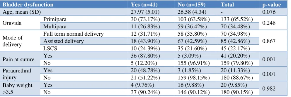
Table 1: Univariate association of risk factors with bladder dysfunction.
Bladder dysfunction Yes (n=41) No (n=159) Total p-value
Age, mean (SD) 27.97 (5.01) 26.58 (4.34) - 0.076
Gravida Primipara 30 (73.17%) 103 (63.58%) 133 (65.52%) 0.248 Multipara 11 (26.83%) 59 (36.42%) 70 (34.48%)
Mode of delivery
Full term normal delivery 12 (31.71%) 58 (35.80%) 70 (34.98%)
0.867 Assisted delivery 18 (43.90%) 67 (42.59%) 85 (42.86%)
LSCS 10 (24.39%) 35 (21.60%) 45 (22.17%)
Pain at suture Yes 36 (87.80%) 5 (3.09%) 41 (20.20%) 0.001 No 5 (12.20%) 155 (96.91%) 159 (79.80%)
Paraurethral injury
Yes 20 (48.78%) 3 (1.85%) 20 (11.33%)
0.001 No 21 (51.22%) 159 (98.15%) 180 (88.67%)
Baby weight >3.5
Yes 4 (9.76%) 16 (9.88%) 20 (9.85%)
0.982 No 37 (90.24%) 146 (90.12%) 180 (90.15%)
p=value 0.001%.
Figure 1: Association of paraurethral tear with bladder dysfunction.
Table 2: Association of mode of delivery with bladder dysfunction.
Mode of delivery
Bladder dysfunction
Yes (n=41) No (n=159) Total
FTND 13 (31.71%) 58 (35.80%) 71 (34.98%) Assisted
delivery 18 (43.90%) 69 (42.59%) 87 (42.86%) LSCS 10 (24.39%) 35 (21.60%) 45 (22.17%) Total 41 (100%) 162 (100%) 200 (100%)
Pearson chi2 value=0.2845; df=2, p-value=0.867.
A total 48.78% of the cases had para urethral tear with p value of 0.001%, hence a statistically significant variable (Figure 1).
The incidence of voiding dysfunction was higher in assisted vaginal deliveries though not statistically significant (Table 2).
Significant association of pain at suture site and bladder dysfunction is found with p value of 0.001 derived from Fishers test. with incidence of voiding dysfunction of about 87% in patients with pain at the episiotomy site (Table 3).
Table 3: Association of pain at suture site with bladder dysfunction.
Pain at suture site
Bladder dysfunction
Yes (n=41) No (n=159) Total
Yes 36 (87.80%) 5 (3.09%) 41 (20.20%) No 5 (12.20%) 155 (96.91%) 159 (79.80%) Total 41 (100%) 159 (100%) 200 (100%)
Pearson chi2 value=145.70; df=1Fisher exact p-value=0.001.
Interpretation of Table 4
• Independent of other factors, multipara compared to primipara had less odds of bladder function (not statistically significant)
• Independent of other factors, females who have undergone for LSCS compared to FTND had higher odds of bladder dysfunction (not statistically significant)
• Independent of other factors, females with pain at suture site had significantly higher odds of bladder dysfunction
• Independent of other factors, females with baby weight >3.5 kg compared to baby weight ≤3.5 kg had higher odds of bladder dysfunction (not statistically significant).
Significant association was found in patients with pain at suture site.
48.78 51.22
1.85
98.15
0. 25. 50. 75. 100. 125.
Yes No
Per
ce
n
ta
g
e
Paraurethral injury
Table 4: Multivariate (independent) association of risk factors with bladder dysfunction.
Variables Comparison OR (SE) 95% CI z-value p-value
Age - 1.12 (0.08%) (0.97, 1.30) 1.56 0.119
Gravida (ref group = primi) Multipara versus primi 0.38 (0.32%) (0.07, 1.96) -1.16 0.245
Mode of delivery (ref group = FTND)
Assisted delivery versus
FTND 0.89 (0.80%) (0.15, 5.19) -0.13 0.895 LSCS versus FTND 6.68 (6.82%) (0.90, 49.38) 1.86 0.063 Pain at suture site (ref group
= no) Yes versus no 295.26 (278.08%) (46.61, 1870.22) 6.04 0.001* Paraurethral injury (ref group
= no) Yes versus no 5.19 (5.71%) 0.60, 44.91 1.50 0.135 Baby weight >3.5 kg (ref
group = no) Yes versus no 4.32 (5.21%) (0.41, 46.11) 1.21 0.226
*significant p-value; OR = Odds ratio; SE = standard error; CI = confidence interval.
Figure 2: Intrapartum events contributing to bladder dysfunction.
The various intrapartum events contributed to voiding dysfunction in the study. About 13 out of 41 cases had significant intrapartum events (Figure 2).
Voiding dysfunction affected the quality of life in all patients in the form of pain and discomfort, difficulty in feeding baby, delayed discharge.
Management strategies included
• Conservative management which includes adequate analgesia, encouragement to pass urine, hot sitz baths, alkalinisers
• Intervention: catheterization done after delivery; re-catheterization done after removal catheter.
A total 93% of patients who were having voiding dysfunction were managed conservatively, and only 7% had to undergo intervention in the form of re-catheterization, 2 patients had to be catheterized for 24 hours and 1 patient with post void residual volume of 500 ml was catheterized for 7 days.
DISCUSSION
In the prospective observational study to look for the incidence in a group of 200 post-partum patients, authors found that the voiding dysfunction was relatively common with an incidence of 20.20%. However, authors also observed that it goes unattended unless there is high index of clinical suspicion.
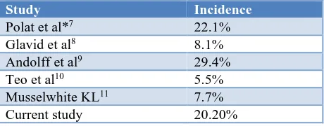
The incidence reported in the literature varies between 0.05-37% (Table 5).
Table 5: Comparison of incidence of voiding dysfunction with literature.
Study Incidence
Polat et al*7 22.1%
Glavid et al8 8.1%
Andolff et al9 29.4%
Teo et al10 5.5%
Musselwhite KL11 7.7%
Current study 20.20%
This was contradicting with the study done by Ismail et al. in which the prevalence of PPUR in their study population was 29.4% and much higher than that of this study.12
These differences may be due to the fact that most patients with urinary retention have no symptoms and hence remain undiagnosed.
Following parameters were studied • Parity
• Prolonged second stage of labor
• Obstructed labor
• Mode of delivery
• Baby weight
• Para urethral tear pain at suture site.
prolonged 2 nd stage of labor , 23
obstructed labor , 23
precipitate labor , 7.6 atonic PPH , 7.6 eclampsia ,
15.38 deep transverse
arrest , 7.6 intra op complication , 7.3
episiotomy haematoma ,
None of the symptomatic patients in the study were detected to have urinary tract infection.
In this study, significant association was found with para urethral tear and pain at the suture site.
Perineal trauma and pain: urethral over activity may ensue from pain, thus giving rise to functional obstruction. In this study out of 41 cases, 1 case of episiotomy hematoma was found to have voiding dysfunction. It resolved with analgesia.
Many of the studies in literature did not address this parameter of pain. This is important as authors can provide adequate postpartum analgesia to reduce the risk of postpartum urinary retention (PPUR). Salemnic et al, performed study on 200 primigravida who delivered vaginally and claimed that there was a relationship between PPUR and mediolateral episiotomy.13 Also,
adequate encouragement and analgesia in the form of oral NSAIDS have been found to have significant recovery of symptoms.
Instrumental delivery, prolonged labor, and episiotomy may impair postpartum voiding in two ways, neurologically or mechanically. Neurologically, instrumental delivery might damage the pudendal nerves that affect the reflexes and voluntary mechanism in normal micturition by causing inadequate relaxation of external urethral sphincter and voiding difficulty.14
A study by Mussel white et al, however revealed that conducted by Glavind and Bjork.8
In contrast, Yip et al and Glavind et al, reported that perineal trauma increased the risk of PPUR was increased by perineal tears including sphincter rupture.4,8
Mode of delivery
In this study the association was evaluated with full term vaginal delivery, vs assisted vaginal delivery versus LSCS. The incidence of voiding dysfunction was 43.9% compared to other mode of delivery (31% and 24%). However no statistically significant association was found in this study.
Glavind et al, found postpartum voiding dysfunction in 12 out of 1649 women with incidence of 0.7%.8
Operative vaginal delivery was performed in 33% of the control group, compared with 8% of the total study group.
Similarly, in Ramsay and Torbett’s study, abnormal voiding parameters were found in 74% of women after
forceps delivery, compared with 36% of women who had spontaneous vaginal delivery.2
In this study, no significant association was found with parity and birth weight. Among 40 cases, 30 (73.17%) of them were primigravida and rest 11 (26%) were multigravida. In this study, birth weight and parity had no statistical association with the incidence of voiding dysfunction. Only 9% of the cases with birth weight of >3.5 kg had voiding dysfunction. Nulliparity is usually perceived as a risk factor in other studies. Nulliparous women are exposed to pelvic floor tenderness and pudendal nerve damage in the course of vaginal birth.15
Hamberg et al, conducted a retrospective analysis of all cases with PPUR between 2003 and 2008 including variables like age, weight, height, body mass index, fetal birth weight and head circumference.16 The incidence of
voiding dysfunction in this study was low being 0.06%. The majority had those risk factors. However no statistical significance was found with respect to fetal weight, but fetal head circumference was larger than 36 cm in four of six cases.
Prolonged 2nd stage of labor
The nulliparous women with second stage lasting >3 hours and multiparous with >2 hours are stated to have prolonged second stage of labor as per NICE clinical guidelines.17 for Intrapartum care for healthy women and
babies. Prolonged bearing down efforts increases the risk of voiding dysfunction. In this study out of 41 patients, 2 of them had prolonged second stage (>2 hours) and 2 of them had obstructed labor. Pelvic nerve damage leading to neurologic impairment of the bladder is one of the proposed mechanisms. Some studies reported the association of prolonged labor with PPUR.18
Similarly, in a study conducted by Kekre et al, it was reported that PPUR rates were higher in patients with a prolonged second stage of labor.19 There was a
relationship between PPUR and longer second stage of birth and mediolateral episiotomy due to levator muscle injury as described by Marsoosi et al.20
Instrumental delivery, prolonged labor, and episiotomy may impair postpartum voiding in two ways, neurologically or mechanically. Neurologically, instrumental delivery might damage the pudendal nerves and affect the reflexes and voluntary mechanism in normal micturition.19 Similarly, perineal trauma might
result in mechanical obstruction due to vaginal hematoma or edema. Higher incidence of postpartum urinary retention in this group was reported in many studies.17
Effect on daily activities of a postnatal women
This increases the mental stress and morbidity for the nursing mothers. The early identification of the symptoms and adequate analgesia would give good results for the patient and prevent covert dysfunction and further sequalae.16,20 Very few studies in the literature
have analyzed this part of the disease.21
Management
In this study majority of the cases, (93%) were managed conservatively, only 3 cases i.e., 7% had to undergo intervention in the form of re catheterization. 2 patients had to be catheterized for 24 hours and 1 patient for 7 days. Post void residual volume was done for this patient and it was 500 ml immediate postpartum.
Management recommendations in the literature may vary from author to author, but the majority have supported the following recommendations in suspected PPUR6,22
• No patient should be left for more than 6 hours without voiding or being catheterized for residual volumes
• Timed voiding every 4th hourly in the immediate
postpartum period
• If no symptomatic improvement by conservative measures, volumes should be measured.
According to RCOG guidelines, the residual volume if >500 ml, the catheter should be kept in situ for 24 hours.6
After the diagnosis of urinary retention, a urine sample should be analyzed and sent for culture, as the presence of infection can contribute to and prolong voiding dysfunction. If a urinary tract infection is detected, prompt antibiotic treatment is required.
CONCLUSION
• In this prospective observational study, the postpartum voiding dysfunction was found to be relatively common with incidence of 20.20%.
• Out of risk factors like parity, mode of delivery, prolonged second stage of labor, pain at suture site, baby weight and para urethral tears, statistically significant association was found for pain at suture site and para urethral tears.
• Primiparity, instrumental delivery, prolonged 2nd
stage of labor was found be independent risk factors.
• The early identification and treatment can reduce the pain and discomfort. Majority of the cases resolve with conservative management and nursing plays a key role in early identification of symptoms.
Recommendations
• Midwives/nursing staff and physiotherapists play a significant role in early identification of symptoms.
Hence, they need to be trained regarding the good bladder care practices intra and postpartum
• Every obstetric unit should have a protocol for the physiotherapist for information regarding fluid management, bladder retraining, good voiding techniques and correct defecation dynamics is important and one with ongoing voiding dysfunction should be referred to urologist/urogynecologist.
Funding: No funding sources Conflict of interest: None declared Ethical approval: Not required
REFERENCES
1. Ramsay IN, Torbet TE. Incidence of abnormal voiding parameters in the immediate postpartum period. Neurourol Urodyn. 1993;12:179-83.
2. Ching-Chung L, Shuenn-Dhy C, Ling-Hong T, Ching-Chang H, Chao-Lun C, Po-Jen C. Postpartum urinary retention: assessment of contributing factors and long-term clinical impact. Aust N Z J Obstet
4. Yip SK, Sahota D, Pang MW, Chang A. Postpartum urinary retention. Acta Obstet Gynecol Scand. 2004;83(10):881-91.
5. Lim JL. Post-partum voiding dysfunction and urinary retention. Aust New Zealand J Obstet Gynaecol. 2010;50:502-5.
6. Kearney R, Cutner A. Postpartum voiding dysfunction. Obstet Gynaecol. 2008;10(2):71-4. 7. Polat M, Şentürk MB, Pulatoğlu Ç, Doğan O, Kılıççı
Ç, Budak MŞ. Postpartum urinary retention: Evaluation of risk factors. J Turkish Soc Obstet Gynecol. 2018;15(2):70-4.
8. Glavind K, Bjørk J. Incidence and treatment of urinary retention postpartum. Int Urogynecol J Pelvic Floor Dysfunct. 2003;14(2):119-21.
9. Andolf E, Iosif CS, Jorgensen C, Rydhstrom H. Insidious urinary retention after vaginal delivery: prevalence and symptoms at follow-up in a population-based study. Gynecol Obstet Invest. 1994;38:51-3.
10. Teo R, Punter J, Abrams K, Mayne C, Tincello D. Clinically overt postpartum urinary retention after vaginal delivery: a retrospective case–control study. Int Urogynecol J Pelvic Floor Dysfunct. 2007;18:521-4.
postpartum urinary retention. Am J Obstet Gynecol 2007;196(5):472.e1-472.e5.
12. Ismail SIMF, Emery SJ. The prevalence of silent postpartum retention of urine in a heterogenous cohort. J Obstet Gynaecol (Lahore). 2008;504-7. 13. Salemnic Y, Gold R, Toov JH, Jaffa A, Gordon D,
Lessing J, et al. Prevalence, obstetric risk factors and natural history of asymptomatic postpartum urinary retention after first vaginal delivery - a prospective study of 200 primipara women. J Urol. 2012;187(Suppl):788.
14. Andolf E, Iosif CS, Jorgensen C, Rydhstrom H. Insidious urinary retention after vaginal delivery: prevalence and symptoms at follow-up in a population-based study. Gynecol Obstet Invest. 1994;38:51-3.
15. Rogers RG, Leeman LL. Postpartum Genitourinary Changes. Urol Clin N Am. 2007;34:13-21.
16. Humburg J, Troeger C, Holzgreve W, Hoesli I. Risk factors in prolonged postpartum urinary retention: an analysis of six cases. Arch Gynecol Obstet. 2011;283(2):179-83.
17. NICE clinical guidelines. Intrapartum care for healthy women and babies. Clinical guideline no: CG190. Published date. Last updated; 2017.
18. McKinnie V, Swift SE, Wang W, Woodman P, O’Boyle A, Kahn M, et al. The effect of pregnancy and mode of delivery on the prevalence of urinary
and fecal incontinence. Am J Obstet Gynecol. 2005;193(2):512-7.
19. Kekre AN, Vijayanand S, Dasgupta R, Kekre N. Postpartum urinary retention after vaginal delivery. Int J Gynecol Obstet. 2011;112(2):112-5.
20. Marsoosi V, Jamal A, Eslamian L, Oveisi S, Abotorabi S. Prolonged second stage of labor and levator ani muscle injuries. Glob J Health Sci. 2014;7(1):267-73.
21. A critical literature review of the incidence of postpartum urinary retention. Available at: https://www.researchgate.net/publication/312176301 _A_Critical_Literature_Review_of_the_Incidence_o f_Postpartum_Urinary_Retention. Accessed on 24th
March 2020.
22. The epidemiology and pathophysiology of neurogenic bladder. Available at: https://www.ajmc.com/journals/supplement/2013/ace 012_jul13_ngb/ace012_jul13_ngb_ginsberg1_s191/. Accessed on 24th March 2020.