pISSN 2320-1770 | eISSN 2320-1789
Research Article
Jaundice during pregnancy: maternal and fetal outcome
Jayanthi Krishnamoorthy
1, Anuradha Murugesan
2*
INTRODUCTION
Jaundice affects a small percentage of pregnant women, yet it takes a major toll on health of both mother and foetus especially in developing countries like India. Jaundice in pregnancy carries a grave prognosis for both the fetus and the mother, and is responsible for 10% of maternal deaths.1 It could be peculiar to the pregnancy such as acute fatty liver of pregnancy, recurrent
cholestatic jaundice in pregnancy and jaundice
complicating toxemia of pregnancy. It can be concurrent with pregnancy such as due to infective pathology like viral hepatitis or due to gallstones or it could be due to drugs administered during pregnancy. The present study
analyses the cause of the disease, altered liver function, maternal and fetal morbidity and mortality and preventive measures in jaundice complicating pregnancy. This study will be helpful in better understanding and improving the maternal and perinatal outcome in jaundice complicating pregnancy.
METHODS
The study was conducted in Institute of obstetrics and gynaecology, Egmore, Chennai, India from 2011 to 2012. This prospective study of maternal and fetal outcome included 51 pregnant women with jaundice admitted in Institute of obstetrics and gynaecology during the study
1Department of Obstetrics and Gynaecology, Government Tiruvannamalai Medical College, Tiruvannamalai,
Tamilnadu, India
2
Department of Obstetrics and Gynaecology, SRM Medical College, Kattankulathur, Tamilnadu, India
Received: 17 June 2016
Accepted: 05 July 2016
*Correspondence:
Dr.Anuradha Murugesan,
E-mail: anuthanigai@yahoo.com
Copyright: © the author(s), publisher and licensee Medip Academy. This is an open-access article distributed under the terms of the Creative Commons Attribution Non-Commercial License, which permits unrestricted non-commercial use, distribution, and reproduction in any medium, provided the original work is properly cited.
ABSTRACT
Background: Jaundice affects a small percentage of pregnant women, yet it takes a major toll on health of both mother and fetus especially in developing countries like India. Jaundice in pregnancy carries a grave prognosis for both the fetus and the mother, and is responsible for 10% of maternal deaths. The aim of the study was to find out the effect of jaundice during pregnancy on maternal and fetal outcome.
Methods: 51 pregnant women with jaundice during pregnancy attending the Institute of obstetrics and gynaecology,
Egmore, Chennai between 2011-2012.
Results: 51 patients had jaundice during pregnancy. The incidence of jaundice was 0.29%.74% of patients was between 20-29 years of age. Maximum numbers of cases were Primigravida. The most common cause of jaundice was Viral Hepatitis. Maternal mortality was 7.8%.The common maternal complications were atonic postpartum haemorrhage 9.8%, hepatic encephalopathy 7.87%, disseminated intravascular coagulation 5.88% and hepatorenal failure 4%. Perinatal mortality was 35.5%.
Conclusions: Jaundice in pregnancy has adverse fetomaternal outcome. Improvement in health awareness, education
and regular antenatal checkups, early referrals result in early diagnosis and treatment of jaundice during pregnancy thus reducing maternal and fetal mortality and morbidity.
period. Elaborate history and thorough general, systemic and obstetric examination were carried out. Liver function tests like serum bilirubin total, direct and indirect, total proteins, albumin and globulin, serum transaminases, serum alkaline phosphatase, clotting time, bleeding time and ultrasonogram, complete hemogram, reticulocyte count, coagulation profile, viral markers study including HBs Ag, Anti HAV IgM, Anti HCV Ab, Anti HEV IgM were done in all patients. Dark field microscope examination and IgM (ELISA) were done for leptospirosis. Maternal outcome was noted in terms of the mode of termination of pregnancy, maternal morbidity and mortality. Fetal outcome was assessed by perinatal morbidity and mortality.
RESULTS
Total number of antenatal admissions during this period was 17,890. Total number of patients with jaundice was 51. The incidence of jaundice complicating pregnancy during this period in the hospital was 0.29%. The patients in the study group were in the age range from 18 years to 36 years. Nearly 74% of the jaundiced patients were between 20 and 29 years. The incidence of jaundice was more common in low socio-economic groups. About 92.1 of cases belonged to class IV and V. Maximum numbers of cases were Primigravida 54.9% and second gravida were 35.29%. Out of 51 cases, 41 cases (80.39%) presented with jaundice during III trimester (Table 1).
Table 1: Demographic profile.
Age group (years) No. of cases Percentage
15-19 5 9.8
20-29 38 74.51
30-40 8 15.69
S.E class
III 4 7.84
IV 24 47.06
V 23 45.1
Gravidity
Primigravida 28 54.9
Multi gravida 23 45.1
Period of gestation
I Trimester 2 3.92
II Trimester 8 15.69
III Trimester 41 80.39
3 Patients had history of contact with jaundice. 2 patients had history of blood transfusion among which one was HBsAg positive. 5% of patients were utilising safe water for drinking purpose and 95% were ignorant about safe water. On analysing the presenting symptoms 86.27% had high coloured urine. Nausea and vomiting were present in 70.6% of patients. Other predominant symptoms were fever, loss of appetite and upper abdominal pain. Jaundice was present in all the cases. Other signs were hepatomegaly, splenomegaly, scratch marks and Ascites (Table 2).
Table 2: Clinical features.
Symptoms No. of cases Percentage
High coloured urine 44 86.27
History of fever 20 39.21
Loss of appetite 23 45.09
Nausea and vomiting 36 70.58
Upper abdominal pain 18 35.29
Itching 3 5.88
Clay stools 3 5.88
Abdominal distension 7 19.6
Clinical signs
Jaundice 51 100
Hepatomegaly 9 17.64
Splenomegaly 4 7.8
Scratch marks 3 5.88
Ascites 4 7.84
92.15% of patients showed positive for bile pigments and bile salts in the urine. 19.6% patients were positive for Protein. The level of S. bilirubin varied widely between 2.8 to 18.4 mg/ dl. 7.84% of patients had high S. bilirubin more than 16 mg/dl. The serum transaminase level was below 100 IU/L in 13.72% of patients, 5.88% patients had level more than 500 IU/L. S. alkaline phosphatase was more than 200 U/L in 37.25% (Table 3).
Table 3: Liver function tests.
Serum bilirubin (mg%) No.of cases Percentage
2-5 16 31.37
6-10 21 41.17
11-15 10 19.60
16-20 4 7.84
SGPT (IU/L)
<200 23 45.09
200-500 25 49.01
>500 3 5.88
ALP (IU/L)
<400 40 78.42
400-800 9 17.64
>800 2 3.92
Viral hepatitis was the commonest etiology in 50.98%. Out of this, hepatitis E was detected in 14 cases, hepatitis B in 10 cases. HELLP syndrome was the next common etiology in 13.72% Acute fatty liver of pregnancy and chronic liver disease and portal hypertension were the cause in 7.84% each. Hemolytic jaundice due to hereditary spherocytosis was seen in one patient. The cause was undetected in 2 cases (Table 4).
LSCS. Out of 45 patients, 23 patients (45%) had term delivery, 22 patients (43%) had preterm delivery (Table 5).
Table 4: Etiology of jaundice.
Diagnosis No. of cases Percentage
Viral hepatitis 26 50.98
HELLP syndrome 7 13.72
Intrahepatic cholestasis
of pregnacy 3 5.88
Acute fatty liver of
pregnacy 4 7.84
Chronic liver disease and
portal hypertension 4 7.84
Blood reaction 1 1.96
Leptospirosis 2 3.92
Hyperemesis gravidarum 1 1.96
Hemolytic jaundice (hereditary
spherocytosis)
1 1.96
Undetected 2 3.92
Table 5: Pregnancy outcome.
Pregnancy outcome No.of cases Percentage
Abortion 1 1.96
Preterm delivery 22 43.13
Term delivery 23 45.09
35% of patients developed complications. In four patients hepatic encephalopathy was seen. Atonic PPH was seen in 5 patients. DIC was seen in 3 patients (Table 6).
Table 6: Maternal outcome.
Complications No. of cases Percentage
DIC 3 5.88
Abruption 2 3.92
Atonic PPH 5 9.8
Hepatic encephalopathy 4 7.87
Hepatorenal failure 2 3.92
Oesophageal varices 2 3.92
Total 18 35
Maternal Mortality 4 7.8
Maternal mortality was 7.8% (4 out of 51 patients) due to jaundice. Among 4 deaths two were due to acute fatty liver of pregnancy, one died of HELLP syndrome and one due to rupture of esophageal varices (Table 7).
Among 4 cases two had S.bilirubin more than 10 gm/dl. Perinatal mortality was 35.5%. Of these 80% was due to prematurity. There were 45 births (83.8%). Of these 22 were preterm and 23 were term babies (Table 8).
Poor fetal outcome was seen with HELLP syndrome 31.25%, hepatitis E 25%, hepatitis B 12.5%, and
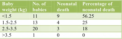
intrahepatic cholestasis of pregnancy 12.5%. 24 babies were below 2.5 kg and among them there was 80% mortality (Table 9).
Table 7: Causes of maternal mortality.
Diagnosis No.of
cases
Maternal
mortality Percentage
HELLP syndrome 7 1 25
Portal hypertension 4 1 25
Acute fatty liver of
pregnancy 4 2 50
Table 8: Fetal outcome.
Outcome Number of cases Percentage
Term live babies 21 46.6
Term IUD babies 2 4.44
Preterm live babies 12 26.6
Preterm still Birth /
IUD 10 22.2
Born alive and dead
(all preterm) 4 8.8
[image:3.595.313.550.392.468.2]Perinatal mortality 16 35.5
Table 9: Influence of baby weight on fetal outcome.
Baby weight (kg)
No. of babies
Neonatal death
Percentage of neonatal death
<1.5 11 9 56.25
1.5-2.5 13 4 25
2.5-3.5 20 3 18
>3.5 1 0 0
DISCUSSION
Total antenatal admissions during the study period were 17890 of which 51 patients had jaundice and the incidence is 0.29%. About 92% patients belonged to lower socioeconomic class and 86% were consuming unsafe water. Begum N et al studied about the seroprevalence (IgG Anti HEV) of subclinical HEV infection in pregnant women and reported that exposure to hepatitis E was more in lower socio economic class.2
The maximum incidence of jaundice was in 3rd trimester and the complications were also high during that period. Harshad et al, Shukla et al and other studies have stated that maximum incidence of jaundice was in III trimester and morbidity and mortality were also higher during III trimester.3,4
cholestasis of pregnancy and hyperemesis had only 2-3 fold elevation.3
Viral hepatitis was the cause in 51% cases comparable to the study by Shukla et al who reported 57% and Harshad et al reported 47% cases of viral hepatitis.3,4 13.72% of cases had HELLP syndrome in present study. Rathi U et al reported 52.3% of cases with liver dysfunction due to preeclampsia and HELLP.5 4 cases had chronic liver disease. Among them 3 had non-cirrhotic portal hypertension (NCPH). Agarwal et al studied 50 pregnant patients with NCPH and reported that in 56% patients NCPH was detected first during pregnancy.6 In India it is commonly due to non-cirrhotic portal fibrosis and extra hepatic portal vein obstruction. But in western countries portal hypertension is mostly due to cirrhosis. Intra hepatic cholestasis of pregnancy was diagnosed in 3 patients; one had history of jaundice in her mother during her antenatal period. Study had one case of haemolytic jaundice due to hereditary spherocytosis which underwent therapeutic abortion due to haemolytic crisis. Pajor et al studied 19 pregnancies with hereditary spherocytosis, concluded that pregnancy precipitated hemolytic anaemia and maternal and fetal
outcome was favourable after splenectomy.7
Leptospirosis was diagnosed in 2 cases by MSAT. Shalini et al reported a case of leptospirosis with Jaundice, coagulopathy and intra uterine death.8 One was due to hyperemesis gravidarum. Matsubara’s et al reported that Jaundice in hyperemesis is due to biliary sludge and it is relieved by hydration.9
In the present study 7.8% patients died, 35 % patients developed complications and 58% had uneventful recovery. 9.8% patients had atonic PPH. 5.8% had DIVC, 7.8% had hepatic encephalopathy. Abruption, hepatorenal failure, esophageal varices was seen in 3.9 % each. Jain S et al reported 52 patients with fulminant hepatic failure and concluded that renal dysfunction was the indicator of poor prognosis in patients with fulminant hepatic failure.10 Rathi U et al reported 3 cases of AFLP and among them 2 cases died of DIVC and multiorgan failure.5 Third patient died of HELLP syndrome, had severe hypertension, proteinuria, ascites delivered a dead born baby, died of DIVC and hepatorenal failure. Rathi U et al reported 25% mortality due to preeclampsia associated liver dysfunction.5 The fourth patient was a case of non-cirrhotic portal hypertension with grade III esophageal varices died due to massive hematemesis at her second trimester. West brook et al reported one death in pregnancy due to variceal bleeding.11
Mortality due to viral hepatitis was not seen in the present study though many patients of hepatitis E had severe morbidity. Study by Jayanthi et al, observed that mortality rate of hepatitis E infection in southern India was very low 3-4% compared to high mortality 30-100%
seen in studies from Northern India.12 Study by Harshad et al, reported that mortality was 41% in
pregnancy associated liver disease and 7.5% in viral
hepatitis and concluded mortality due to hepatitis E was low.3
Preterm deliveries were 48.8% (26.6% live birth and 22.2% intra uterine deaths). The higher incidence of preterm delivery was supported by Kumar et al 66.6% and Harshad et al 32% is due to high fever, increased cytokine release, disturbed hormonal status and debilitating effects of viremia of hepatitis.13,3 The perinatal mortality in present study was 35.5% comparable to Rathi U et al who reported 35.4% and Kumar et al reported 26.5%.5,13 Among 16 Neonatal deaths, HELLP syndrome constitutes 31.2%, hepatitis E 25%, hepatitis B 12.5%, and intrahepatic cholestasis of pregnancy 12.5%. According to Williamson et al the poor fetal outcome in intrahepatic cholestasis of pregnancy was due to the toxic bile acid level in the fetus causing fetal arrhythmia.14 One intrauterine death was seen with chronic liver disease. Westbrook et al reported 26% of fetal loss with chronic liver disease.11 53.3% babies were below 2.5 kg in present study and among them there was 80% mortality. Shukla et al reported 30.8% mortality in low birth weight babies.4
CONCLUSION
Although liver dysfunction is infrequently seen in pregnancy, it can result in severe maternal and fetal compromise. Viral hepatitis is the most common cause of jaundice in pregnancy. Generating public awareness about the various routes of transmission of the different types of infective hepatitis, improving sanitary conditions and habits, imparting health education and knowledge of preventive measures, routine and regular antenatal check-ups and viral markers as a part of routine antenatal screening can help in reducing the burden of jaundice in pregnancy. Jaundice in pregnancy should be managed as a team with collaboration of obstetrics, internal medicine, gastroenterology, anaesthesia and critical care so that early diagnosis and aggressive management can prevent and reduce fetomaternal morbidity and mortality.
Funding: No funding sources Conflict of interest: None declared
Ethical approval: The study was approved by the Institutional Ethics Committee
REFERENCES
1. Tripti N, Agarwal S. Fetomaternal outcome in
jaundice during pregnancy. Obstet Gynecol India. 2005;10:424-7.
2. Begum N, Devi SG, Husain SA, Kumar A, Kar P.
Seroprevalence of subclinical HEV infection in pregnant women from north India: a hospital based study. Indian J Med Res. 2009;130:709-13.
3. Harshad D, Walter KK, Ross D, Lakshmi P.
4. Shukla S. Prospective study on acute viral hepatitis in pregnancy; seroprevalence, and fetomaternal outcome of 100 cases. J Biosci Tech, 2011;2(3):279-86.
5. Rathi U, Bapat M, Rathi P, Abraham P. Effect of liver disease on maternal and fetal outcome-a
prospective study. Indian J Gastroentero.
2007;26:59-63.
6. Aggarwal N, Sawhney H, Vasishta K, Dhiman RK,
Chawla Y. Non-cirrhotic portal hypertension in pregnancy. Int J Gynaecol Obstet. 2001;72(1):1-7.
7. Pajor A, Lehoczky D, Szakács Z. Pregnancy and
hereditary spherocytosis report of 8 patients and a review. Arch Gynecol Obstet. 1993;253(1):37-42.
8. Gainder S, Singla R, Dhaliwal L, Suri V.
Leptospirosis as a cause of intrauterine fetal demise: short report of rare presentation materno-fetal. Med Archives Gyneco Obst. 2010;281(6):1061-3.
9. Matsubara S, Kuwata T, Kamozawa C, Sakamoto Y,
Suzuki M, Tamada K. Connection between
hyperemesis gravidarum, jaundice or liver
dysfunction, and biliary sludge. Trop Gastroenterol. 2000;21(3):118-20.
10. Jain S, Pendyala P, Varma S, Sharma N, Joshi K, Chawla Y. Effect of renal dysfunction in fulminant
hepatic failure. Indian J Med Res. 2009;130(6):709-13.
11. Westbrook RH, Yeoman AD, Ogrady JG. Model for
end-stage liver disease score predicts outcome in
cirrhotic patients during pregnancy. Clin
Gastroenterol Hepatol. 2011;9(8):694-9.
12. Rasheeda CA, Udayakumar N, Jayanthi V. Diseases
in pregnancy and its influence on maternal and fetal mortality-a prospective study from Chennai, southern India. Eur J Gastroenterol Hepatol. 2008;20:362-6.
13. Kumar A, Beniwal M, Kar P, Sharma JB, Murthy
NS. Hepatitis E in pregnancy. Int J Gynaecol Obstet. 2004;85(3):240-4.
14. Williamson C, Miragoli M, Kadir S. Bile acid
signaling in fetal tissues: implications for
intrahepatic cholestasis of pregnancy. Dig Dis. 2011;29(1):58-61.