pISSN 2320-1770 | eISSN 2320-1789
Original Research Article
The correlation of visual inspection, Pap-smear and
immunocytochemistry of human papilloma virus in detection
of cervical cytology
Anuradha Khemka*, Shubhada Jajoo
INTRODUCTION
Carcinogenicity presents as a major challenge to scientists and society. Our health services are adequately equipped with facilities and expertise for diagnosis and management of cancers. The technology is available in the country; the need is to network the centers in public and private sectors with strict quality control. . There should be adequate screening programs to detect pre-cancerous conditions. Mortality and morbidity due to cancers are very high but remains undetected. The focus should be to identify the preventable risk factors and understand the mechanism of carcinogenesis. There
should be adequate screening programs to detect pre-cancerous condition and preventable causes involving
clinical, epidemiological and modern molecular
techniques.
Cervical cancer ranks fifth in the world and is the second cause of death in developing countries like India and China. There are almost 365.71 million women who are above age of 15 years and are at risk of developing malignancy. Worldwide India alone accounts for about 1/3rd load of death from cervical cancer with current estimates of 1,32,000 new cases per year and 74,000 deaths annually.1 At any time women are at risk of Department of Obstetrics and Gynecology, Datta Meghe Institute of Medical Sciences, Wardha, Maharashtra, India
Received: 23 October 2016
Accepted: 23 November 2016
*Correspondence:
Dr.Anuradha Khemka,
E-mail: anuradha.khemka@yahoo.in
Copyright: © the author(s), publisher and licensee Medip Academy. This is an open-access article distributed under the terms of the Creative Commons Attribution Non-Commercial License, which permits unrestricted non-commercial use, distribution, and reproduction in any medium, provided the original work is properly cited.
ABSTRACT
Background: Carcinogenicity presents as a major challenge to scientists and society.. Cervical cancer ranks fifth in the world and is the second cause of death in developing countries like India and China. The rate of death due to cervical cancer is greatest in India. At any time women are at risk of harboring HPV infection, which has been seen to cause cervical cancer. Clinical judgment should be used in spite of all high quality modalities available for screening and diagnosis, as initiated by WHO (World health organization) the Down Staging of cervical cancer, VIA, VILI, Pap smear introduced by George Papanicolaou in 1940s , HPV testing by Immunocytochemistry (ICC) and HPV DNA , Colposcopy , are other modalities to screen cervix. The study aims to evaluate the correlation between Pap smear, visual inspection and Immunocytochemistry of Human papilloma virus (HPV).
Methods: All 100 consecutive women were subjected to visual inspection of cervix, Pap smear and immunocytochemistry of HPV. Pap smear was done by conventional method.
Results: it was seen that out 100 consecutive cervico-vaginal, ears for Pap and Immunocytochemistry, only 33 were abnormal, 24 were ASCUS, 6 were LSIL, 2 were HSIL, 1 was SCC and 67 were NILM out of which 3 were reactive for ICC. And all 33 were reactive for ICC.
Conclusions: The p16 immunostaining performed on conventional smears can become alternative or addition to l HPV DNA tests. The Bethesda System (TBS) 2001 for reporting cervical cytology diagnosis is of high value in detecting abnormal cervical cytology. It is further concluded that the ancillary screening for HPV in NILM category seems to be not advisable as percentage of detection of HPV in this category is low.
harboring HPV infection, which has been seen to cause cervical cancer. The primary prevention from Human papilloma virus (HPV) can be attained by complete abstinence of sexual activity and mass immunization against high risk from Human papilloma virus (HPV). For a developing country like India to screen large population we need measures, which are economical, acceptable, and feasible to both clinicians and women. Clinical judgment should be used in spite of all high quality modalities available for screening and diagnosis, as initiated by WHO (World health organization) the Down Staging of cervical cancer. The cervical cytology by Pap-smear and HPV testing are important advents in the screening of cervical cancer. Therefore such a study would provide us with valuable information about diagnosis of HPV infection along with cervical cytology and visual inspection as a better factor in evaluation of precancerous conditions of cervix leading to invasive carcinoma. The HPV is recognized as major cause of cervical cancer.
Infection of the cervical epithelium with oncogenic types of Human Papilloma virus (HPV) is essential for the development of precursor lesion and cervical cancer.2 The main problem is to identify from the large number of HPV positive patients the ones who are at risk of progressive disease. HPV genotyping can establish the presence of high risk HPV, subclinical and clinical relevant infections. Neither cytological tests nor HPV DNA typing can indicate whether there will be remission or progression to invasive disease. Therefore it is necessary to identify some markers to offer information, concerning the HPV status and progressive risk. So it becomes prudent to know the immunocytochemical expression of biomarker p16INK4a and HPV L1 capsid protein in detection of HPV infection. HPV L1 capsid protein is expressed in the early productive phase of cervical carcinogenesis and is lost in later phases when p16 INK4a gets overexpressed.3 Hence for women’s preventive health care, screening plays a central role. Now we enter the CO-testing era of clinical evaluation,
Cytology and Human Papilloma Virus testing
(Immunocytochemistry) at the same setting.
The study aims to evaluate the correlation between Pap smear, visual inspection and Immunocytochemistry of Human papilloma virus (HPV).
Objectives
To establish correlation between results of Pap smear, Visula inspection and immunocytochemistry.
To study the correlation of socio-demography with Pap smear and immunocytochemistry of Human Papilloma Virus (HPV).
METHODS
The study was conducted on 100 consecutive patients visiting Acharya Vinoba Bhave Rural Hospital (AVBRH)
Gynecology outpatient department (OPD). Visual inspection, Pap smear and immunocytochemistry for HPV were done at the same sitting. The design chosen for this study was prospective type. All women who gave consent for study were selected. Married women, age above 25 years was the inclusion criteria. Women below 24 years, post hysterectomy and already diagnosed cases of cancer with radiotherapy were not included. Cervix was examined by naked eyes, per speculum and per vaginum examination was performed and readings were noted in pre-designed proforma. Cervical cytology was collected by conventional technique and slide was prepared for Papanicolaou smear. Simultaneously Ayres disposable spatula was used for collection of cells from the transformation zone. The end of spatula was used to shed cervical cells to a solution of 2ml normal saline in a pre-labelled glass vial, and immediately taken to
cytopathology for cytospin preparation for
immunocytochemistry (ICC) of HPV. Papanicolaou smear was reports as per The Bethesda system and ICC slides were stained. Monoclonal mouse antibody (LOT NO- 10064645, DAKO, Code M3528) was used for immunocytochemistry that is Immunoreactive with HPV infected issue. The immunostaining was performed as detailed by manufacturer’s instructions.
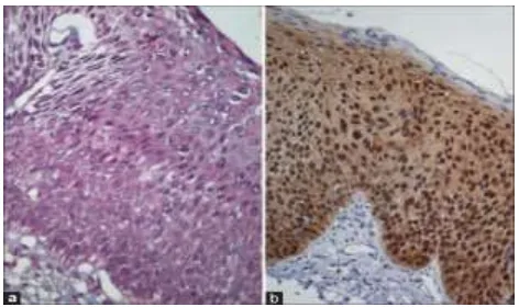
Figure 1: (a) Abnormal cervical cells stained by Papanicolaou method; (b) Cervical cells when immunostained for p16INK4a by cytospin for Human
Papilloma Virus. When seen under light microscope staining of nucleus and cytoplasm brown was
suggestive of infection with HPV.
Statistical analysis
Data are reported as number (percentage) and mean (Standard deviation). Association of outcome and exposure was tested with t-test and exposure to more than two categories with the non-parametric trend Pearson chi-square test. P<0.05 was considered significant.
RESULTS
Table 1 shows the 100 women enrolled for the study, The Mean age was 41.96 years with standard deviation of 10.98, majority of the women were from rural area being 57%, nearly half of the study population was from villages and rest were from urban area 43%. Forty-three women were home-makers (43%), 50 were farm labourers (50%) and 7 were clerk (7%). The mean income in rupees of the study population was seven thousand three hundred forty rupees with SD of 4524.91.
Table 1:Distribution of socio-demographic data.
Mean age in years (standard deviation SD)
41.96 (10.98)
Type of residence (%) N=100
Urban 43 (43%)
Mean income in rupees (standard deviation SD)
7430 (4524.91)
Table 2:The data of 100 women who underwent
clinical evaluation of cervix by visual inspection, Pap smear and immunocytochemistry. subjected for clinical evaluation of cervix, cervical cytology by conventional method (Pap smear), and immunocytochemistry of Human Papilloma virus (HPV). On visual inspection there were 46% with unhealthy cervix, rest 54% were healthy. There were 67% patients who were negative for intraepithelial malignancy (NILM), 24% (24) patients had atypical cells of undetermined significance (ASCUS), 6% (6) had low-grade squamous intraepithelial lesion (LSIL), 2% (2) had high-grade squamous intraepithelial lesion (HSIL) and only 1% (1) women had squamous cell carcinoma.
36 % patients reacted positive for immunostaining (ICC) for HPV and 64% were negative.
Table 3:The percentage of abnormal cervical
cytology.
Abnormal cervical cytology (N=33)
Squamous cell carcinoma (SCC) 1
Out of 100 cervical cytology done 33 were abnormal, out of which 72.7 % was ASCUS, 18.18% were LSIL, 6.06% were HSIL and 3.03% were SCC. Maximum no. of cases was of atypical squamous cells of undetermined significance.
Table 4:The correlation between the
socio-demographic data and immunocytochemistry for HPV.
with a significant p value of 0.002.that is immunostaining was seen in majority of farm labourers. The mean income of the women who were positive was rupees 5944, and women who were negative for HPV had mean income of rupees 8265.62 with significant p value 0.0131 by t-test i.e. HPV infection was more in low socio- economic class women.
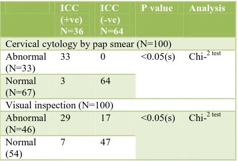
Table 5:The correlation between visual inspection of
cervix, cervical cytology by Pap smear and immunocytochemistry.
Cervical cytology by pap smear (N=100) Abnormal interventions done in present study visual inspection of cervix, cervical cytology and immunocytochemistry for HPV. Out of 100 women only 33 had abnormal Pap 29 were positive for HPV ,54 had normal looking cervix and 47 of them were negative for ICC with significant p value of <0.05,by chi-square test. The clinical evaluation of cervix is a very important step in diagnosing pathology at an early stage. To screen NILM category with HPV did prove to be beneficial.
DISCUSSION
In our study we included 100 consecutive women attending AVBRH outdoor patient department. The over expression of p16-INK4a by immunocytochemistry was compared with cervical cytology by Papanicolaou test (conventional method) and visual inspection of cervix.
Many studies on p16 cytology have been published, most of them have used convenience samples or consecutive specimens of a particular cytological diagnosis either taking specifically abnormal cervical smears or retrospectively investigating HPV in higher grades of cervical dysplasia. Table below summarizes 9 large studies incorporating almost 1,510 specimens (Kate C et al.4 About 5% of the normal cytology specimens, 32% of
the atypical squamous cells of undetermined significance (including also ASC-US and ASC-H), 37% of the low-grade squamous intraepithelial lesion (LSIL), 93% of the HSIL, and 99% of the cancer specimens were found to be positive in p16 cytology which was comparable with our present data where out of 33 abnormal cervical cytology, (24/33) 72.7% were ASCUS, (6/33)18.08% were LSIL, (2/33) 6.06% were HSIL and (1/33) 3.03% were SCC but all the abnormal cervical cells tested positive for p16 reactivity.(Table below). These data are summarized different cytology platforms and systems for p16 cytology assessment were used. Sahebali et al. determined p16-positive cell counts in consecutive slides from a routine cytology laboratory.5 Bibbo et al analysed a series of HSIL result and correlated it with biopsy.6 In most cases of low grade and high-grade dysplasia p16 was positive.
In our study we focused on the feasibility of p-16 INK4a immunostaining in consecutive 100 cervico-vaginal samples processed by conventional technique in parallel with clinical evaluation of uterine cervix. There are few
studies that have compared p16-INK4a
immunocytochemistry analysis with conventional Pap smear and visual inspection of uterine cervix. During the last decade technological advances in sample collection and processing have been introduced and implemented in cancer screening programs. But we cannot attempt to replicate the strategies of the developed countries. The burden of cervical malignancy in late stage in most developed countries is about 10 to 12 percent, where as in India it is 70 percent. Our highest priority is therefore to identify disease earlier as stage 1B where the cure rate can be significantly high. Pap smear is considered to be the mainstay of cervical cancer screening programs worldwide, but the interpretation of cytological tests still has limitations with great inter-observer and intra-observer variability ranging from 43% to 68%.
Stoler et al demonstrated the subjective differences in morphology of cervical cytology. It is common specifically with ASCUS, LSIL categories.12 In our study we also encountered different morphology of cervical cytology by The Bethesda system 2001 and we found that 33 out 100 were abnormal with conventional smear, 24 were ASCUS (72.7%), 6 were LSIL (18.1%), 2 were HSIL (6.06%) and 1 was SCC (3.03%) with 67 were Negative for intraepithelial malignancy (Table 3 and pie-chart).
Deshpande and Nene BM et al in a rural area in Maharashtra looked at the profile of cervical cytology in women attending their health center and total of 186 women were enrolled.13 The mean age of the women in the study was 35.3 years which was comparable to our study with mean age of 41.9 (Table 1).
screening was (34.5 years+/- 9.23) which was comparable with our study where mean age was 41.9 years with SD 10.98. Majority of the women in his study were home-makers (80.4%) and 59.6% were from low
socio-economic class (59.6%). In our study also the abnormal cervical cytology and HPV reactivity was seen in women with low income per family per month (Table 1, 4).
Table 6: Comparison of various studies.
Author Platform NILM ASCUS LSIL HSIL SCC
Bibbo6 LBC - (14/19)74% (25/26)97% -
Saqui7 LBC (5/28)(18%) - (20/24)74% (9/10)90% (1/1)100%
Pientonng14 Smear in PBS - (17/30)57% (10/30)33% (28/30)98% (29/30)100%
Yoshida8 Pap slides - (5/38)13% (7/12)58% (33/33)100% (12/12)100%
Pientong9 Smear in PBS 0/30(0%) (21/40)53% (19/35)54% (29/30)97% (30/30)100%
Bose et al10 Conventional smear (3/23)13% (10/34)29% (7/34)21% (13/16)81% -
Table 7: Comparison of various studies.
Meyer et al11 LBC 9/235(5%) (9/49)32% (24/57)42% (43/53)81% -
Wentzensen4 LBC - (37/137)27% (21/88)24% - -
Ekalaksananan Conventional
smear
(11/148)7% (12/19)63% (3/5)60% (12/12)100% (2/2)100%
Present study Conventional
smear
67/100(67%) 24/100(24%) 6/100(6%) 2/100(2%) 1/100(1%)
Abnormal cervical cytology and HPV reactivity was seen in farm labourers. Twenty-four women out of 50 were farm labourers were positive for immunostaining .Very few women were professional and none of them were reactive to immunostaining of HPV (Table 6).7 The findings are in accordance that HPV is seen in low socio economic class (Table 4).
In a study done by Sauvaget C et al in western India they concluded that the prevalence of HPV infection was more in women from low socioeconomic class, low education level and manual workers.15 In our study (Table 1) and the mean income was seven thousand three hundred forty (7340 rupees) with SD (4525.91). The mean income of women who had abnormal cervical cytology and tested positive for immunostaining was rupees five thousand nine hundred forty four per family (5944.44) with SD (12.14). It was seen that the HPV infection was present in women of low socio economy (Table 4).
The strength of our study was that we gave the benefit of index test and reference test to all 100 women. There by doing a blind comparison between the methods to screen cervix. For a country like India with burden of cervical cancer, huge exploratory studies are needed which does not have cost as a barrier.
Funding: No funding sources Conflict of interest: None declared
Ethical approval: The study was approved by the Institutional Ethics Committee
REFERENCES
1. WHO I. WHO/ICO Information Centre on HPV and
Cervical Cancer (HPV Information Centre). Human Papillomavirus and Related Cancers in Kenya Summary Report. 2010;2010.
2. Sartwell PE. On the methodology of investigations of etiologic factors in chronic diseases”—Further
comments. Journal of Chronic Diseases.
1960;11(1):61-3.
3. Hilfrich R, Hariri J. Prognostic relevance of HPV L1
capsid protein detection within mild to moderate dysplastic lesions of the cervix uteri in combination with a second biomarker p16. Anal Quant Cytol Histol. 2008;30(2):78-82.
4. Cuschieri K, Wentzensen N. Human papillomavirus
mRNA and p16 detection as biomarkers for the improved diagnosis of cervical neoplasia. Cancer
Epidemiology Biomarkers and Prevention.
2008;17(10):2536-45.
5. Sahebali S, Depuydt CE, Boulet GA, Arbyn M,
Moeneclaey LM, Vereecken AJ, et al.
Immunocytochemistry in liquid‐based cervical
sectional study. International Journal of cancer. 2006;118(5):1254-60.
6. Bibbo M, Klump WJ, DeCecco J, Kovatich AJ.
Procedure for immunocytochemical detection of P16INK4A antigen in thin-layer, liquid-based specimens. Acta Cytologica. 2002;46(1):25-9. 7. Saqi A, Pasha TL, McGrath CM, Yu GH, Zhang P,
Gupta P. Overexpression of p16INK4A in liquid‐ based specimens (SurePath™) as marker of cervical dysplasia and neoplasia. Diagnostic Cytopathology. 2002;27(6):365-70.
8. Yoshida T, Fukuda T, Sano T, Kanuma T, Owada N,
Nakajima T. Usefulness of liquid‐based cytology specimens for the immunocytochemical study of p16 expression and human papillomavirus testing. Cancer Cytopathology. 2004;102(2):100-8.
9. Pientong C, Ekalaksananan T, Kongyingyoes B,
Kritpetcharat O, Swadpanich U, Pengsa P, et al. Immunocytochemical staining of p16INK4a protein from conventional Pap test and its association with
human papillomavirus infection. Diagnostic
Cytopathology. 2004;31(4):235-42.
10. Bose S, Evans H, Lantzy L, Scharre K, Youssef E. p16INK4A is a surrogate biomarker for a subset of human papilloma virus‐associated dysplasias of the uterine cervix as determined on the Pap smear. Diagnostic Cytopathology. 2005;32(1):21-4.
11. Meyer JL, Hanlon DW, Andersen BT, Rasmussen
OF, Bisgaard K. Evaluation of p16INK4a expression in ThinPrep cervical specimens with the CINtec
p16INK4a assay. Cancer Cytopathology.
2007;111(2):83-92.
12. Stoler MH, Schiffman M. Interobserver
reproducibility of cervical cytologic and histologic interpretations: realistic estimates from the ASCUS-LSIL Triage Study. Jama. 2001;285(11):1500-5.
13. Nene B, Deshpande S, Jayant K, Budukh AM, Dale
P, Deshpande D, et al. Early detection of cervical cancer by visual inspection: A population‐based study in rural India. International Journal of Cancer. 1996;68(6):770-3.
14. Chhabra S, Bhavani M, Mahajan N, Bawaskar R.
Cervical cancer in Indian rural women: trends over two decades. Journal of Obstetrics and Gynaecology. 2010;30(7):725-8.
15. Pientong C, Ekalaksananan T, Swadpanich U,
Kongyingyoes B, Kritpetcharat O, Yuenyao P, et al. Immunocytochemical detection of p16INK4a protein
in scraped cervical cells. Acta Cytologica.
2003;47(4):616-23.
16. Sauvaget C, Nene BM, Jayant K, Kelkar R, Malvi SG, Shastri SS, et al. Prevalence and determinants of high-risk human papillomavirus infection in middle-aged Indian women. Sexually Transmitted Diseases. 2011;38(10):902-6.
17. Aswathy S, Quereshi MA, Kurian B, Leelamoni K. Cervical cancer screening: Current knowledge and practice among women in a rural population of Kerala, India. The Indian Journal of Medical Research. 2012;136(2):205.