Page 1 of 12
Ultrasonographic hemodynamics for prediction of poor liver
regeneration induced by severe portal vein stenosis in rats
Lin Ma1, Kefei Chen2, Lulu Yang1, Hong Wang1, Qiang Lu1, Yan Luo1
1Department of Ultrasound, 2Department of Liver and Vascular Surgery, West China Hospital of Sichuan University, Chengdu 610041, China Contributions: (I) Conception and design: Y Luo; (II) Administrative support: Q Lu, Y Luo; (III) Provision of study materials: L Ma, K Chen, L Yang, H Wang; (IV) Collection and assembly of data: L Ma, L Yang; (V) Data analysis and interpretation: L Ma; (VI) Manuscript writing: All authors; (VII) Final approval of manuscript: All authors.
Correspondence to: Yan Luo. Department of Ultrasound, West China Hospital of Sichuan University, 37 Guoxue Lane, Chengdu 610041, China. Email: luoyand@hotmail.com.
Background: Insufficient portal vein blood flow, such as portal vein stenosis (PVS), plays a significant influence on liver regeneration. Early prediction of poor liver regeneration induced by severe PVS is critical. Ultrasound serves as a first-line imaging technique in diagnosing PVS based on the changes of portal vein hemodynamics. However, there is still no consensus on the criteria for evaluating the degree of PVS. Moreover, which degree of PVS can induce poor liver regeneration still is unclear. Therefore, it is essential to determine the stenosis degree that leads to significantly poor liver regeneration and to evaluate the value of ultrasonographic hemodynamics for predicting poor liver regeneration induced by severe PVS.
Methods: Rats were randomly subjected to sham operation rats group (SOR), PH group (group A), and PVS groups with mild, moderate, or severe stenosis flowing PH (groups B-D). PH group was set up a model of 70% hepatectomy, and PVS groups were produced by different degrees of partial portal vein ligation following PH. In the SOR group and PH group, the portal vein diameter (PVD) and portal vein velocity (PVV) were measured by Ultrasound at preoperative and postoperative 1, 3, 7, and 14 d. In PVS groups, PVD and PVV at the stenotic (PVDs, PVVs) and pre-stenotic (PVDpre, PVVpre) sites were also detected on
1, 3, 7, and 14 d after surgery, calculating the diameter stenosis ratio (DSR) and accelerating blood flow velocity ratio (AVR). Rats were sacrificed at 1, 3, 7, and 14 d post-surgery, and the expression of proliferating cell nuclear antigen (PCNA) and the liver regeneration rate (LRR) at 14 d were evaluated. The PVVs, DSR,
and AVR in the different groups were analyzed combined with the status of liver regeneration, and receiver operating characteristic (ROC) analysis was also applied to assess the value of PVVs, DSR, and AVR in
diagnosing severe PVS and the resulting poor liver regeneration.
Results: Seventy-two rat models of different degrees of PVS were successfully set up following 70% PH. The stenosis ratios (SRs) of each PVS group were 45.16%±3.44%, 59.21%±3.83%, and 69.56%±2.16%, respectively. Poor liver regeneration appeared to be significant when PVS was greater than 65% (group D), of which the LRR at 14 d was significantly lower compared to PH group (group A) and PVS groups with SR ≤50% (group B) and SR >50–65% (group C), respectively (all P<0.05). Meanwhile, PCNA expression of group D was significantly lower compared to group C at 1 d and groups A-C at 3 d (all P<0.05). Differences were also detected at 3 d between groups A and B and groups A and C (both P<0.05). Among PVS groups, PVVs accelerated dramatically, with significant differences demonstrated between group D and groups B and
C at 1 d, as well as group B and groups C and D at 3 d (all P<0.05). At 1, 3, and 7 d, DSR of groups C and D were significantly higher than that of group A (all P<0.05). At 1 and 3 d, AVR of group D was significantly higher than that of groups B and C (all P<0.05). ROC analysis showed the AUC of PVVs at 1 d in diagnosing
severe PVS was 0.84, while at 3 d, it was unable to differentiate from mild-moderate or severe PVS by PVVs
Introduction
Because of the severe shortage of donor’s livers, living donor liver transplantation (LDLT) has undergone rapid development with the advancement in modern surgical techniques (1-3). Liver regeneration post-LDLT is a vital process of liver recovery, and the portal vein blood
flow, accounting for 70–80% of the hepatic blood supply,
provides a fundamental basis and a prerequisite for successful liver regeneration and survival. Insufficient
portal vein blood flow, such as portal vein stenosis (PVS),
is a significant complication after LDLT, especially in children. Mild PVS is conventional but does not affect liver function and regeneration. In patients with severe PVS, poor regeneration may occur, complicated with portal hypertension, small-for-size syndrome, or even liver failure. Early diagnosis and prompt treatment are critical (4-8).
As a non-invasive, cost-effective, and non-radioactive technique with bedside availability, ultrasound (US) serves
as a first-line imaging modality to diagnose PVS in the early
postoperative period and long-term follow-up. In general, PVS is suggested if the portal vein is regional stenosis, with blood flow aliasing and acceleration at the stenotic site. Most of the researchers believe that evident PVS can be diagnosed if the stenotic diameter is <2.5–3.5 mm
or the diameter stenosis ratio (DSR)>50%, blood flow
velocity (PVV) at the stenotic site (PVVs) >150 cm/s or the
accelerating velocity ratio (AVR) ≥3 (9-11). However, to date, unlike the criteria for grading carotid artery stenosis (12), there is still no consensus on the ultrasound diagnostic criteria for evaluating the degree of PVS. Moreover, which degree of PVS could induce poor liver regeneration still is unclear. In the clinic, the treatments are usually
provided when evident PVS was detected, accompanied by apparent symptoms such as abnormal transaminase level, blood coagulation disorders, portal hypertension, and so on. But in that case, irreversible damages of hepatocytes may already occur, and consequently, the grafts would be in the state of poor regeneration for a long-time even treatments are applied. Liver regeneration can be assessed by pathological and immunohistochemical tests such as the expression of proliferating cell nuclear antigen (PCNA), a kind of nucleoprotein only synthesized and expressed in proliferative cells, but these examinations are invasive and more costly (13,14). Therefore, there is a growing demand for the correct assessment of the degree of PVS, early predicting the possible poor liver regeneration induced by severe PVS noninvasively.
Due to the diverse causes of hepatic diseases and the limitation in ethics, most research studies liver regeneration based on experimental animals. A rat model of 70% partial hepatectomy (PH) is a widely accepted liver regeneration research model, which is famous for its regeneration induction without causing fulminant hepatic failure (15). Partial ligation of the portal vein is commonly used for producing the PVS model (16). Therefore, this study used a combination of 70% PH and portal vein partial ligation to set up (PH + PVS) rat models. Based on the assessment of liver regeneration in the different degrees of PVS, the degree of PVS inducing poor liver regeneration was determined. Moreover, a comparative analysis was performed to evaluate the value of Ultrasound
parameters PVVs, DSR, and AVR for predicting poor liver
regeneration induced by severe PVS. These findings will supply experimental evidence for further investigating PVS diagnosis and grading in the clinic.
Conclusions: Poor liver regeneration could be significantly induced when PVS was greater than 65%. Ultrasound can well prove the changes of portal vein hemodynamics in different degrees of PVS in rats. The parameters PVVs could be regarded as a valid index for diagnosing PVS but were not applicable for
evaluating the stenosis degree. Comparatively, the parameters DSR and AVR, especially AVR, proved to be useful for differentiating severe PVS (>65%) in the early postoperative period, predicting the resulting poor liver regeneration.
Keywords: Liver regeneration; portal vein stenosis (PVS); ultrasound; hemodynamics; portal vein diameter (PVD); portal vein blood flow velocity (PVV)
Submitted Feb 11, 2020. Accepted for publication Mar 18, 2020. doi: 10.21037/atm.2020.04.21
Methods
Study subjects
One hundred and two healthy male Sprague-Dawley rats (200–400 g, 7–14 weeks of age, SPF grade) were purchased from Chengdu Dashuo Biotechnology Co., Ltd., and acclimatized for at least 7 days to laboratory conditions with a constant temperature and a 12 h light-dark cycle at the animal experiment center of West China Hospital.
The rats were randomly divided into five groups as follows: sham operation rats (SOR group, n=6), PH group (group A, n=24), and PVS groups with mild, moderate, and severe PVS after PH (group B, C, and D, n=24 respectively). Also, rats in A–D groups were further divided into four subgroups, based on the time after surgery (1, 3, 7, and 14 d), with 6 rats in each subgroup. The animal ethics committee approved all the rats and procedures of West China Hospital, Sichuan University. Rats were kept warm and had free access to food and water throughout the experiment.
Construction of rat models
Construction of 70% PH rat model
Rats were placed in the supine position after ether anesthesia, and then a 70% hepatectomy model was constructed
based on methods described by Higgins et al. (15).
Briefly, the middle lobe and left lateral lobe (about 68% of
the total volume of the liver) were excised and weighed.
Construction of PVS model after PH
After PH, the portal vein was separated. A microvascular caliper was used to measure the portal vein diameter (PVD) at the site of pre-ligation, which was below the porta hepatis, and about 3 mm above the joint of the splenic vein
and superior mesenteric vein. Based on pre-defined stenosis
degrees, the diameter of the stenotic segment was calculated
from the formula SR = (1 − Dstenosis/PVD) × 100%, and then
the corresponding gauge of a medical needle was selected. The separated portal vein and the needle were ligated together by 3-0 silk suture, and afterward, the needle was removed slowly. The diameter of the stenotic segment of the portal vein was equaled to the outer diameter of the needle. Different degrees of PVS was induced using this method. In the end, 32,000 units penicillin and 5 mL NaCl (0.9%) was injected intraperitoneally in each rat before closing the abdominal cavity.
For the SOR group, neither hepatectomy nor portal vein
ligation was performed during surgery. For the PH group (group A), only 70% hepatectomy was conducted. For the PVS groups (groups B–D), different degrees of PVS were conducted following PH.
Ultrasound examination
Ultrasound was used to check the portal vein hemodynamic changes by a Philips iu22 ultrasound machine with a 5.0–12.0 MHz linear transducer. The rats were ether-anesthetized and placed in supine positions. The abdomen was shaved with an electric hair remover to minimize ultrasound attenuation. Ultrasound examination was performed by the same experienced physician, with rats supporting stable anesthesia.
In the SOR group and PH group (group A), PVD and PVV were measured by grayscale and Doppler ultrasound at preoperative and postoperative 1, 3, 7, and 14 d. In PVS groups (groups B–D), the PVD and PVV at the stenotic (PVDs, PVVs) and pre-stenotic (PVDpre, PVVpre) sites were
measured, and then the DSR and AVR were subsequently
calculated as follows: DSR = (PVDpre − PVDs)/PVDpre
× 100%, and AVR = (PVVs − PVVpre)/PVVpre. When
measuring the velocities, based on the vessel diameter and direction, the Doppler sample volume should be adjusted to 0.5 mm, and the angle of insolation should remain constant
at less than 60°. PVVs should be detected at the stenotic
site where the blood flow disturbance could be seen. All observation data were measured three times and averaged as the resultant values.
Evaluation of liver regeneration
Assessment of the expression of PCNA
The rats in A-D groups were sacrificed at 1, 3, 7, and 14 d
post-surgery, and the liver tissue specimens (2.0×1.0×1.0 cm3)
were obtained. Then, the liver tissues were fixed in 10% neutral formalin, embedded in paraffin, made into slices at 5 μm. Immunohistochemical staining was performed to
assess PCNA expression under high magnification based on the paraffin sections, and hepatocytes with a brown-yellow
nucleus were considered positive for staining. Besides, about 100 mg of liver tissue was also harvested when rats were sacrificed. The adipose tissue and connective tissue on the surface were removed as much as possible, and then the liver tissue was placed in cryogenic vials and stored in liquid nitrogen. Western blotting was performed for
SOR group, the rats were sacrificed at 14 d post-surgery, with its liver tissues obtained for PCNA examination.
Assessment of liver regeneration rate (LRR)
The residual liver was excised and weighed when rats
sacrificed at 14 d post-surgery. LRR was calculated based on
the equation: LRR = D/E×100%, in which D represents the
liver weight per 100 g of the body weight when sacrificed,
and E represents the preoperative liver weight per 100 g of the body weight, which was calculated by (the excised liver weight/0.68) (17).
Statistical analysis
The analysis of variance (ANOVA) test was employed to study the differences in LRR, PCNA expression, and ultrasound parameters PVD, PVV, DSR, and AVR among
groups. The least significant difference (LSD) test was used
to compare the results pairs of groups with even variance, and the Dunnett-t3 test was used to test the data with uneven variance. Differences were considered significant if the P value <0.05. The receiver operating characteristic (ROC) curve was drawn, and the area under the curve (AUC), Std. Error, asymptotic Sig. (b), asymptotic 95%
confidence interval, best cut-off, sensitivity, and specificity
were calculated to evaluate the value of PVVs, DSR, and
AVR in diagnosing severe PVS, predicting the resulting poor liver regeneration. All statistical analyses were performed utilizing SPSS 25.0 and GraphPad Prism 8.
Results
Model construction
In this study, 72 rat models of different degrees of PVS were successfully set up following 70% PH. The SRs of each PVS group were 45.16%±3.44%, 59.21%±3.83%, and 69.56%±2.16%, respectively. In each PVS group, no statistical difference of SRs was observed at postoperative 1, 3, 7, 14 d (all P>0.05, Figure 1).
LRR
The residual liver weight of the rats increased with time after PH. The LRR of 70% PH group and mild PVS group with
SR ≤50% on the 14th day both reached above 90%, which
means the regenerated liver weight per 100 g bodyweight almost recovered to its original weight before surgery. Poor liver regeneration could be significantly induced when PVS was greater than 65% (group D), with the LRR of less than
80%. That was significantly lower compared to 70% PH
group (group A) and PVS groups with SR ≤50% (group B) and
SR >50–65% (group C), respectively (all P<0.05, Figure 2).
The expression of PCNA
Immunohistochemical staining of PCNA demonstrated that only a few positive cells, with brown-stained nuclei, were seen in the SOR group. In the 70% PH group, the positive expression of cells increased at 1 d post-surgery, presenting
100
90
80
70
60
50
40
30
SR of the portal vein (%)
1 d 3 d 7 d 14 d 1 d 3 d 7 d 14 d 1 d 3 d 7 d 14 d B (Mild PVS)
45.16%±3.44% 59.21%±3.83% 69.56%±2.16% C (Moderate PVS) D (Severe PVS)
Days after operation
in the portal areas. At 3 d, the levels of PCNA-positive expression were still relatively high, with more expression in the central areas. At 7 d, the number of PCNA-positive cells
decreased (Figure 3), while at 14 d, it was comparable to the
levels in normal liver tissues. In PVS groups, the expression of PCNA showed similar trends with time. The negative reaction was observed in karyokinesis.
According to western blot analysis, PCNA expression appeared to be extremely low in the SOR group. In groups A-D, the expression had a similar trend with time. In brief, PCNA expression increased at 1 d after surgery, peaked at 3 d, and then gradually decreased at 7 and 14 d. At 3 d, the expression of PCNA in groups B and C were the highest, followed by group A, and then group D. More precisely, significant differences were detected between groups A and B and between groups A and C (both P<0.05). The
expression of group D was significantly lower compared to
group C at 1 d and groups A, B, and C at 3 d (all P<0.05). At 7 and 14 d, there were no statistical differences among groups (all P>0.05, Figures 4 and 5).
Ultrasound examination
PVD and PVV in 70% PH group
In the 70% PH group, the residual liver and blood flow in
the portal vein could be proved clearly by ultrasound. PVD started to increase at 1 d, peaked at 3 d, decreased gradually at 7 d, and recovered to the normal levels at 14 d. PVD at 3 d was significantly higher than 14 d PVD (P<0.05). In contrast, PVV decreased to the lowest level at 3 d, increased gradually at 7 d, and then recovered similarly to normal
levels at 14 d. PVV at 3 d was significantly lower than 1d
and 14 d PVV (both P<0.05). The results are shown in
Figure 6.
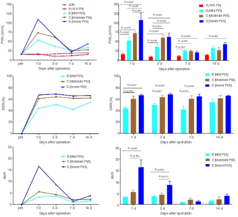
Ultrasound PVVs, DSR, and AVR in PVS groups
PVVs:gray-scale ultrasound could prove the portal vein
stenosis clearly, and Doppler ultrasound showed blood flow disturbance at the stenotic site with accelerated flow velocity
(Figure 7). After surgery, PVVs increased dramatically to the
peak at 1 d, and then gradually decreased at 3 and 7 d. The
Figure 2 LRR in the distinct groups at 14 d post-surgery. LRR, liver regeneration rate.
Stenosis ratio of portal vein (%)
140
120
100
80
60
40
20
0
140
120
100
80
60
40
20
0
Liver r
egeneration rate (%)
Liver r
egeneration rate (%)
0% 50% 65% P=0.007
70% PH SR ≤50% SR >50−65% SR >65−99% Days after operation
0.00 0.000.000.000.000.0040.0040.0045.4546.6747.1050.0053.8557.1460.0060.0060.0063.6068.0068.0068.0070.3771.4373.33
P=0.004 P=0.045
Figure 3 Immunohistochemical staining of PCNA in SOR group and PH group (×400). (A) SOR group; (B) PH group at 1 d; (C) PH group
at 3 d; (D) PH group at 7 d, the negative reaction was observed in karyokinesis (arrow). PCNA, proliferating cell nuclear antigen; SOR, sham operation rats; PH, partial hepatectomy.
PVV of 70% PH group was significantly lower compared to
PVVs of groups B–D at 1, 3, and 7 d and PVVs of group D
at 14 d (all P<0.05). Among PVS groups, PVVs of group D
was significantly higher than that of groups B and C at 1 d,
and PVVs of group Cand D were significantly higher than
that of group B at 3 d (all P<0.05).
DSR: DSR in PVS groups were by the stenosis degree.
Briefly, the DSR of group D was the highest, followed by SOR A B C D
Figure 4 The expression of PCNA detected by western blot at 3 d post-PH in the distinct groups. PCNA expression in the SOR group
was extremely low, and the expression of group D was significantly lower compared to groups A, B, and C. PCNA, proliferating cell nuclear antigen; PH, partial hepatectomy; SOR, sham operation rats.
Figure 5 The expression of PCNA in the distinct groups. PVS, portal vein stenosis; PH, partial hepatectomy; PCNA, proliferating cell nuclear
antigen. 1.50
1.25
1.00
0.75
0.50
0.25
0.00
1.50
1.25
1.00
0.75
0.50
0.25
0.00
PCNA PCNA
1 3 7 14 A (PH) B (Mild PVS) C (Moderate PVS) D (Severe PVS)
A (70% PH) B (Mild PVS) C (Moderate PVS) D (Severe PVS) P=0.029
P<0.001
P<0.001 P<0.001 P<0.001 P=0.003
Days after operation Days after operation
1 d 3 d 7 d 14 d
Figure 6 PVD and PVV in SOR group and PH group (&, P<0.05 vs. SOR group; *, P<0.05 vs. 3 d). PH, partial hepatectomy; PVD, portal
vein diameter; SOR, sham operation rats; PVV, portal vein velocity. 2.5
2.0
1.5
1.0
25
20
15
10
5
PVD (mm) PVV (cm/s)
pre 1 d 3 d 7 d 14 d Days after operation
70% PH SOR
& &
&
* * *
& &
70% PH SOR
group C, and then group B. At 1, 3, 7 d post-surgery, DSR of group C and D were significantly higher than that of
group A (all P<0.05), while at 14 d, no significant difference
was observed among PVS groups (all P>0.05).
AVR: at 1 d post-surgery, AVR reached the peak,
and then gradually decreased to the lowest levels until
7–14 d. AVR of group D was significantly higher than that
of groups B and C at 1 and 3 d after surgery (all P<0.05), while there were no statistical differences among PVS groups at 7 and 14 d (all P>0.05).
All the results of PVVs, DSR, and AVR in the distinct
groups are showed in Table 1 and Figure 8.
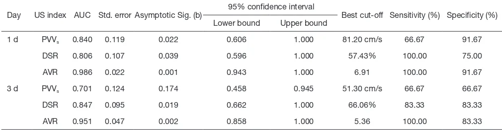
ROC analysis
In this study, poor liver regeneration could be significantly
induced by severe PVS (SR >65–99%). According to the comparative analysis of portal vein hemodynamic changes
in different degrees of PVS, ultrasound parameters PVVs,
DSR, and AVR of severe PVS group were significantly different from mild or moderate PVS groups, especially at 1 and 3 d post-surgery. Therefore, the ROC analysis
was applied to assess the value of PVVs, DSR, and AVR
in differentiating severe PVS from mild-moderate PVS at 1 and 3 d, with mild-moderate PVS, defined as negative
Figure 7 Ultrasound images in severe PVS rat. (A) Gray-scale ultrasound can clearly prove the stenotic site in the portal vein (arrow); (B)
color Doppler ultrasound showed blood flow disturbance at the stenotic site (arrow); (C) spectral Doppler show decelerated flow velocity at the pre-stenotic site; (D) spectral Doppler showed accelerated flow velocity at the stenotic site. PVS, portal vein stenosis.
A
B
C
D
Table 1 Ultrasound parameters PVVs, DSR and AVR of the distinct groups at the same time point
US index Group 1 d 3 d 7 d 14 d
PVVs A (70% PH) 16.57±2.36 9.95±1.52 11.60±1.65 14.85±1.62
B (mild PVS) 53.92±9.44a 37.17±6.31a 26.12±1.36a 33.93±5.65
C (moderate PVS) 73.50±4.07a 61.73±8.18a,b 25.27±3.07a 27.83±8.25
D (severe PVS) 109.67±18.42a,b,c 63.90±10.69a,b 21.75±3.28a 43.97±3.94a
DSR (%) B (mild PVS) 44.40±2.22 50.72±2.39 42.30±6.19 54.97±5.13
C (moderate PVS) 61.16±4.73b 64.39±3.03b 61.57±4.84b 62.07±5.34
D (severe PVS) 67.54±1.75b 69.16±1.84b 65.74±3.73b 66.50±1.43
AVR B (mild PVS) 3.21±0.32 3.67±0.58 0.92±0.21 1.53±0.41
C (moderate PVS) 5.54±1.00 4.32±0.77 2.10±0.73 2.24±1.07
D (severe PVS) 16.58±3.23b,c 8.65±1.62b,c 1.31±0.45 3.81±0.74
a, P<0.05 vs. group A; b, P<0.05 vs. group B; c, P<0.05 vs. group C. PVV
s, portal vein velocity at the stenotic stie; DSR, diameter stenosis
results and severe PVS defined as a positive diagnosis.
As is shown in Figure 9 and Table 2, at postoperative
1 d, the AUC of PVVs in diagnosing severe PVS was 0.84
(P<0.05 vs. AUC =0.50), and the best cut-off points were
81.20 cm/s, with a specificity up to 91.67% and a sensitivity
of 66.67%. While at 3 d, it was unable to differentiate from
severe or mild-moderate PVS by PVVs (P>0.05 vs. AUC
=0.50). Comparatively, the AUC of DSR and AVR at 1 and 3 d in differentiating severe PVS from mild-moderate PVS
were all greater than 0.80 (P<0.05 vs. AUC =0.50), much
better in AVR (AUC >0.95). The best cut-off points of DSR at 1 and 3 d were 57.43% and 66.06%, with the sensitivity
and specificity respectively 100%, 75% at 1 d, and 83.33%,
83.33% at 3 d. About AVR, the best cut-off points at 1 and
3 d were 6.91 and 5.36, with the sensitivity and specificity
respectively 100%, 91.67% at 1 d, and 100%, 83.33% at 3 d.
Discussion
The 70% partial hepatectomy (PH) model developed
150 Days after operation Days after operation
Days after operation
Figure 8 Ultrasound parameters PVVs, DSR, and AVR in the distinct groups. SOR, sham operation rats; PH, partial hepatectomy; PVS,
Figure 9 ROC curves of PVVs, DSR, and AVR in differentiating severe PVS from mild-moderate PVS at 1 and 3 d post-surgery. ROC,
receiver operating characteristic; PVVs, portal vein velocity at the stenotic site; DSR, diameter stenosis ratio; AVR, accelerating velocity
ratio; PVS, portal vein stenosis. 100
50
0
Sensitivity (%)
0 50 100 Day 1
100%-Specificity%
PVVs DSR AVR
PVVs DSR AVR
100%-Specificity% Day 3
0 50 100
Sensitivity (%)
100
50
0
Table 2 Results of ROC analysis in differentiating mild-moderate and severe PVS
Day US index AUC Std. error Asymptotic Sig. (b) 95% confidence interval Best cut-off Sensitivity (%) Specificity (%) Lower bound Upper bound
1 d PVVs 0.840 0.119 0.022 0.606 1.000 81.20 cm/s 66.67 91.67
DSR 0.806 0.107 0.039 0.596 1.000 57.43% 100.00 75.00
AVR 0.986 0.022 0.001 0.943 1.000 6.91 100.00 91.67
3 d PVVs 0.701 0.124 0.174 0.458 0.945 51.30 cm/s 66.67 66.67
DSR 0.847 0.095 0.019 0.662 1.000 66.06% 83.33 83.33
AVR 0.951 0.047 0.002 0.858 1.000 5.36 100.00 83.33
ROC, receiver operating characteristic; PVS, portal vein stenosis; PVVs, portal vein velocity at the stenotic stie; DSR, diameter stenosis
ratio; AVR, accelerating velocity ratio.
by Higgins et al. is the classical model used for liver
regeneration research (15). It is easy to construct and have high survival rates. Partial ligation of the portal vein is commonly used to induce portal vein stenosis (PVS) model (16). In this study, portal vein partial ligation was performed using different gauges of medical needles to induce rat models with different degrees of PVS following 70% PH. This (PH + PVS) model is easy to construct, stable, and ease of hemodynamic monitoring (18).
Liver regeneration occurs shortly after PH in rats, including regeneration of hepatocytes and reconstruction of hepatic tissue structures. Hepatocytes are in the G0 phase under normal physiological conditions. After PH, an orderly proliferation of residual liver cells can be triggered by injury-related factors. Hepatocytes start to
enter the cell cycle firstly, with DNA synthesis peaking at
1 d, and afterward, cell division peaking at 3 d. After 1–2
cell cycles, the liver weight can return to a normal level at 7–14 d. The reconstruction of hepatic tissue structures depends on the proliferation of hepatic non-parenchymal cells, such as Kupffer cells and sinusoidal endothelial cells. In these cells, DNA synthesis generally peaks after 48 h post-PH. Proliferating cell nuclear antigen (PCNA) is a kind of nucleoprotein only synthesized and expressed in proliferative cells, and to aid in the assessment of the cell proliferation status in liver regeneration. In our study, the expression of PNCA after PH increased to the peak at 3 d, and the weight of regenerated liver recovered to near normal level at 14 d, following the regularity of liver regeneration (13,14,19-21).
hepatocyte proliferation status and the changes of hepatic sinusoidal blood flow resistance in liver regeneration. After PH, the total blood volume to the whole liver was infused into the residual liver tissues. The reduction of sinusoidal capillary beds and the resulting blood flow resistance augmentation caused portal hypertension, with an increase in PVD and a decrease in PVV. At 3 d,
blood flow resistance in the portal vein augmented further
with peaks of hepatocytes mitosis and hepatic sinusoidal construction, resulting in PVD increasing to its highest and PVV decelerating to its lowest. At 7 and 14 d, with the completion of liver regeneration and restoration of liver volume, portal hypertension was alleviated, and so, the PVD and PVV gradually recovered to normal levels. Also, the changes in the blood flow volume of the portal vein
during liver regeneration influenced PVD and PVV.
Enough blood flow in the portal vein is critical for liver
regeneration. When severe PVS occurs, the blood supply to the liver can be reduced dramatically, leading to poor liver regeneration or even liver atrophy (4-8). However, which degree of PVS can induce poor liver regeneration still is unclear. In this study, poor liver regeneration was
significantly induced when PVS was greater than 65%, with
the LRR at postoperative 14 d respectively lower compared
to 70% PH group and PVS groups with SR ≤50% and
SR >50–65%, as well as the expression of PCNA at 3 d.
The levels of liver regeneration were not significantly low
when PVS >50–65%, which may because the reduction of portal vein blood flow had a negative correlation with liver regeneration in a particular range, with more active liver regeneration than non-PVS rats. Additionally, unlike the carotid artery, of which the mild, moderate and severe stenosis were respectively defined as <50% stenosis,
50–69% stenosis, and ≥70 stenosis to near occlusion (12),
there is no explicit standard for grading PVS to date. In this study, it was found that the rats could hardly survive when PVS was greater than 75%. Based on the liver regeneration
in different degrees of PVS, the severe PVS was defined as
SR >65–99%, which could induce significantly poor liver regeneration. Logically, the mild and moderate PVS were
defined as SR ≤50% and SR >50–65%, respectively.
Imaging techniques play a vital role in the diagnosis of PVS. Angiography is the gold standard, but it is invasive and complex. Computed tomography (CT) causes radiation effects, and magnetic resonance imaging (MRI) is more costly (22,23). Ultrasound has been considered the primary noninvasive imaging modality to detect PVS, and the availability at the bedside and the absence of radiation
hazards make it an ideal first-line examination. PVS can be proved by Ultrasound, with regional stenosis of PVD and blood flow aliasing and acceleration at the stenotic
site. The PVV at the stenotic site (PVVs), the DSR,
and the accelerating velocity ratio (AVR) are the most
common measure to find PVS. Stenoses greater than 50%
are considered hemodynamically significant. The more
severe the stenosis is, the higher the PVVs becomes (9-11).
However, there is still no consensus on the ultrasound criteria for grading PVS, especially in diagnosing the severe
PVS, which can induce significantly poor liver regeneration.
In this study, the differences in PVVs, DSR, and AVR of
mild, moderate, and severe PVS varied by time. At 1 d
post-surgery, PVVs and AVR of severe PVS were significantly
higher compared to mild and moderate PVS. At 3 d, PVVs
of moderate and severe PVS were both higher than that of mild PVS, and AVR of severe PVS still supported at a relatively higher level compared to mild and moderate PVS. At 7 and 14 d, there were no statistical differences among PVS groups.Similarly, DSR of moderate and severe PVS was higher than that of mild PVS at 1, 3, and 7d, while at 14 d, no significant difference was observed among PVS groups. For these changes, besides the stenosis degree was a critical affected factor, the changes of blood flow resistance in different levels of liver regeneration may also influence the portal vein hemodynamics, as well as the
blood flow volume. With the progress of liver regeneration,
the differences between PVVs, DSR, and AVR among
PVS groups gradually diminished. Therefore, Ultrasound grading of PVS might better be performed in the early postoperative period, especially within three days, as
showed in our experimental study. Also, PVVs accelerated
significantly higher than PVV of non-PVS rats,but the
differences among PVS groups fluctuated dramatically with time.
Moreover, based on ROC analysis, it was unable to
differentiate from mild-moderate or severe PVS by PVVs
at 3 d. Therefore, the PVVs could be regarded as a useful
index for diagnosing PVS but were not applicable for evaluating the stenosis degree. Comparatively, the DSR and AVR proved to be useful in diagnosing severe PVS (SR >65%) and predicting the resulting poor liver regeneration, especially AVR (AUC >0.95). At 1 d, AVR >6.91 was proposed as the diagnostic criterion for severe PVS, the sensitivity and specificity were respectively up to 100% and 91.67%. Similarly, at 3 d, AVR >5.36 was regarded as the diagnostic criterion for severe PVS, the sensitivity and
Conclusions
Poor liver regeneration could be significantly induced when
PVS was greater than 65%. Ultrasound can well show the changes in portal vein hemodynamics in different degrees
of PVS in rats. The parameters PVVs could be regarded as
a useful index for diagnosing PVS but were not applicable for evaluating the stenosis degree. Comparatively, the parameters DSR and AVR, especially AVR, proved to be useful for differentiating severe PVS (>65%) in the early postoperative period, predicting the resulting poor liver regeneration.
Acknowledgments
Funding: This study was supported by the National Natural Science Foundation of China (No. 81671702 and No. 81701702).
Footnote
Conflicts of Interest: All authors have completed the ICMJE
uniform disclosure form (available at http://dx.doi. org/10.21037/atm.2020.04.21). The authors have no
conflicts of interest to declare.
Ethical Statement: The authors are accountable for all aspects of the work in ensuring that questions related to the accuracy or integrity of any part of the work are appropriately investigated and resolved. The animal ethics committee approved all the rats and procedures of West China Hospital, Sichuan University (No. 2019054A).
Open Access Statement: This is an Open Access article distributed in accordance with the Creative Commons Attribution-NonCommercial-NoDerivs 4.0 International License (CC BY-NC-ND 4.0), which permits the non-commercial replication and distribution of the article with the strict proviso that no changes or edits are made and the original work is properly cited (including links to both the formal publication through the relevant DOI and the license). See: https://creativecommons.org/licenses/by-nc-nd/4.0/.
References
1. Tulla KA, Jeon H. Living Donor Liver Transplantation: Technical Innovations. Gastroenterol Clin North Am 2018;47:253-65.
2. Bozkurt B, Dayangac M, Tokat Y. Living Donor Liver
Transplantation. Chirurgia (Bucur) 2017;112:217-28. 3. Kim SH, Park J, Park SJ. Impact of ABO-incompatibility
on hepatic artery thrombosis in living donor liver transplantation. Ann Transl Med 2019;7:625. 4. YF Cheng, TL Huang, TY Chen, et al. Liver Graft
Regeneration in Right Lobe Adult Living Donor Liver Transplantation. Am J Transplant 2009;9:1382-8. 5. Perkins JD. Incidence of portal vein complications
following liver transplantation. Liver Transpl 2008;14:1813-5.
6. Pérez-Saborido B, Pacheco-Sánchez D, Barrera-Rebollo A, et al. Incidence, management, and results of vascular complications after liver transplantation. Transplant Proc 2011;43:749-50.
7. Youssef D, Niazi A, Alkhouri N. Long Term
Complications in Pediatric Liver Transplant Recipients: What Every Pediatrician Should Know. Curr Pediatr Rev 2016;12:209-21.
8. Taniguchi M, Shimamura T, Todo S, et al. Small-for-size Syndrome in Living-Donor Liver Transplantation Using a Left Lobe Graft. Surg Today 2015;45:663-71.
9. Mullan CP, Siewert B, Kane RA, et al. Can Doppler
sonography discern between hemodynamically significant and insignificant portal vein stenosis after adult liver
transplantation. AJR Am J Roentgenol 2010;195:1438-43. 10. Hsu HW, Huang TL, Cheng YF, et al. Sonographic
evaluation of post-transplantation portal vein stenosis in pediatric living-donor liver transplant recipients with left-liver grafts. Transplant Proc 2016;48:1162-5.
11. Ma L, Lu Q, Luo Y. Vascular complications after adult living donor liver transplantation: Evaluation with ultrasonography. World J Gastroenterol 2016;22:1617-26. 12. Grant EG, Benson CB, Moneta GL, et al. Carotid artery
stenosis: gray-scale and Doppler US diagnosis--Society of Radiologists in Ultrasound Consensus Conference. Radiology 2003;229:340-6.
13. Ren YS, Qian NS, Tang Y, et al. Beneficial effects of
splenectomy on liver regeneration in a rat model of massive hepatectomy. Hepatobiliary Pancreat Dis Int 2012;11:60-5.
14. Okay E, Simsek T, Subasi C, et al. Cross effects of resveratrol and mesenchymal stem cells on liver
regeneration and homing in partially hepatectomized rats. Stem Cell Rev Rep 2015;11:322-31.
in alcoholic men due in part to portal hypertension: a rat model. Gastroenterology 1980;78:81-91.
17. Jian-Liang Qiao, Juan Sun, Jun Li et al. Liver Dual Arterial Blood Supply Maintains Liver Regeneration: Analysis of Signaling Pathways in Rats. Mol Med Rep 2018;17:979-87.
18. Yang L, Luo Y, Ma L, et al. Establishment of a novel rat model of different degrees of portal vein stenosis following 70% partial hepatectomy. Exp Anim 2016;65:165-73. 19. Koniaris LG, McKillop IH, Schwartz SI, et al. Liver
regeneration. J Am Coll Surg 2003;197:634-59.
20. Nobuoka T, Mizuguchi T, Oshima H, et al. Portal blood
flow regulates volume recovery of the rat liver after
partial hepatectomy: molecular evaluation. Eur Surg Res 2006;38:522-32.
21. Garnol T, Kucera O, Stankova P, et al. Does simple steatosis affect liver regeneration after partial hepatectomy in rats? Acta medica (Hradec Kralove) 2016;59:35-42. 22. Singh AK, Nachiappan AC, Verma HA, et al. Postoperative
imaging in liver transplantation: what radiologists should know. Radiographics 2010;30:339-51.
23. Roberts JH, Mazzariol FS, Frank SJ, et al. Multimodality imaging of normal hepatic transplant vasculature and graft vascular complications. J Clin Imaging Sci 2011;1:50.
Cite this article as: Ma L, Chen K, Yang L, Wang H, Lu Q,