Volume 4, No. 2, Jan-Feb 2013
International Journal of Advanced Research in Computer Science
REVIEW ARTICLE
Available Online at www.ijarcs.info
ISSN No. 0976-5697
A Review: Image Segmentation using Active Contours
SheenamAdhlakha*, Arvind Selwal
Department of Computer Science & Engineering
Ambala College of Engineering & Applied Research, Devsthali, Ambala -133101 Ambala City, India
*
[email protected], [email protected]
Abstract: Image segmentation is the process of partitioning an image into into multiple segments or its constituent regions and objects, so as to change the representation of an image into something that is more meaningful and easier to analyze. Several general-purpose algorithms and techniques have been developed for image segmentation. Firstly, this paper describes the segmentation and use of active contours to detect the boundaries of the object whose boundaries are not defined. Then it describes the use of active contours in medical image segmentation.
Keywords: Active contours, energy minimization, segmentation, energy function, deformable models.
I. INTRODUCTION
An image may be defined as a two-dimensional function, f(x, y), where x and y are spatial (plane) coordinates, and the amplitude of f at any pair of coordinates (x, y) is called the intensity or gray level of the image at that point [1]. Digital image processing refers to processing digital images by means of a digital computer. A digital image is composed of a finite number of elements, each of which has a particular location and value. These elements are referred to as picture elements, image elements, and pixels. Digital image processing encompasses processes whose inputs and outputs are images and, in addition, encompasses processes that extract attributes from images, up to and including the recognition of individual objects [1]. In order to understand images and extract information or objects, a method is needed, image segmentation fulfil these requirements. Thus, image segmentation is the first step in image analysis. Some time image denoising is done before the segmentation to avoid from the false contour selection for segmentation to segment the image without loss of information for medical diagnosing purpose is a challenging job.
Segmentation procedures partition an image into its constituent parts or objects. In general, autonomous segmentation is one of the most difficult tasks in digital image processing. A rugged segmentation procedure brings the process a long way toward successful solution of imaging problems that require objects to be identified individually [2]. Section III describes the various segmentation techniques, Section IV gives the introduction to active contours and rest of the paper gives its working and application of active contours in medical image segmentation.
II. IMAGE SEGMENTATION
Image segmentation refers to the process of partitioning a digital image into multiple segments i.e. set of pixels, pixels in a region are similar according to some homogeneity criteria such as colour, intensity or texture, so as to locate and identify objects and boundaries in an image [1]. The goal of image segmentation is to cluster pixels into salient image regions, i.e., regions corresponding to
individual surfaces, objects, or natural parts of objects. Practical application of image segmentation range from filtering of noisy images, medical applications (Locate tumours, cyst, Measure tissue volumes, Computer guided surgery, Diagnosis, Treatment planning, Study of anatomical structure), Locate objects in satellite images (roads, forests, etc.), Face Recognition, Fingerprint Recognition[2], Object recognition, Occlusion boundary estimation within motion or stereo systems, image compression, image editing, The choice of a segmentation technique over another and the level of segmentation are decided by the particular type of image and characteristics of the problem being considered.
III. SEGMENTATION TECHNIQUES
Most of the image segmentation algorithms are based on one of the two basic properties of intensity values: discontinuity and similarity.
a. Detecting Discontinuities:
It means to partition an image based on abrupt changes in intensity [1], this includes image segmentation algorithms like edge detection.
b. Detecting Similarities:
It means to partition an image into regions that are similar according to a set of predefined criterion [1], this includes image segmentation algorithms like thresholding, region growing, region splitting and merging.
A. Segmentation Based on Edge Detection:
This method attempts to resolve image segmentation by detecting the edges or pixels between different regions that have rapid transition in intensity are extracted [1] and linked to form closed object boundaries. The result is a binary image [2]. Based on theory there are two main edge based segmentation methods- gray histogram and gradient based method [3].
we approximately substitute the curves of object and background with two conic Gaussian curves [3], whose intersection is the valley of histogram. Threshold T is the gray value of intersection point of that valley.
b. Gradient Based Method: Gradient is the first derivative for image f(x, y), when there is abrupt change in intensity near edge and there is little image noise, gradient based method works well [4]. This method involves convolving gradient operators with the image. High value of the gradient magnitude is possible place of rapid transition between two different regions. These are edge pixels, they have to be linked to form closed boundaries of the regions.
Edge detection methods requires a balance between detecting accuracy and noise immunity in practice, if the level of detecting accuracy is too high, noise may bring in fake edges making the outline of images unreasonable and if the degree of noise immunity is too excessive [4], some parts of the image outline may get undetected and the position of objects may be mistaken. Thus, edge detection algorithms are suitable for images that are simple and noise-free as well often produce missing edges or extra edges on complex and noisy images [2].
B. Thresholding Method:
Thresholding technique is based on imagespace regions i.e. on characteristics of image [3]. Thresholding operation convert a multilevel image into a binary image i.e., it choose a proper threshold T, to divide image pixels into several regions and separate objects from background. Any pixel (x, y) is considered as a part of object if its intensity is greater than or equal to threshold value i.e., f(x, y) ≥T, else pixel belong to background [1].
Limitation of thresholding method is that, only two classes are generated, and it cannot be applied to multichannel images. In addition, thresholding does not take into account the spatial characteristics of an image due to this it is sensitive to noise [3], as both of these artifacts corrupt the histogram of the image, making separation more difficult.
C. Region Based Segmentation Methods:
Compared to edge detection method, segmentation algorithms based on region are relatively simple and more immune to noise [3]. Edge based methods partition an image based on rapid changes in intensity near edges whereas region based methods, partition an image into regions that are similar according to a set of predefined criteria [1].
a. Region Growing: Region Growing is a procedure that groups pixels or sub regions into larger regions based on predefined criteria for growth. The basic approach is to start with a set of “seed” points and from these grow regions by appending to each seed those neighbouring pixels that have predefined properties similar to the seed [1].
b. Region Splitting and Merging: Rather than choosing seed points, user can divide an image into a set of arbitrary unconnected regions and then merge the regions [3] in an attempt to satisfy the conditions of reasonable image segmentation. Region splitting and merging is usually implemented with theory based on quad tree data.
D. Segmentation Method Based on Partial Differential Equation:
Using a PDE based method & solving the PDE equation by a numerical scheme one can segment the image. Image segmentation based on PDEs is mainly carried out by active contour model or snakes. This method was first introduced by Kass et al in 1987 [4]. Kass developed this method to find familiar objects in presence of noise and other ambiguities. The central idea of snake is transforming a segmentation problem into a PDE framework.
That is, the evolution of a given curve, surface or image is handled by PDEs and the solution of these PDEs is what we look forward to various methods for image segmentation are - snake, level set and Mumford-shah model[2].
IV. ACTIVE CONTOURS
Active contours or snakes are computer generated curves [5] that move within the image to find object boundaries under the influence of forces of curve and image itself. The basic idea in active contour models or snakes is to evolve a curve, subject to constraints from a given image, in order to detect objects in that image as shown in Figure 1. For instance, starting with a curve around the object to be detected, the curve moves toward its interior normal and has to stop on the boundary of the object. These curves can be initialized in the image anywhere irrespective of the position of the object.
The name deformable models or snakes first appeared in work by Terzopoulos and his collaborators, the ideas of deforming an elastic template date back much further to the work of Fischler and Elschlager's spring loaded template(1973) and Widrow's rubber mask technique(1973).
However, the popularity of deformable models to date is mostly credited to the work of Snakes by Kass, Witkin, and Terzopoulos (1987). Since the publication of Snakes, deformable models have grown to be one of the most active research areas in the boundary mapping community[2].
Active contour model, also called snakes, is a framework for delineating an object outline from a possibly noisy 2D image. This framework attempts to minimize an energy associated to the current contour as a sum of an internal and external energy. The external energy is supposed to be minimal when the snake is at the object boundary position. The most straightforward approach consists in giving low values when the regularized gradient around the contour position reaches its peak value. The internal energy is supposed to be minimal when the snake has a shape which is supposed to be relevant considering the shape of the sought object. The most straightforward approach grants high energy to elongated contours (elastic force) and to bended/high curvature contours (rigid force), considering the shape should be as regular and smooth as possible.
A new model for active contours to detect objects in a given image, based on techniques of curve evolution, Mumford–Shah functional for segmentation and level sets is proposed. This model can detect objects whose boundaries are not necessarily defined by gradient. The original idea by Kass, Witkin and Terzopoulos [5] is to initialize a curve in an image and let this curve move until it adapts to the contour of a searched object.The motion of the curve is driven by the image itself. The driving force is obtained by defining a potential in the image that shall be small near objects contours. The objective is to minimize an energy which can be seen as a particular case of the minimal partition problem. In the level set formulation, the problem becomes a “mean-curvature flow”-like evolving the active contour, which will stop on the desired boundary.
V. ENERGY FUNCTION
Chan-Vese Energy function [5] is given by the following equation 1
e. C1 = average intensity of the image inside C. f. C2 = average intensity of the image outside C. µ, υ, λ₁, λ₂ are the parameters whose values lie between 0 and 1[5].The objective is to minimize the E, when E will be minimum C= C₀ and image will be segmented.
Active contours can be classified as parametric active contours and geometric active contours according to their representation and implementation. Geometric active contours have many advantages over parametric active contours, such as computational simplicity and the ability to change curve topology during deformation[6]. Geometric active contours were in use now a days, which provide solution to tackle the problem of topological changes required in curve evolution. In [5] Chan-Vese approach involves geometric active contour model (based upon Mumford – Shah Functional [5] ). The model begins with a
contour in the image plane defining an initial segmentation and then contour is evolved according to evolution equation. The Basis of Chan- Vese algorithm is a “Fitting Energy Functional”. The goal of algorithm is to minimize this fitting energy for a given image and corresponding Φ will define segmentation.
The problem with Chan-Vese algorithm is that it suits to a class of images by setting the parameters in the Partial Differential Equation. It runs slow for real time processing e.g. for video tracking ,as much time is consumed in reinitialization step for Φ .But the technique is suitable for medical image analysis where processing power is not of great importance. Chan-Vese implemented active contours to find segmentation of the image through various methods. Active contours without edges, Active contour without edges for vector image, Active contours with multi-phases. Each one is having different speed and quality. These methods could be implemented on gray scale images as well as on coloured images. Image without noise can be treated as a gray image. Otherwise it can be treated as a vector image for better de-noising ability indicating more calculations and complexity. The contour ultimately segments the image into foreground and background. Chan Vese algorithm evolves this contour via a level set method [5].
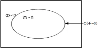
A. Level Set Function:
The function Φ (I,i,j) (the level set function where(i, j) are co-ordinates in the image and t is time). The segmentation is given by two regions {Φ >0} and {Φ<0}. The boundary of the shape is then the zero level set as in Figure 2. In the piecewise-smooth MS algorithm [8], the evolution of curves coupled with diffusion, so the image might have become very homogeneous in certain iterations. Furthermore, the evolution speed of curves is reduced greatly. In order to speed up convergence, a further modification is done to adjust the location of the curves by an additional force during evolution of curves. The aim of modification is to update the level set function Φ to a better location [8].
Active contours were slow in processing. A method introduced the segmentation problem to an ordinary Differential equation ignoring the length and area terms and setting λ1 = λ2. Th is method is known as Fast Hybrid k -means Level Set Algorithm for Segmentation. The method was significantly faster than Chan-Vese algorithm. Disadvantage is that it does not work well for Noisy Images [7].
Another attempt for fast segmentation is peicewise – smooth Mumford Shah functional. Here curve evolution is driven by an external additional force at each time step, which makes algorithm to converge faster. It is known as Fast Segmentation for the Piecewise Smooth Mumford Shah Functional [8].
Basic steps of Chan-Vese algorithm are:
a. Initialize Φ, the initial curve in the image.
b. Compute C1 and C2., the average intensities inside
the curve and outside of it.
c. Calculate the energy function from equation (1). d. Check whether the solution is stationary, energy is
minimum. If not, and repeat [5].
VI. MEDICAL IMAGE SEGMENTATION
Medical imaging is a group of examinations, techniques and processes that use different types of physical effects to visualize anatomical structures and pathological changes inside the human body [1]. Recently, it is one of the fastest growing fields of medical examinations. Development of medical imaging techniques contributed to a significant increase in the effectiveness of detection and treatment of various lesions and pathological changes. Radiological examinations allow for an accurate detection of the lesion. Proper analysis of the disease is crucial to make decision of the treatment. However, manual assessment of pathological changes is always subjective and time-consuming. There is also a risk that such assessment can be cursory and incorrect. Therefore, development of tools for an automatic detection and analysis of pathological changes is one of the most challenging tasks of a present day medical image processing and analysis.
Image segmentation remains a difficult task, however, due to both the tremendous variability of object shapes and the variation in image quality. In particular, medical images are often corrupted by noise and sampling artifacts, which can cause considerable difficulties when applying classical segmentation techniques such as edge detection and thresholding. As a result, these techniques either fail completely or require some kind of post processing step to remove invalid object boundaries in the segmentation results. Segmentation of mouse brain using active contours is shown in Figure 3, Figure 4, and Figure 5.
Figure. 3 Initial Contour in brain image[9].
Figure. 4 Chan-Vese Contours [9]
Figure.5 Final Contour [9]
To address these difficulties, deformable models or active contours have been extensively studied and widely used in medical image segmentation, with promising results. Deformable models are curves or surfaces defined within an image domain that can move under the influence of internal forces, which are defined within the curve or surface itself, and external forces, which are computed from the image data. The internal forces are designed to keep the model smooth during deformation. The external forces are defined to move the model toward an object boundary or other desired features within an image. By constraining extracted boundaries to be smooth and incorporating other prior information about the object shape, deformable models offer robustness to both image noise and boundary gaps and allow integrating boundary elements into a coherent and consistent mathematical description. Such a boundary description can then be readily used by subsequent applications [10].
VII. CONCLUSION
Active contour is popular in computer vision, and led to several developments in 2D and 3D. It is an energy minimizing, deformable curve influenced by image forces that pull it towards object contours. They are greatly used in applications like object tracking, shape recognition, segmentation, edge detection, stereo matching, automatic crop spraying, traffic monitoring, audio-visual speech analysis and medical diagnosis. Active contours make effective use of specific prior information about objects and this makes them inherently efficient algorithms.
VIII. REFERENCES
[1] Rafael C. Gonzalez, Richard E. Woods, “Digital Image Processing”, 2nd ed., Beijing: Publishing House of Electronics Industry, 2007.
[2] Rajeshwar Dass, Priyanka, Swapna Devi, “Image segmentation techniques”, International Journal of Electronics & Communication Technology, Vol. 3, Issue 1,pp 66-67,2012.
[3] W. X. Kang, Q. Q. Yang, R. R. Liang,“The Comparative Research on Image Segmentation Algorithms”, IEEE Conference on ETCS, pp. 703-707, 2009.
[5] Tony F. Chan and Luminita A Vese, “Active Contours Without Edges”,IEEE Transactions on Image Processing,Vol. 10 No.2, Feb 2001.
[6] Chenyang Xu, Anthony Yezzi, Jr., Jerry L. Prince, “On the relationship between parametric and geometric active contours”, in Proc. of 34th Asilomar Conference on Signals, Systems, and Computers, pp. 483-489, October 2000.
[7] F.Gibon,R.Fedkin, “Fast Hybrid k-means Level Set Algorithm for Segmentation”,Proceedings of the 4th Annual Hawaii International Conference on Statistics & Mathematics, Stanford Technical Report,Nov. 2002.
[8] Yingjie Zhang, “Fast Segmentation for the Piecewise Smooth Mumford Shah Functional”, International Journal of Signal Processing, 2006.
[9] Ahmad Almhdie, Frédéric Szeremeta, Roger Lédée, “CHAN-VESE based method to segment mouse brain MRI images: Application to cerebral malformation analysis in TRISOMY 21”, 17th European Signal Processing Conference (EUSIPCO 2009).
[10] Chenyang Xu, Dzung L. Pham,Jerry L. Prince, “ Image segmentation using deformable models” [Online].Available:http://citeseerx.ist.psu.edu/viewdoc/do wnload?doi=10.1.1.62.8216&rep=rep1&type=pdf.
![Figure.5 Final Contour [9]](https://thumb-us.123doks.com/thumbv2/123dok_us/700705.1077931/4.595.77.238.633.772/figure-final-contour.webp)
