Mirko Pham, MD Philipp Bäumer, MD Hans-Michael Meinck,
MD
Johannes Schiefer, MD Markus Weiler, MD Martin Bendszus, MD Henrich Kele, MD
Correspondence to Dr. Pham:
mirko.pham@med.uni-heidelberg.de
Editorial, page 554
Supplemental data at www.neurology.org
Anterior interosseous nerve syndrome
Fascicular motor lesions of median nerve trunk
ABSTRACT
Objective:We sought to determine lesion sites and spatial lesion patterns in spontaneous anterior interosseous nerve syndrome (AINS) with high-resolution magnetic resonance neurography (MRN). Methods:In 20 patients with AINS and 20 age- and sex-matched controls, MRN of median nerve fascicles was performed at 3T with large longitudinal anatomical coverage (upper arm/elbow/ forearm): 135 contiguous axial slices (T2-weighted: echo time/repetition time 52/7,020 ms, time of acquisition: 15 minutes 48 seconds, in-plane resolution: 0.2530.25 mm). Lesion classifica-tion was performed by visual inspecclassifica-tion and by quantitative analysis of normalized T2 signal after segmentation of median nerve voxels.
Results:In all patients and no controls, T2 lesions of individual fascicles were observed within upper arm median nerve trunk and strictly followed a somatotopic/internal topography: affected were those motor fascicles that will form the anterior interosseous nerve further distally while other fascicles were spared. Predominant lesion focus was at a mean distance of 14.665.4 cm proximal to the humeroradial joint. Discriminative power of quantitative T2 signal analysis and of qualitative lesion rating was high, with 100% sensitivity and 100% specificity (p,0.0001). Fascicular T2 lesion patterns were rated as multifocal (n517), monofocal (n52), or indetermi-nate (n51) by 2 independent observers with strong agreement (kappa50.83).
Conclusion:It has been difficult to prove the existence of fascicular/partial nerve lesions in spon-taneous neuropathies using clinical and electrophysiologic findings. With MRN, fascicular lesions with strict somatotopic organization were observed in upper arm median nerve trunks of patients with AINS. Our data strongly support that AINS in the majority of cases is not a surgically treat-able entrapment neuropathy but a multifocal mononeuropathy selectively involving, within the main trunk of the median nerve, the motor fascicles that continue distally to form the anterior interosseous nerve.Neurology®2014;82:598–606
GLOSSARY
AIN5anterior interosseous nerve;AINS5anterior interosseous nerve syndrome;AUC5area under the curve;CI5 con-fidence interval;DTI5diffusion tensor imaging;FDP5flexor digitorum profundus;FDPII5index finger;FDPIII5middle finger;FPL5flexor pollicis longus (thumb);IVIg5IV immunoglobulin;MRC5Medical Research Council;MRN5magnetic resonance neurography;NCS5nerve conduction studies;PQ5pronator quadratus (forearm);ROC5receiver operating characteristic;ROI5region of interest.
Spontaneous anterior interosseous nerve syndrome (AINS) is an uncommon peripheral neuropathy
of unclear etiology.
1Except for fine articular branches at the wrist, the anterior interosseous nerve
(AIN) is an almost purely motor branch of the median nerve important for thumb and hand
function.
2It leaves the median nerve trunk at forearm level, immediately distally to the
pronator-teres muscle, and innervates the flexor pollicis longus (FPL), pronator quadratus (PQ), and flexor
digitorum profundus (FDP) muscle to the index and middle finger.
2,3AINS presents with
spon-taneous acute weakness of distal phalanx flexion of the thumb (FPL) and/or index finger (FDPII),
middle finger (FDPIII), and forearm pronation (PQ). The severity and completeness of these motor
symptoms vary substantially, as described originally.
4Typically, no sensory abnormalities are
From the Departments of Neuroradiology (M.P., P.B., M.B.) and Neurology (H.-M.M., M.W.), Heidelberg University Hospital; Clinical Cooperation Unit Neurooncology (M.W.), German Cancer Research Center (DKFZ), Heidelberg; Department of Neurology (J.S.), RWTH University Hospital Aachen; and Center for Neurology and Clinical Neurophysiology Neuer Wall (H.K.), Hamburg, Germany.
detected by clinical or electrophysiologic
exam-ination. However, pain of different quality,
intensity, and location may occur.
1,5Usually median nerve conduction studies
(NCS) are normal in AINS and thus unhelpful
for lesion localization. EMG reveals typical
pat-terns of muscle denervation compatible with a
lesion of the AIN itself or, alternatively, of its
motor fascicles located further proximally within
the median nerve trunk. These fascicles continue
distally in an ordered fashion of functional
grouping to form the AIN. In fact, a more
prox-imal lesion site has been suggested previously.
6,7However, it has been difficult to obtain evidence
of a more proximal lesion because NCS/EMG
may not differentiate it from a lesion to the AIN
itself. This study used high-resolution magnetic
resonance neurography (MRN) to determine
lesion sites and spatial lesion patterns of AINS
and estimated its accuracy in discriminating
between AINS and controls.
METHODS Between April 2009 and March 2013, 24 consec-utive patients with symptoms of AINS were referred to the Department of Neurology, Heidelberg University Hospital, Germany, or the Center for Neurology and Clinical Neurophysiology Neuer Wall, Hamburg, Germany. Twenty of 24 patients consented to undergo MRN and were scheduled prospectively (figure e-1 on the
Neurology®Web site at www.neurology.org). Nonspontaneous AINS
following trauma was not included. Twenty age- and sex-matched controls without symptoms or signs of median neuropathy or risk factors for neuropathy such as diabetes, alcoholism, or infectious diseases underwent the same imaging protocol.
Standard protocol approvals, registrations, and patient consents.The study was approved by the institutional ethics committee (S-057/2009). All subjects gave written informed con-sent to participate.
Clinical and electrophysiologic examination.All patients underwent clinical/electrophysiologic examinations performed by board-certified neurologists with at least 10 years of experience in clinical neurophysiology (H.K. or H.M.M.). Motor strength was recorded for FPL, FDPII, and FDPIII using the Medical Research Council (MRC) rating scale. Complete AINS was defined as weakness (MRC #4) of FPL and FDPII/III. PQ was not evaluated quantitatively. Incomplete AINS was defined as weakness (MRC#4) of FPL or FDPII. In all patients, motor and sensory NCS were performed on ipsilateral median/ulnar nerves and contralateral median nerve with surface stimulation at standard sites including the axilla.8Compound muscle action potentials were recorded following supramaximal stimulation from abductor pollicis brevis and abductor digiti minimi muscle. Sensory nerve action potentials were obtained antidromically over second, third, and fifth digit. Furthermore, median and ulnar nerve F-waves and cortical median somatosensory evoked potentials were measured. Using concentric needle EMG, FPL, PQ, or FDPII was examined for signs of denervation.
MRN.All participants underwent MRN at Heidelberg University Hospital. MRN acquired nerve T2 signal at high spatial resolution
and, at the same time, provided large proximal to distal coverage along the upper extremity. Participants were examined prone at 3T magnetic field strength (Magnetom VERIO, Siemens, Erlangen/ Germany) using an 8-channel phased-array extremity coil. Three contiguous slabs were recorded with a 2D fat-saturated T2-weighted turbo spin echo pulse sequence (echo time/repetition time 52/7,020 ms, time of acquisition 5 minutes 16 seconds, in-plane resolution 0.2530.25 mm, slice thickness 3 mm, interslice gap 0.3 mm, 45 slices). The 3T MRI combined with the employed extremity coil provided sufficient signal to resolve fascicles within median nerve. At lower field strength, such detail may not be achievable. Slab position was as follows: 1) distally: central forearm to humeroradial joint, 2) centrally: humeroradial joint to central upper arm, 3) proximally: central upper arm to axilla.
Image and statistical data analysis.Images were rated quali-tatively with regard to the following items by 2 investigators (M.P., P.B.) blinded to all participant data.
1. Lesion determination: Dichotomous ratings on presence vs absence of lesions as evident by increased T2 signal of median nerve fascicles were obtained independently from both raters. 2. Lesion localization: Anatomical site of predominant lesion focus, i.e., the slice position (with reference to humeroradial joint) with strongest increase in T2 signal of fascicles, was determined by consensus.
3. Fascicular involvement: Dichotomous consensus ratings were obtained on whether increased T2 signal involved the entire nerve cross-section or only a partial area of nerve cross-section (fascicular lesion).
4. Longitudinal lesion pattern: Dichotomous ratings were ob-tained independently from both raters on multifocality vs monofocality. Multifocality: slices with increased fascicular T2 signal alternated with normal slices. Monofocality: single lesion focus over contiguous slices with normal T2 signal proximal and distal to it.
Further steps of quantitative analysis were undertaken. In con-trols, a region of interest (ROI) was defined by manual segmenta-tion of the median nerve (ROImedian_control). ROImedian_controlwas
derived in each control at 14.6 cm (tolerance of60.6 cm) proximal to the humeroradial joint. This distance corresponded to the mean distance in patients of the predominant lesion focus proximal to the humeroradial joint (14.665.4 cm, table e-1).
In patients, the median nerve was segmented on the slice harbor-ing the predominant lesion focus. Two ROIs within the segmented median nerve were defined: 1) ROImedian_lesioncomprised the area
within nerve cross-section at the dorsal and radial/lateral aspect consistently showing increased T2 signal. 2) ROImedian_no_lesion
com-prised the remainder of nerve cross-section excluding ROImedian_lesion.
For each subject, the mean T2 signal of the medial head of the biceps muscle was determined to calculate normalized median nerve T2 values as follows:
Control subjects:
T2median_control5ROImedian_control=ROImuscle
Patients (lesioned fascicles):
T2median_no_lesion5ROImedian_no_lesion=ROImuscle
Patients (normal-appearing fascicles):
T2median_lesion5ROImedian_lesion=ROImuscle
(FMRIB Software Library v 5.0).9Before registration, images were flipped horizontally from right to left in patients with right sided symptoms to obtain comparability with patients affected contralaterally. Then, spatial maps of mean normalized T2 values were rendered for patients. Statistical analyses were calculated with STATA 12 (StataCorp LP, College Station, TX).
RESULTS Clinical findings.Mean age of patients was 46.4 611.1 years (15 male/5 female) and 45.3 6 11.3 in controls (15 male/5 female). Mean duration between symptom onset and clinical/electrophysio-logic examinations was 22 days (range 2–94) and 3.4 months (range 12 days to 9 months) between symptom onset and MRN. Detailed findings are given in table e-1. Complete AINS was observed in 15 and incomplete AINS in 5 patients. Sensory test-ing, sensory and motor NCS including F-waves, evoked potentials, and EMG of biceps/triceps were normal in all patients. EMG of FPL, FDPII, or PQ showed denervation in all patients. Pain before or at symptom onset was reported by 13 of 20 patients and varied with regard to quality, anatomical distribution, and time of onset relative to onset of motor symp-toms. In the majority of these patients, pain was
experienced as sharp or burning sensation at the medial aspect of elbow or upper arm.
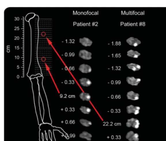
Imaging findings and statistical image analysis.Figure 1 illustrates a complete array of T2 source images show-ing the predominant lesion focus of each patient.
Lesion determination and localization.T2 lesions of the median nerve were rated as present in all patients and absent in all controls (sensitivity 100%, specificity 100%, interrater agreement: Cohen kappa 5 1). The positions of predominant lesion foci are given in table e-1. Their spatial distribution is illustrated in figure 1. Mean distance of predominant lesion focus was 14.665.4 cm proximal to the humerora-dial joint.
Fascicular involvement.In all patients but none of the controls, median nerve T2 lesions were present and involved only a partial area of the nerve cross-section (Cohen kappa51). The exact location of T2 signal increase was at the dorsal and radial/lateral aspect of the nerve cross-section. As illustrated on a somatotopic map of median nerve fascicles (figure 2), this lesion area corresponded precisely to the somatotopic/
Figure 1 Fascicular lesions of median nerve at upper arm level in anterior interosseous nerve syndrome
topographic internal arrangement of a particular group of motor fascicles within the median nerve trunk at upper arm level: this fascicle group will form the AIN, which does not emerge from the median nerve trunk epineurium until further distally at forearm level.
Spatial lesion patterns and lesion extension.The longi-tudinal pattern of fascicular lesions was rated monofo-cal for patients 1 and 2. For patient 3, no agreement was obtained (indeterminate). Patients 4–20 were rated multifocal (Cohen kappa50.83,p,0.001). Figure 3 illustrates the 2 different longitudinal lesion patterns on contiguous slices. Patients with monofo-cality were not discernable from patients with multi-focality by presence of painful symptoms, type of onset, or by other clinical/electrophysiologic findings (table e-1). In none of the patients did T2 lesions extend to the proximal extreme of coverage (axilla).
Quantitative analysis of fascicular median nerve lesions.
The mean normalized median nerve T2 value of 20 controls was T2median_control51.1960.05. A similar
value was found in patients for normal-appearing median nerve fascicles: T2median_no_lesion 5 1.39 6
0.08 (p 5 0.104). In patients, however, the mean T2median_lesion 5 2.57 6 0.13 of lesioned fascicles
within the median nerve trunk was significantly higher compared with controls (T2median_lesion vs
T2median_control;p,0.0001); it was also significantly
higher compared with normal-appearing fascicles of patients (T2median_lesion vs T2median_no_lesion; p ,
0.0001). Receiver operating characteristic (ROC) analysis of T2median_lesionvs T2median_controlcalculated
an area under the curve (AUC) of 1.00 (95% confidence interval [CI] 1.00–1.00), corresponding to sensitivity and specificity of 100% at a cutoff of $1.7 of normalized T2 signal. ROC analysis of T2median_lesion vs T2median_no_lesion (lesioned fascicles
vs normal-appearing fascicles of patients) revealed AUC50.98 (95% CI 0.94–1.00). Empirical values of normalized T2 signal for lesioned fascicles of patients are plotted against controls in figure 4.
Follow-up.In the majority of patients with multifocal-ity, the administration of corticosteroids was elected as primary intervention. Their clinical response varied widely, with satisfactory recovery occurring in only some patients (table e-1). IV immunoglobulins (IVIg) were available for 4 patients, none of whom re-sponded to corticosteroids, and were administered ac-cording to the ICE Study scheme.10 Upon IVIg,
satisfactory improvement was observed in 2 of these 4 patients.
In patient 1 with monofocality, symptoms per-sisted after corticosteroids had been administered over 1 month (FPL 0). The monofocal proximal T2 lesion was discussed with the patient as a novel finding of unclear significance. It was mentioned that few cases had been described with torsion of motor median nerve fascicles at upper arm level, and that interfascic-ular neurolysis in some cases was followed by
Figure 2 Somatotopy of fascicular T2 median nerve lesion on individual level, group level, and atlas
On the left, the T2-weighted source image of the median nerve of patient 15 is shown for the site of predominant lesion focus (17.1 cm proximal to humeroradial joint). Anatomical orientation is given by labeling ventral/dorsal/medial/lateral con-tours. In the middle, a spatial map of the patient group mean normalized T2 signal is shown. This map was rendered after segmentation and intersubject image registration. On the right is a somatotopic/topographic internal map of fascicles of the median nerve trunk. This schematic drawing was obtained by Jabaley et al.31from tracing extraneural median nerve
branches from distally to intraneural proximal fascicles within the median nerve trunk on 20-mm-thick cuts after intraneural microsurgical dissection and histologic photographing (modified from Jabaley et al.31with permission). On this map, the red
improvement.11–13The patient opted for this surgical
procedure. Epineurotomy was performed at the site of T2 lesion. Markedly swollen fascicles at the dorsal and lateral aspects of median nerve emerged with torsion around their longitudinal axis (figure 5). Careful mobi-lization and detorsion was performed. Follow-up until 8 months did not reveal any improvement and showed persistence of denervation with lack of voluntary activ-ity in EMG of FPL and PQ. After 8 months, a positive Hoffmann-Tinel sign at the mid-forearm level was noticed. At 15 months, recovery to MRC 4 (from 0) was noted for FPL. In the second patient (2) with monofocality, administration of corticosteroids was paralleled by satisfactory improvement.
DISCUSSION The internal longitudinal organization of peripheral nerve fascicles was first studied in com-prehensive fashion by Sunderland,14who used surgical
dissection to trace fascicles from distal to proximal. In this work, he described some degree of plexiform exchange between fascicles at proximal nerve levels. Other authors illustrated this finding as “intraneural chaos,”15 which reflects that a concept of fascicular
somatotopy—the meaningful grouping of nerve
fascicles with regard to their function—has long been negated for the peripheral nervous system.16Later, when
the longitudinal course of fascicles became traceable on histology, it could be established that somatotopy is well-preserved despite some plexiform exchange between fascicles.15 The clinical implications of
fascicular somatotopy have been reviewed in detail by Stewart,16who emphasized that fascicular nerve lesions
represent a major pitfall for lesion localization: typical symptom patterns that resemble the functional territory of a peripheral nerve trunk appear only if all fascicles at the lesion site are compromised. However, selective fascicular injury may result in symptoms not following expected distributions. For example, if only certain fascicles supplying distal muscles are selectively injured at a more proximal site, the lesion would be expected erroneously to involve a further distal nerve branch to this muscle group. To prove the existence of fascicular nerve lesions has been challenging because it is difficult if not impossible to localize and objectify these lesions by clinical/electrophysiologic findings.
In the case of AINS, the view of a neuropathy of the AIN itself at forearm level, or its terminal branches, prevails especially among authors from surgical disci-plines. Consequently, entrapment, e.g., by a fibrous band, has been favored as principal mechanism and surgical release at forearm level advocated.17–24
Com-peting views see AINS not as entrapment neuropathy, but as a disease of immune-mediated inflammatory origin.1,4,5,25Certain similarities with neuralgic
shoul-der amyotrophy support an immune-mediated etiol-ogy.5 Six of the original 136 patients reported by
Parsonage and Turner5 had weakness of FPL and
FDPII, one of them without weakness of the shoulder girdle. Later, England and Sumner25raised awareness
that definite lesion localization remains difficult in Parsonage-Turner syndrome. From the distribution of symptoms in 9 well-documented cases, they con-cluded that lesion sites involve peripheral nerve branches rather than the brachial plexus and suspected involvement of the AIN in 4 patients.25
Improved lesion localization and determination of spatial lesion patterns would permit us to better understand the etiology of AINS and, in particular, to understand if and at which anatomical site AINS is potentially treatable by surgery. We prospectively investigated a relatively large sample of 20 patients with AINS and obtained detailed clinical/electrophys-iologic data.
At the core of our study was MRN, providing large longitudinal coverage including upper arm, elbow, and forearm levels. With this protocol we sought to determine lesion sites and longitudinal lesion patterns by T2 signal analysis of median nerve fascicles. Increased T2 signal has been shown to indicate nerve injury of mechanical and nonmechanical origin, e.g., Figure 3 Monofocality and multifocality as 2 principal lesion patterns of
anterior interosseous nerve syndrome
in focal entrapment,26,27 after trauma,28in multifocal
motor neuropathy,29and also in metabolic
polyneuro-pathies such as diabetic polyneuropathy.30
In AINS, we consistently found a strictly organized somatotopic/topographic internal fascicular lesion pat-tern within the median nerve at upper arm level: affected were those motor fascicles forming the AIN, which exits from the median nerve trunk further dis-tally at forearm level. Other median nerve fascicles seemed to be spared. Our interpretation that this fas-cicular T2 lesion pattern corresponds to an exclusive or, at least, predominant involvement of the motor fas-cicles forming the AIN is based on its close resem-blance to the position of the anterior interosseous fascicles as mapped by Jabaley et al.31and on visual
tracing of T2 lesion fascicles from proximally within the median nerve trunk to distally into the AIN itself, which was reliably recognizable at the given spatial resolution. We acknowledge limitations of both meth-ods, e.g., interindividual variability and potential inac-curacy of visual rating. Therefore, we propose as future research aim to track lesion fascicles from proximal to distal by diffusion tensor imaging (DTI). However, so far, nerve DTI has not been implemented in humans at the submillimeter isotropic resolution needed to resolve fascicles. We further acknowledge that we can-not answer whether the extent of fascicular involve-ment differed between patients with complete and
incomplete AINS, because it was beyond the limit of spatial resolution to determine the exact number of involved fascicles. It is noteworthy that fascicular lesions in some cases of AINS were detectable also by high-resolution ultrasound; however, lesion contrast and thus diagnostic performance of fascicular hypoe-chogenicity seem to be inferior to nerve fascicle increase of T2 signal (figure e-2).
In addition, the longitudinal lesion pattern was ana-lyzed on contiguous slices. Multifocality was found in the majority of patients and monofocality in only 2. Interestingly, there were no differences between these 2 distinct lesion patterns with regard to symptoms or clinical/electrophysiologic findings. The responses to therapeutic intervention were markedly heterogeneous in patients with both lesion patterns. This observation is in accordance with reported variable outcomes after therapeutic intervention in AINS and also with evi-dence that spontaneous recovery may occur in a sub-stantial portion of patients.18,22,32
Monofocality in our cohort was rare. In one patient, surgical exploration with dissection of median nerve trunk epineurium (epineurotomy) revealed fas-cicular torsion precisely at the lesion site, which was localized by imaging (figure 5). After interfascicular neurolysis and detorsion, clinical recovery was observed in this patient. The rationale to offer such individually tailored surgical therapy guided by a novel imaging Figure 4 T2 signal analysis of median nerve trunk fascicles at upper arm level
sign was based on several case reports of median nerve fascicular torsion.11–13,33 Nagano and colleagues11,33
reported the largest series to date and described an
“hourglass-like fascicular constriction”between 2 and 7.5 cm above the elbow in 22 patients. Nagano33
re-ported good recovery after interfascicular neurolysis in 21 of 22 patients but stressed that“we do not know whether this recovery was spontaneous or due to the neurolysis.”Fascicular torsion as a causative factor for AINS will remain difficult to prove. Some plausible explanations for its occurrence have been offered, partic-ularly high mobility of AIN fascicles promoting torsion during elbow flexion,13 or initial inflammation and
edema followed by intraneural adhesions, which increase traction forces on anterior interosseous fascicles.33
The histopathologic alterations underlying the observed T2 lesions remain unclear because biopsies at the lesion site are unethical. It is also difficult to explain by which pathophysiologic mechanisms proxi-mal lesions are associated with functional compromise. It seems attractive to speculate that the accumulation of multifocal proximal injury is involved in the mani-festation of symptoms and may result in further distally located functional or structural compromise, as believed to occur in other neuropathies such as diabetic polyneuropathy30,34,35or the noncompressive
polyneu-ropathy associated with type 2 neurofibromatosis.36
The significance of our results may be 2-fold. This study is the first to provide strong diagnostic evidence
by imaging for the existence of fascicular/partial nerve lesions in a spontaneously occurring neuropathy. The existence of fascicular nerve lesions had been assumed before but could not be objectified so far by NCS/ EMG studies.6,16 Invasive near-nerve recordings or
even intraneural fascicular stimulation by needle mi-croneurography would be necessary to detect selective fascicular conduction abnormalities.37,38Both
techni-ques are not readily available in humans and have not been reported in AINS. Noninvasive stimulation with surface electrodes for motor or sensory NCS typically remain normal and therefore nonlocalizing in AINS. EMG detects denervation in muscles sup-plied by the AIN; however, this finding is nonlocaliz-ing: it cannot discriminate between injury to the AIN itself and a more proximal lesion of anterior interos-seous fascicles within the median nerve trunk.
As second major implication, the predominance of lesions at upper arm level in all patients strongly sup-ports that AINS is not an entrapment neuropathy of the AIN itself nor of its branches, at least in our cohort. The observation of selective fascicular lesions following motor somatotopy clearly suggests that AINS is a motor fascicular neuropathy of the median nerve trunk. The observation of multifocality in the majority of patients argues in favor of an immune-mediated inflammatory origin and against any surgi-cal treatment options either at forearm or upper arm level, at least in these multifocal cases.
Figure 5 Fascicular torsion of median nerve motor fascicles as rare cause of anterior interosseous nerve syndrome
AUTHOR CONTRIBUTIONS
Dr. Pham: study design, analysis and interpretation of data, acquisition of data, writing of manuscript. Dr. Bäumer: acquisition of data, interpreta-tion of data, revising manuscript for intellectual content. Dr. Meinck: acquisition of data, interpretation of data, revising manuscript for intel-lectual content. Dr. Schiefer: interpretation of data, revising manuscript for intellectual content. Dr. Weiler: interpretation of data, revising man-uscript for intellectual content. Dr. Bendszus: study supervision, study design, interpretation of data, revising manuscript for intellectual content. Dr. Kele: study design, acquisition of data, analysis and interpretation of data, writing of manuscript.
ACKNOWLEDGMENT
The authors thank Dr. Thomas Dombert for documentation of the intra-operative field of patient 1 and for his permission to use the 2 intraoper-ative photographs of figure 5; and Prof. Dr. Sabine Heiland, Division of Experimental Radiology, Department of Neuroradiology, Heidelberg Uni-versity Hospital, for assistance in setting up the MRN protocol and help in revising the manuscript.
STUDY FUNDING
This study was funded by a project grant from the German Osteoarthritis Foundation (P215-A482) and supported by a postdoctoral fellowship from the Medical Faculty of Heidelberg University (P.B.).
DISCLOSURE
M. Pham receives project grants from the German Osteoarthritis Foun-dation (Deutsche-Arthrose-Hilfe e.V.: P215-A482) and the EFSD/ JDRF/Novo Nordisk European Programme in Type 1 Diabetes Research. P. Bäumer receives a postdoctoral fellowship from the Medical Faculty of Heidelberg University, Germany. H. Meinck, J. Schiefer, and M. Weiler report no disclosures. M. Bendszus receives a project grant from the German Osteoarthritis Foundation (Deutsche-Arthrose-Hilfe e.V.: P215-A482). H. Kele reports no disclosures. Go to Neurology. org for full disclosures.
Received July 25, 2013. Accepted in final form October 10, 2013.
REFERENCES
1. Stevens JC. Median neuropathy. In: Dyck PJ, Thomas PK, eds. Peripheral Neuropathy, vol 1, 2, 4th ed. Philadelphia: Elsevier; 2005:1453–1454.
2. von Lanz T, Wachsmuth W. Praktische Anatomie: Arm, vol I/3, 2nd ed. Heidelberg: Springer; 1959.
3. Sunderland S. The innervation of the flexor digitorum profundus and lumbrical muscles. Anat Rec 1945;93: 317–321.
4. Kiloh LG, Nevin S. Isolated neuritis of the anterior inter-osseous nerve. Br Med J 1952;1:850–851.
5. Parsonage MJ, Turner JW. Neuralgic amyotrophy: the shoulder-girdle syndrome. Lancet 1948;1:973–978. 6. Wertsch JJ, Sanger JR, Matloub HS. Pseudo-anterior
inter-osseous nerve syndrome. Muscle Nerve 1985;8:68–70. 7. Wertsch JJ. AAEM case report #25: anterior interosseous
nerve syndrome. Muscle Nerve 1992;15:977–983. 8. Kimura J. Peripheral Nerve Diseases: Handbook of Clinical
Neurophysiology, 1st ed. Philadelphia: Elsevier; 2006. 9. Smith SM, Jenkinson M, Woolrich MW, et al. Advances
in functional and structural MR image analysis and implementation as FSL. Neuroimage 2004;23(suppl 1): S208–219.
10. Hughes RA, Donofrio P, Bril V, et al. Intravenous immune globulin (10% caprylate-chromatography purified) for the treatment of chronic inflammatory demyelinating polyradic-uloneuropathy (ICE Study): a randomised placebo-controlled trial. Lancet Neurol 2008;7:136–144.
11. Nagano A, Shibata K, Tokimura H, et al. Spontaneous anterior interosseous nerve palsy with hourglass-like fascicular constriction within the main trunk of the median nerve. J Hand Surg Am 1996;21:266–270. 12. Yasunaga H, Shiroishi T, Ohta K, et al. Fascicular torsion
in the median nerve within the distal third of the upper arm: three cases of nontraumatic anterior interosseous nerve palsy. J Hand Surg Am 2003;28:206–211. 13. Haussmann P, Patel MR. Intraepineurial constriction of
nerve fascicles in pronator syndrome and anterior interos-seous nerve syndrome. Orthop Clin North Am 1996;27: 339–344.
14. Sunderland S. Nerves and Nerve Injuries. Edinburgh: Churchill Livingstone; 1978.
15. Brushart TM. Central course of digital axons within the median nerve ofMacaca mulatta. J Comp Neurol 1991; 311:197–209.
16. Stewart JD. Peripheral nerve fascicles: anatomy and clinical relevance. Muscle Nerve 2003;28:525–541.
17. Mackinnon SE, Dellon AL. Surgery of the Peripheral Nerve. New York: Thieme; 1988.
18. Lake PA. Anterior interosseous nerve syndrome. J Neurosurg 1974;41:306–309.
19. Spinner M. The anterior interosseous nerve syndrome, with special attention to its variations. J Bone Joint Surg Am 1970;52:84–94.
20. Fearn CB, Goodfellow JW. Anterior interosseous nerve palsy. J Bone Joint Surg Br 1965;47:91–93.
21. Farber JS, Bryan RS. The anterior interosseous nerve syn-drome. J Bone Joint Surg Am 1968;50:521–523. 22. Vichare NA. Spontaneous paralysis of the anterior
inter-osseous nerve. J Bone Joint Surg Br 1968;50:806–808. 23. Collins DN, Weber ER. Anterior interosseous nerve
syn-drome. South Med J 1983;76:1533–1537.
24. Nigst H, Dick W. Syndromes of compression of the median nerve in the proximal forearm (pronator teres syn-drome; anterior interosseous nerve syndrome). Arch Orthop Trauma Surg 1979;93:307–312.
25. England JD, Sumner AJ. Neuralgic amyotrophy: an increasingly diverse entity. Muscle Nerve 1987;10: 60–68.
26. Baumer P, Dombert T, Staub F, et al. Ulnar neuropathy at the elbow: MR neurography–nerve T2 signal increase and caliber. Radiology 2011;260:199–206.
27. Subhawong TK, Wang KC, Thawait SK, et al. High res-olution imaging of tunnels by magnetic resonance neurog-raphy. Skeletal Radiol 2012;41:15–31.
28. Filler AG, Howe FA, Hayes CE, et al. Magnetic resonance neurography. Lancet 1993;341:659–661.
29. Van Asseldonk JT, Franssen H, Van den Berg-Vos RM, et al. Multifocal motor neuropathy. Lancet Neurol 2005; 4:309–319.
30. Pham M, Oikonomou D, Baumer P, et al. Proximal neu-ropathic lesions in distal symmetric diabetic polyneuropathy: findings of high-resolution magnetic resonance neurography. Diabetes Care 2011;34:721–723.
31. Jabaley ME, Wallace WH, Heckler FR. Internal topogra-phy of major nerves of the forearm and hand: a current view. J Hand Surg Am 1980;5:1–18.
33. Nagano A. Spontaneous anterior interosseous nerve palsy. J Bone Joint Surg Br 2003;85:313–318.
34. Dyck PJ, Karnes JL, O’Brien P, et al. The spatial distri-bution of fiber loss in diabetic polyneuropathy suggests ischemia. Ann Neurol 1986;19:440–449.
35. Dyck PJ, Lais A, Karnes JL, et al. Fiber loss is primary and multifocal in sural nerves in diabetic polyneuropathy. Ann Neurol 1986;19:425–439.
36. Baumer P, Mautner VF, Baumer T, et al. Accumulation of non-compressive fascicular lesions underlies NF2 polyneu-ropathy. J Neurol 2013;260:38–46.
37. Hallin RG, Wu G. Fitting pieces in the peripheral nerve puzzle. Exp Neurol 2001;172:482–492.
38. Hallin RG. Microneurography in relation to intraneural topog-raphy: somatotopic organisation of median nerve fascicles in humans. J Neurol Neurosurg Psychiatry 1990;53:736–744.
www.neurology.org Offers Important Information to
Patients and Their Families
TheNeurology®Patient Page provides:
• A critical review of ground-breaking discoveries in neurologic research that are written especially for patients and their families
• Up-to-date patient information about many neurologic diseases
• Links to additional information resources for neurologic patients
AllNeurologyPatient Page articles can be easily downloaded and printed, and may be reproduced to distribute for educational purposes. Click on the‘Patients’link on the home page (www.neurology.org) for a complete index of Patient Pages.
Earn 20 CME Credits Toward MOC with New
NeuroPI
SMModules
Choose from the latest lineup of quality modules to join the AAN’s exclusive performance improve-ment programs designed to help you address both the Performance in Practice (PIP) and Continu-ing Medical Education (CME) components of Maintenance of Certification (MOC).
• NEW!Distal Symmetric Polyneuropathy (DSP)includeseight quality measures, addressing
accurate and appropriate evaluation/monitoring of DSP and associated symptoms to guide treat-ment options, patient safety, and best practices to assist patients in managing their pain and improving quality of life
• Acute Stroke addresses six quality measures, including deep vein thrombosis prophylaxis
(DVT) for ischemic stroke or intracranial hemorrhage, discharged on antiplatelet therapy, dys-phagia screening, rehabilitation service considerations, and more
• Dementiaincludes10 quality measuresaddressing underuse of effective services and patient-centered care strategies, and patient safety issues
Learn about all of the other available modules and purchase yours today:
DOI 10.1212/WNL.0000000000000128
2014;82;598-606 Published Online before print January 10, 2014
Neurology
Mirko Pham, Philipp Bäumer, Hans-Michael Meinck, et al.
Anterior interosseous nerve syndrome: Fascicular motor lesions of median nerve trunk
This information is current as of January 10, 2014
rights reserved. Print ISSN: 0028-3878. Online ISSN: 1526-632X.
1951, it is now a weekly with 48 issues per year. Copyright © 2014 American Academy of Neurology. All ® is the official journal of the American Academy of Neurology. Published continuously since
Services
Updated Information &
http://n.neurology.org/content/82/7/598.full
including high resolution figures, can be found at:
Supplementary Material
128.DC3
http://n.neurology.org/content/suppl/2014/10/31/WNL.0000000000000 128.DC2
http://n.neurology.org/content/suppl/2014/01/10/WNL.0000000000000 128.DC1
http://n.neurology.org/content/suppl/2014/01/10/WNL.0000000000000
Supplementary material can be found at:
References
http://n.neurology.org/content/82/7/598.full#ref-list-1
This article cites 34 articles, 3 of which you can access for free at:
Citations
http://n.neurology.org/content/82/7/598.full##otherarticles
This article has been cited by 5 HighWire-hosted articles:
Subspecialty Collections
http://n.neurology.org/cgi/collection/peripheral_neuropathy Peripheral neuropathy
http://n.neurology.org/cgi/collection/mri MRI
http://n.neurology.org/cgi/collection/all_neuromuscular_disease All Neuromuscular Disease
http://n.neurology.org/cgi/collection/all_clinical_neurophysiology All clinical neurophysiology
http://n.neurology.org/cgi/collection/all_clinical_neurology All Clinical Neurology
following collection(s):
This article, along with others on similar topics, appears in the
Permissions & Licensing
http://www.neurology.org/about/about_the_journal#permissions
its entirety can be found online at:
Information about reproducing this article in parts (figures,tables) or in
Reprints
http://n.neurology.org/subscribers/advertise
Information about ordering reprints can be found online:
rights reserved. Print ISSN: 0028-3878. Online ISSN: 1526-632X.
1951, it is now a weekly with 48 issues per year. Copyright © 2014 American Academy of Neurology. All ® is the official journal of the American Academy of Neurology. Published continuously since