R E V I E W
Diagnostic Accuracy Of Fecal Occult Blood Tests
For Detecting Proximal Versus Distal Colorectal
Neoplasia: A Systematic Review And Meta-Analysis
This article was published in the following Dove Press journal:
Clinical Epidemiology
Ming Lu 1,*
Xiaohu Luo 2,*
Ni Li 1
Hongda Chen 1
Min Dai 1
1National Cancer Center/National
Clinical Research Center for Cancer/ Cancer Hospital, Chinese Academy of Medical Sciences and Peking Union
Medical College, Beijing 100021, People’s
Republic of China;2Department of
Toxicant Occupational Disease Testing Laboratory, Xuzhou Cancer Hospital,
Xuzhou 221000, People’s Republic of
China
*These authors contributed equally to this work
Objective:We conducted a systematic review and meta-analysis aimed at evaluating the
differences of diagnostic performance of fecal occult blood tests (FOBTs) in detecting advanced colorectal neoplasms located in the proximal versus distal colorectum.
Methods: PubMed, Embase, Cochrane Library, and Web of Science were searched for eligible
articles published before August 17, 2018. Two independent reviewers conducted study assessment and data extraction. Diagnosis-related indicators of FOBT for detecting proximal and distal colorectal neoplasms were summarized, and further stratified by the type of FOBT (guaiac-based FOBT (gFOBT) and immunochemical FOBT (iFOBT)). Pooled sensitivities and specificities were calcu-lated using a random effect model. Summary receiver operating characteristic curves were plotted and area under the curves were calculated.
Results: Overall, 31 studies meeting the inclusion criteria were included in this review. For
gFOBT, we found no site-specific difference (proximally vs distally located) of pooled sensitivities observed in the colorectal cancer (CRC), advanced adenomas, and advanced neoplasms groups. As for iFOBT, pooled sensitivities for detecting CRC located in the distal colon/rectum were compar-able with that in the proximal colon (proximal vs distal, 0.67, 95% CI 0.62–0.72 vs 0.72, 95% CI 0.68–0.75), while higher pooled sensitivities for detecting advanced adenomas and advanced neoplasms located in the distal colon/rectum than for detecting those in the proximal colon were observed for iFOBT with the values of 0.24 (95% CI 0.22–0.25) vs 0.32 (95% CI 0.30–0.34) and 0.25 (95% CI 0.23–0.28) vs 0.38 (95% CI 0.36–0.40), respectively. Summary receiver operating characteristic curve analyses showed similar patterns for both types of FOBT regarding the diagnostic accuracy for detecting colorectal neoplasms according to the anatomical sites of the colorectum.
Conclusion: iFOBT had higher sensitivity for detecting advanced adenomas and advanced
neoplasia located in the distal colon/rectum than that for those in the proximal colon.
Keywords:sensitivity, colorectal cancer, screening test
Introduction
With an estimated 1,849,518 new cases and 880,792 deaths occurring in 2018, colorectal cancer (CRC) is the third most commonly diagnosed cancer and fourth most common cause
of cancer-related deaths worldwide.1Current practices have suggested that screening for
CRC is effective in reducing CRC mortality.2,3The fecal occult blood test (FOBT) is a
non-invasive CRC screening test that has been widely used in many screening programs and is
recommended by current guidelines for CRC screening.4,5Overall, there are two types of
FOBTs which use different mechanisms to identify fecal occult blood. The traditional one is Correspondence: Min Dai; Hongda Chen
National Cancer Center/National Clinical Research Center for Cancer/Cancer Hospital, Chinese Academy of Medical Sciences and Peking Union Medical
College, Beijing 100021, People’s Republic
of China
Tel +86-10-8778-7394
Email daimin2002@hotmail.com; hongda.chen@cicams.ac.cn
Clinical Epidemiology
Dove
press
open access to scientific and medical research
Open Access Full Text Article
Clinical Epidemiology downloaded from https://www.dovepress.com/ by 118.70.13.36 on 20-Aug-2020
the guaiac-based FOBT (gFOBT) for detecting peroxidase activity of heme. Another type of FOBT is immunochemical FOBT (iFOBT, also referred to as FIT) that uses antibodies to
specifically detect human hemoglobin in stool, which make
fecal immunochemical tests less likely to result in false-positive results due to the ingredients. The iFOBT has been shown to have better analytical and clinical sensitivity and higher detec-tion ability in terms of CRC and its precursors, improved
com-pliance, and cost-effectiveness, compared to gFOBT.6
Previous randomized controlled trials have demonstrated that FOBT-based screening could reduce the mortality
asso-ciated with CRC.7,8In a FOBT-based CRC screening, patients
with positive FOBT results are scheduled to undergo subse-quent colonoscopies for clinical diagnosis. Therefore, the
diag-nostic performance of FOBT would strongly affect the efficacy
of screening. In a meta-analysis including 19 studies the pooled sensitivity of FOBT for CRC was relatively high at 79% (95%
CI, 69–86%), with corresponding pooled specificity of 94%.9
However, some studies have reported that the diagnostic per-formance of FOBT varied according to the anatomical site of
the colorectum.10,11Even with screening colonoscopy, which
is considered as the gold standard for CRC screening, a much higher protective effect for distal colon/rectum than proximal
colon was observed for colonoscopy-based screening.12
Therefore, the gradient in the efficacy of population-based
screening according to the anatomic site of the colorectal
lesions may be larger considering the site-specific sensitivities
for colorectal neoplasms. Two systematic reviews in this area were published several years ago although the focus on this issue was constricted due to their limited sample size and
incomplete study scope of colorectal neoplasia.10,11Based on
these studies, we conducted an updated systematic review including all relevant studies to further evaluate the diagnostic accuracy of FOBT for CRC and advanced adenomas according to different anatomical sites.
We aimed to evaluate the differences of diagnostic perfor-mance of FOBT in detecting colorectal neoplasms located in the proximal versus distal colorectum and to provide important references for designing effective FOBT-based CRC screening programs in the future.
Methods
We conducted a systematic review and meta-analyses
fol-lowing the PRISMA statement.13
Data Sources And Search Process
To identify potential studies reporting data on the diagnostic performance of FOBT for detecting colorectal neoplasms of
the proximal and distal colon/rectum, two independent inves-tigators searched PubMed, Embase, Cochrane Library, and Web of Science to retrieve studies published before August 17, 2018 using the following combined search terms: [color-ectal (or) colon (or) rectum] (and) [cancer (or) neoplasm (or) carcinoma (or) adenoma (or) malignancy] (and) [faecal immu-nochemical test (or) fecal immuimmu-nochemical testing (or) fecal immunochemical test (or) faecal immunochemical testing (or) faecal occult blood test (or) FOBT] (and) [detection (or) screening (or) detecting (or) diagnosis]. Detailed search terms
and retrieval records are provided inTable S1.
Study Selection
An initial search based on the titles and abstracts was conducted to exclude studies that were not relevant to the study topic. In addition, conference abstracts without full texts or studies writ-ten in non-English language were also excluded. For powrit-tential
relevant articles identified in the initial search, a full-text review
was performed using the following inclusion criteria: 1) FOBT results should be reported along with colonoscopy results
avail-able as the gold standard of reference; 2) site-specific diagnosis
information was provided in detail or the diagnosis-related indicators were accessible to be assessed.
Quality Assessment
Potential risks of bias and applicability of the included stu-dies were assessed according to the Quality Assessment of
Diagnostic Accuracy Studies (QUADA-2) scoring system,14
and the detailed protocol was shown in the supplementary
material (Protocol 1).
Data Extraction
Assessment of the included studies was performed indepen-dently by two reviewers (ML and XL) during the whole process. When disagreement occurred, consensus was reached through discussion between the authors or referral to the third reviewer (HC and MD). The following information was extracted: year of publication, country, study setting (clinical setting: participants recruited in hospitals or clinics in an opportunistic manner; screening setting: participants recruited in communities in an organized manner), population characteristics (sample size, age, and sex, etc.), diagnostic outcomes, attributes of FOBTs (type,
brand, and cut-off value), sensitivity, and specificity.
In this review, we only focused on the diagnostic accuracy of FOBT in one single round of testing. For multiple rounds of
FOBT tests, only thefirst-round result was extracted. Sensitivity
was defined as the proportion of FOBT-positive patients among
those who were diagnosed with the outcome of interest. As the
Clinical Epidemiology downloaded from https://www.dovepress.com/ by 118.70.13.36 on 20-Aug-2020
main indicator of diagnostic performance, sensitivity was extracted directly according to the anatomical site (proximal and distal colon/rectum) if reported or was calculated based on
reported information. Specificity referred to the number of
participants with negative FOBT results divided by the number of participants without neoplasms. For quantitative FOBT results with more than one cut-off value reported in the study, the cut-off values recommended by the manufacturer were used. Only patients with targeted tumors located in an isolated side were considered. In this review article, the proximal colon was
defined as colon involving the area proximal to the splenic
flexure while distal colon including distal to the splenicflexure.
Advanced adenoma was defined by at least one adenoma with
any of the following features:≥1 cm in size, tubulovillous or
villous components, and high-grade dysplasia. Advanced neo-plasms included CRC and advanced adenomas.
Data Synthesis And Statistical Analysis
Wefirst summarized the basic characteristics of all the included
studies. Diagnosis-related indicators including sensitivity (true
positive rate) and specificity (true negative rate), stratified by
anatomical location were extracted or calculated and respective 95% CIs were also reported (the Wilson method was used if not reported in the studies). We applied the random effect model to
estimate the pooled sensitivity and specificity. Statistical
hetero-geneity was assessed byI2statistics. Forest plots for the pooled
sensitivities of FOBT for detecting colorectal neoplasms in the proximal and distal colon/rectum were generated. Summary receiver operating characteristic (SROC) curves were plotted
to assess the accuracy of FOBT for detecting proximal and distal
colorectal neoplasms.Ztest was applied to examine the
statis-tical difference of the areas under SROC curves according to the anatomical sites.
Subgroup analyses were also performed to estimate the effect of study setting (clinical setting vs screening setting) and type of iFOBT (qualitative vs quantitative) on the diagnostic performance of FOBT. Potential publication bias was evaluated
for studies using gFOBTand iFOBTseparately by Deeks’funnel
plot.
All the analyses were conducted using Meta-Disc software 1.4 and Stata 12.0. Statistical tests presented were two-sided,
andP-value <0.05 was considered statistically significant.
Results
Literature Search Result
As shown inFigure 1, 9066 studies were identified from the
initial literature search. After removal of 3694 duplicates, afirst
round of title and abstract review was conducted, and 4888 articles unrelated to the review topic, 421 non-English language articles and 11 articles having no full-text were excluded. For the 52 remaining articles, a full-text review was conducted and 21 of them were excluded due to the following reasons: 1)
site-specific diagnostic indicators could not be calculated (n=16); 2)
colonoscopy conducted for participants with positive FOBT results only (n=4); and 3) repeated information contained in the previous articles by the same study group (n=1). Finally, 31 eligible studies were included in this review.
Figure 1Flow chart of search strategy and selection of reports (search until August 20, 2018).
Clinical Epidemiology downloaded from https://www.dovepress.com/ by 118.70.13.36 on 20-Aug-2020
Study Characteristics
Table 1shows the main characteristics of the studies included in
this review.15–45 Overall, most studies were carried out in
Western countries (16 of 31), while only some of them were conducted in East Asia (12 of 31). With regards to the study setting, most of the included studies (20 of 31) were conducted in a screening setting. The sample sizes varied greatly across the studies, ranging from 112 to 21,805. The mean ages of the population in these studies ranged from 48 to 69 years. Regarding the types of FOBT, 2 studies used gFOBT, 21 studies used iFOBT, and 8 studies had results available for both types of FOBT. The brands of the FOBT also varied greatly, with a total number of 24. Among these 31 studies, 24 (77.4%) studies had already excluded patients with incomplete bowel examination
by colonoscopy,17,18,20–29,31–33,35,36,39,41–44while 7 (22.6%)
stu-dies did not report such exclusion.19,20,30,34,37,38,40
Diagnostic Accuracy Of gFOBT
The summaries of the diagnostic performance of FOBT for detecting colorectal neoplasms according to the anatomical locations of all the included studies were shown in detail in Tables S2–S4. Forest plots showing the pooled sensitiv-ities of FOBTs for detecting proximal and distal colorectal
neoplasms are shown inFigures 2and3. For studies using
gFOBT, the pooled sensitivity for detecting CRC located in the proximal colon was comparable with the pooled sensi-tivity of that located in the distal colon/rectum, with values
of 0.60 (95% CI 0.45–0.74) and 0.70 (95% CI 0.57–0.80),
respectively. The diagnostic accuracy of gFOBT for detect-ing advanced adenomas was typically low, and there was no difference between detection of advanced adenomas located
at the proximal colon (0.10, 95% CI 0.07–0.14) and that at
the distal colon/rectum (0.11, 95% CI 0.07–0.15) and for
detection of proximally and distally located advanced
neo-plasms (0.18, 95% CI 0.14–0.22 vs 0.2, 95% CI 0.17–0.24).
The corresponding pooled specificities of gFOBT for CRC,
advanced adenomas, and advanced neoplasms according to the anatomic site were equal, with the values of 0.88 (95%
CI 0.87–0.89), 0.91 (95% CI 0.90–0.92), and 0.95 (95% CI
0.95–0.96), respectively.
Diagnostic Accuracy Of iFOBT
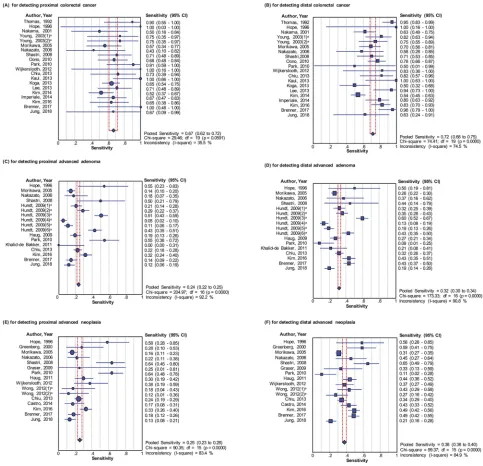
For studies using iFOBT, the sensitivities for detecting color-ectal neoplasms were higher than that with traditional gFOBT. For detecting CRC, comparable sensitivities of iFOBT for detecting CRC located in the proximal colon
and distal colon/rectum were observed (0.67, 95% CI 0.62–
0.72 and 0.72, 95% CI 0.68–0.75, respectively). However,
higher sensitivities were observed for detecting advanced adenomas or advanced neoplasms located in the distal colon/rectum than that for the ones located in the proximal
colon, with the values of 0.24 (95% CI 0.22–0.25) and 0.32
(95% CI 0.30–0.34) for proximally and distally located
advanced adenomas, respectively, and with values of 0.25
(95% CI 0.23–0.28) for proximally located advanced
neo-plasms vs 0.38 (95% CI 0.36–0.40) for distally located
advanced neoplasms. The corresponding pooled specificities
of iFOBT for CRC, advanced adenomas, and advanced neo-plasms were equal in terms of anatomic site, with the values
of 0.95 (95% CI 0.94–0.95), 0.93 (95% CI 0.93–0.93), and
0.94 (95% CI 0.94–0.94), respectively.
Summary Operating Characteristics
Analyses
We further constructed SROC curves to compare the overall diagnostic performance of gFOBT and iFOBT for detecting colorectal neoplasms located in the
proxi-mal or distal colorectum (Figure 4). For gFOBT, the areas
under the SROC curve for CRC, advanced adenomas, and advanced neoplasms located in the distal colon/rectum
were not significantly higher than that for those in the
proximal colon/rectum (CRC, proximal vs distal, 0.853 vs 0.896; advanced adenomas, proximal vs distal, 0.588 vs 0.531; advanced neoplasms, proximal vs distal, 0.683
vs 0.728). For iFOBT, the results confirmed a signifi
-cantly better diagnosis performance for advanced
neo-plasms (proximal vs distal, 0.760 vs 0.822; P=0.02)
located in the distal colon/rectum than for that in the proximal colon, while similar results were not observed for CRC and advanced adenomas (CRC, proximal vs distal, 0.929 vs 0.942; advanced adenomas, proximal vs distal, 0.671 vs 0.733).
Subgroup Analyses
To evaluate the potential effect of two factors including the type of iFOBT (qualitative or quantitative) and the study setting (clinical setting or screening setting), we further
con-ducted subgroup analyses stratified by these factors and the
results are shown inFigures S1A–S3B. Regarding the types
of iFOBT, both qualitative and quantitative iFOBTs had higher sensitivities for detecting colorectal neoplasms located in the distal colon/rectum than for those located in the proximal colon/rectum. Regarding the study setting, higher sensitivities for CRC in the distal colon/rectum than
Clinical Epidemiology downloaded from https://www.dovepress.com/ by 118.70.13.36 on 20-Aug-2020
T able 1 Characteristics Of Included Studies In This Re vie w Ref. Author , Y ear Study P eriod Countr y/District Study Setting a Study P opulation No . Of P opulation FOBT Brand b T otal c Male (%) Mean Age (Rang e) CRC AA F ree Of AN gFOBT iFOBT 15 Thomas, 1992 N.A. UK 0 332 64 69 (29 – 86) 50 21 d 261 3 2 16 Hope , 1996 1991.5 – 1992.10 Australia 0 160 40 51 (24 – 89) 3 21 d 136 12 4 17 Gr eenberg, 2000 N.A. US 0 554 44 60 16 39 390 1 18 Lieberman, 2001 1994.2 – 1997.1 P ortla nd 1 2885 97 63 (50 – 75) 24 282 1791 4 19 Nakama, 2001 1990.4 – 1999.3 Japan 1 9952 59 54 64 70 9569 13 20 Sung, 2003 N.A. P eople ’ s Republic of China 1 505 44 57 (50 – 79) 4 102 d 399 4 21 Y oung, 2003 1999.1 – 2001.8 Australia 0 398 52 63 (24 – 90) 36 29 277 1, 6 22 Morika wa, 2005 1983.4 – 2002.3 Japan 1 21,805 72 48 (20 – 91) 79 648 17,480 14 23 Nakazato , 2006 1998.7 – 2002.7 Japan 1 3090 85 53 19 53 2765 N.A. 24 Ahlquist, 2008 2001 – 2007 US 1 2497 72 64 (50 – 80) 39 135 1871 3, 5 25 Shastri, 2008 2003.1 – 2007.5 German y 0 640 41 55 (42 – 88) 55 21 516 11 26 Bjerre gaar d, 2009 2002.9 – 2003.12 Denmark 0 256 42 63 (40 – 94) 8 12 206 5 27 Graser , 2009 N.A. German y 1 285 60 61 (50 – 81) 1 29 194 N.A. 17 28 Hundt, 2009 2006.1 – 2007.12 German y 1 1319 50 63 (31 – 86) 0 405 d 914 3 11,18 – 22 29 Haug, 2010 2006.1 – 2007.13 German y 1 1319 51 63 (31 – 87) 0 405 d 914 15 30 Oono , 2010 2007.6 – 2008.6 Japan 0 1073 52 65 (26 – 89) 91 224 d 758 7 31 Park, 2010 2007 .12 – 2 008.1 1 South K or ea 1 770 51 59 (50 – 75) 13 59 479 4 23 32 Haug, 2011 2006.1 – 2009.11 German y 1 2310 49 N.A. 14 214 2082 e 15 33 Khalid-de Bakk er , 2011 N.A. The Netherlands 1 329 42 55 (50 – 65) 0 38 243 10 34 De Wijk erslooth, 2012 2009.6 – 2010.7 The Netherlands 1 1256 58 60 (50 – 75) 8 113 1135 e 10 35 W ong, 2012 2008.4 – 2009.10 Canada 1 1075 46 56 (50 – 75) 2 67 1006 e 4 14, 16 36 Chiu, 2013 2005.9 – 2010.9 T aiwan 1 18,296 59 60 28 632 14,252 9 ( Continued )
Clinical Epidemiology downloaded from https://www.dovepress.com/ by 118.70.13.36 on 20-Aug-2020
T able 1 (Continued). Ref. Author , Y ear Study P eriod Countr y/District Study Setting a Study P opulation No . Of P opulation FOBT Brand b T otal c Male (%) Mean Age (Rang e) CRC AA F ree Of AN gFOBT iFOBT 37 Kaul, 2013 N.A. UK 0 112 46 66 (34 – 87) 17 0 96 9 38 K oga, 2013 2009.10 – 2012.9 Japan 0 224 60 65 (30 – 84) 117 0 107 8 39 Lee, 2013 2011.3 – 2012.2 T aiwan 1 3139 61 53 (19 – 92) 39 106 2965 5 10 40 Kim, 2014 N.A. South K or ea 0 326 N.A. N.A. 175 0 51 10 41 Imperiale , 2014 2011.6 – 2012.11 US 1 9989 N.A. N.A. 65 758 6281 24 42 Castr o , 2015 2010.1-2011.12 Spain 1 1292 47 58 8 145 1129 e 10 43 Kim, 2016 2013.6 – 2015.5 K ore a 1 3990 54 64 79 376 3566 e 10 44 Br enner , 2017 2008.11 – 2014.9 German y 1 3466 50 62 (50 – 80) 29 354 2397 17 45 Jung, 2018 2010 – 2014 South K or ea 1 12,270 71 N.A. 13 301 9575 10 Notes: a0=clinical setting; 1=scr eening setting. b1=FlexSure OBT ; 2=HemeSelect; 3=Hemoccult blood; 4=Hemoccult blood II; 5=Hemoccult blood Sensa; 6=InSure; 7=Hemo Techt NS-Plus; 8=OC-Hemocatch ; 9=OC-Light; 10=OC-Sensor ; 11=Pre ventID-CC; 12=Monohaem; 13=Imdia-HemSp; 14=Magstream 1000/Hem SP automated system; 15=RID ASCREEN haemoglobin; 16=Hemoccult bl ood ICT ; 17=Sentinel Diagnostics; 18=Bionexia FOBplus; 19=Bionexia Hb/Hp Complex; 20=ImmoC ARE-C; 21=FOB advanced; 22=QuickV ue iFOB; 23=OC-SENSA MICR O; 24=OC FIT -CHEK. cNumbers of participates who actually completed both FOBT and colonoscop y. dPatients with all kind of adenomas wer e included. ePatients with non-advanced adenomas w er e also included. Abbre viations: AA, advanced adenomas; CRC , color ectal cancer ; AN, advanced neoplasia, including color ectal cancer and advanced adenoma.
Clinical Epidemiology downloaded from https://www.dovepress.com/ by 118.70.13.36 on 20-Aug-2020
for that in the proximal colon/rectum were observed, in both clinical and screening setting subgroups, and similar results were observed for advanced neoplasms. As for advanced adenomas, higher sensitivities for distal advanced adenomas were only observed in the screening setting because the sample size was too limited in the clinical setting to calculate the corresponding pooled sensitivities.
Publication Bias And Quality Assessment
Publication bias was assessed by Deeks’ funnel plots
(Figure S4) and we found no strong evidence for
publica-tion bias (Egger regression tests with all p-values >0.05).
The risk of bias assessment for included studies
(Figure S5) suggested the greatest risk of bias occurred
in the“flow and timing”. This is mainly because 5 studies
did not use identical reference standard,15,26,33,37,42while 3
studies failed to include the whole participant group.9,21,40
The greatest concern of applicability came from the
“patient selection” category, where 12 studies potentially
included patients who had a history of CRC,
inflammatory bowel disease and were actively bleeding,
or who had a history of colonoscopy in the preceding 5
years.15–17,19,21,25,30,31,34,37,38,40A summary of risk of bias
and applicability concerns for each study was shown in
Figure S6.
Discussion
In this article, we systematically evaluated the site-specific
diagnostic performance of both gFOBT and iFOBT with a single test for detecting colorectal neoplasms. For gFOBT, the diagnostic accuracy of detecting colorectal lesions located in the two different colon sites were similar, with comparable pooled sensitivities and areas under the SROC curves. For iFOBT, we found that the diagnostic perfor-mance of iFOBT for detecting advanced adenomas and advanced neoplasia varied according to the anatomical sites of the colorectum, with better sensitivities for the detection of these lesions in the distal colon/rectum than in the proximal colon. As the evidences have shown that iFOBT is superior to gFOBT for CRC screening, iFOBT has been one of the most widely used non-invasive tests for CRC screening. However, the different diagnostic accuracy Figure 2Forest plots for the pooled sensitivities of guaiac-fecal occult blood test on the detection of colorectal neoplasms, (A) for detecting proximal colorectal cancer; (B) for detecting distal colorectal cancer; (C) for detecting proximal advanced adenoma; d) for detecting distal advanced adenoma; (E) for detecting proximal advanced neoplasia; (F) for detecting distal advanced neoplasia.
Note:a
Ordinal numbers were applied to mark the studies which contain variable FOBT brands.
Clinical Epidemiology downloaded from https://www.dovepress.com/ by 118.70.13.36 on 20-Aug-2020
for advanced neoplasms in terms of anatomic locations may
strongly affect the efficacy of CRC screening, and the effect
of this technique on long-term reduction of incidence and mortality would be expected to be stronger for distal than for proximal CRC, which should be given attention in population-based CRC screening programs.
Our results were not completely consistent with that of the two published systematic reviews. A systematic litera-ture review performed by Haug et al suggested a lower sensitivity of FOBT for advanced neoplasia in the right
versus left colon.10However, the results may be prone to
be bias due to a lack of rigid statistical analyses. Moreover, the conclusion from a meta-analysis by Hirai et al mentioned a worse diagnostic performance of FOBT
for CRC in the proximal bowel than in the distal bowel,11
but this may not be convincing because of the largely
overlapping confidence intervals of the site-specific
sensi-tivities. For our study, we conducted an updated systematic review with quantitative analysis to evaluate the
site-spe-cific diagnostic accuracy of FOBT not only for CRC, but
Figure 3Forest plots for the pooled sensitivities of immunochemical fecal occult blood test on the detection of colorectal neoplasms, (A) for detecting proximal colorectal cancer; (B) for detecting distal colorectal cancer; (C) for detecting proximal advanced adenoma; (D) for detecting distal advanced adenoma; (E) for detecting proximal advanced neoplasia; (F) for detecting distal advanced neoplasia.
Note:a
Ordinal numbers were applied to mark the studies which contain variable FOBT brands.
Clinical Epidemiology downloaded from https://www.dovepress.com/ by 118.70.13.36 on 20-Aug-2020
Figure 4Comparison of SROCs for detecting colorectal neoplasms located in the proximal and distal colon/rectum, (A) for detecting colorectal cancer using guaiac-fecal occult blood test; (B) for detecting colorectal cancer using immunochemical fecal occult blood test; (C) for detecting advanced adenoma using guaiac-fecal occult blood test; (D) for detecting advanced adenoma using immunochemical fecal occult blood test; (E) for detecting advanced neoplasia using guaiac-fecal occult blood test; (F) for detecting advanced neoplasia using immunochemical fecal occult blood test.
Abbreviations:gFOBT, guaiac-fecal occult blood testing; iFOBT, immunochemical fecal occult blood testing.
Clinical Epidemiology downloaded from https://www.dovepress.com/ by 118.70.13.36 on 20-Aug-2020
also for advanced adenomas. In addition, we performed subgroup analyses to assess the consistency of the results.
Ourfindings showed that both gFOBT and iFOBT had
comparable sensitivities for detecting CRC located in the proximal or distal colorectum, indicating the overall good performance of FOBT in CRC screening. However, for advanced adenoma, iFOBT showed inferior sensitivity for detecting lesions located in the proximal colon than that for lesions in the distal colon/rectum. Advanced adenoma is the most important precursor of CRC. In population-based CRC screening programs, timely detection of advanced adenoma and adoption of appropriate clinical intervention would strongly improve the overall survival of patients and even reduce the likelihood to develop
CRC.46,47 Therefore, the relative lower sensitivity of
iFOBT for detecting proximal advanced adenoma might affect the detection rate of advanced adenoma and limit its overall effectiveness in population-based CRC screening programs. Such an issue should be investigated and resolved in further studies.
To address the lower sensitivity for detecting colorectal neoplasms located in the proximal colon, further measures to optimize the scheme of FOBT-based CRC screening need to be explored. First, tests can be repeatedly performed. Our results were focused on a one-time application of iFOBT. Nevertheless, in a real-world setting, iFOBT-based screen-ing programs are implemented with serial application of
iFOBT over time (ie, every 1–2 years),48,49 and a better
detection of colorectal neoplasms is expected to be achieved
in such a setting.49,50 Second, lower cut-off values for
quantitative iFOBT could be applied. Previous studies have suggested that the sensitivity of iFOBT for advanced adenomas could be improved when the positivity threshold was lowered. It was shown that quantitative iFOBT, if using
a low cut-off value less than 20 μg hb/g feces, may offer
improved sensitivity for the detection of CRC compared
with a cut-off range from 20 to 50 μg hb/g feces.9 Third,
multiple fecal samples could be used for testing. Bleeding in advanced neoplasia cases may be missed with single-stool sampling due to the characteristic of intermittent bleeding. Therefore, multiple separate samples collected on several consecutive days could probably increase the
sensitivity of the test.4,6 A study from the Netherlands
found that 2-sample iFOBT screening using at least 1 posi-tive test as a cut-off was associated with a higher detection rate for advanced neoplasia compared to 1-sample iFOBT
screening.51However, concerns may be raised regarding the
compliance, colonoscopy capacity, and over-screening after
implementation of this screening scheme wherein improve-ments are made based on the mentioned suggestions. Hence, more studies on cost-effectiveness need to be per-formed to explore the balance between expense, medical resources, and yield of screening.
There are several strengths and limitations that need to be taken into consideration when interpreting our results. Strengths of our study include the adoption of rigorous inclusion and exclusion criteria in four widely used medical databases to ensure that all potential stu-dies were included in this review. In addition, we
com-pared the site-specific diagnostic accuracy of FOBT in
detecting multiple outcomes, including CRC and
advanced adenoma, which has not been done in previous review articles. Limitations of our studies include mod-est heterogeneity among the studies under review, due to different study designs, different study populations, and
numerous FOBTs with different cut-off values.
Therefore, we conducted subgroup analyses, which showed overall consistency with our main results that
indicated to be reliable enough not to be influenced by
the heterogeneity. Second, the sensitivity for detecting advanced neoplasms may be under- or overestimated because such an indicator is strongly affected by the proportion of CRC and advanced adenoma cases in the study population. Third, participants younger than 40 years old were enrolled into some studies, which may introduce spectrum bias into our analysis. Fourth, given the limited data on advanced adenoma, its scope was unrestricted and extended to include any colorectal
ade-noma in 2 studies.28,32
Altogether, our study showed that iFOBT had higher sensitivity for detecting advanced colorectal neoplasms, especially advanced adenomas, located in the distal colon/rectum than that for those in the proximal colon. Further efforts should be made to develop customized schemes of CRC screening according to local program needs with enhanced detection of precursors to CRC in the proximal colon and enhanced potential of proximal CRC prevention, while maintaining or even increasing the cost-effectiveness yielded by present screening strategies.
Author Contributions
All authors contributed to data analysis, drafting and revising
the article, gavefinal approval of the version to be published,
and agree to be accountable for all aspects of the work.
Clinical Epidemiology downloaded from https://www.dovepress.com/ by 118.70.13.36 on 20-Aug-2020
Funding
The study was supported by the National Natural Science Foundation (81703309) and CAMS Innovation Fund for Medical Science (2017-I2M-1-006). The sponsor had no role in the study design, in the collection, analysis, and interpretation of data.
Disclosure
The authors report no conflicts of interest in this work.
References
1. Bray F, Ferlay J, Soerjomataram I, Siegel RL, Torre LA, Jemal A. Global cancer statistics 2018: GLOBOCAN estimates of incidence and mortality worldwide for 36 cancers in 185 countries.CA Cancer J Clin.2018;68(6):394–424. doi:10.3322/caac.21492
2. Elmunzer BJ, Hayward RA, Schoenfeld PS, Saini SD, Deshpande A, Waljee AK. Effect of flexible sigmoidoscopy-based screening on incidence and mortality of colorectal cancer: a systematic review and meta-analysis of randomized controlled trials. PLoS Med.
2012;9(12):e1001352. doi:10.1371/journal.pmed.1001352
3. Shaukat A, Mongin SJ, Geisser MS, et al. Long-term mortality after screening for colorectal cancer.N Engl J Med.2013;369(12):1106–1114. 4. Robertson DJ, Lee JK, Boland CR, et al. Recommendations on fecal
immunochemical testing to screen for colorectal neoplasia: a consensus statement by the US multi-society task force on colorectal cancer. Gastroenterology. 2017;152(5):1217–1237 e1213. doi:10.1053/j. gastro.2016.08.053
5. Schreuders EH, Ruco A, Rabeneck L, et al. Colorectal cancer screen-ing: a global overview of existing programmes. Gut. 2015;64 (10):1637–1649. doi:10.1136/gutjnl-2014-309086
6. Tinmouth J, Lansdorp-Vogelaar I, Allison JE. Faecal immunochem-ical tests versus guaiac faecal occult blood tests: what clinicians and colorectal cancer screening programme organisers need to know.Gut.
2015;64(8):1327–1337. doi:10.1136/gutjnl-2014-308074
7. Faivre J, Dancourt V, Lejeune C, et al. Reduction in colorectal cancer mortality by fecal occult blood screening in a French controlled study. Gastroenterology. 2004;126(7):1674–1680. doi:10.1053/j. gastro.2004.02.018
8. Zorzi M, Hassan C, Capodaglio G, et al. Long-term performance of colorectal cancer screening programmes based on the faecal immu-nochemical test. Gut. 2017;67(12):2124–2130. doi:10.1136/gutjnl-2017-314753
9. Lee JK, Liles EG, Bent S, Levin TR, Corley DA. Accuracy of fecal immunochemical tests for colorectal cancer: systematic review and meta-analysis.Ann Intern Med.2014;160(3):171. doi:10.7326/M13-1484 10. Haug U, Knudsen AB, Brenner H, Kuntz KM. Is fecal occult blood
testing more sensitive for left- versus right-sided colorectal neopla-sia? A systematic literature review.Expert Rev Mol Diagn.2011;11 (6):605–616. doi:10.1586/erm.11.41
11. Hirai HW, Tsoi KK, Chan JY, et al. Systematic review with meta-analysis: faecal occult blood tests show lower colorectal cancer detection rates in the proximal colon in colonoscopy-verified diag-nostic studies. Aliment Pharmacol Ther. 2016;43(7):755–764. doi:10.1111/apt.13556
12. Brenner H, Stock C, Hoffmeister M. Effect of screening sigmoido-scopy and screening colonosigmoido-scopy on colorectal cancer incidence and mortality: systematic review and meta-analysis of randomised con-trolled trials and observational studies. BMJ. 2014;348:g2467. doi:10.1136/bmj.g2467
13. Moher DSL, Clarke M, Ghersi D, et al. Preferred reporting items for systematic review and meta-analysis protocols (PRISMA-P) 2015 statement.Syst Rev.2015;4:1. doi:10.1186/2046-4053-4-1
14. Whiting PF, Rutjes AW, Westwood ME, et al. QUADAS-2: a revised tool for the quality assessment of diagnostic accuracy studies.Ann Intern Med. 2011;155(8):529–536. doi:10.7326/0003-4819-155-8-201110180-00009
15. Thomas WM, Hardcastle JD, Jackson J, Pye G. Chemical and immuno-logical testing for faecel occult blood: a comparison of two tests in symptomatic patients.Br J Cancer.1992;65(4):618–620. doi:10.1038/ bjc.1992.125
16. Hope RL, Chu G, Hope AH, Newcombe RG, Gillespie PE, Williams SJ. Comparison of three faecal occult blood tests in the detection of color-ectal neoplasia.Gut.1996;39(5):722–725. doi:10.1136/gut.39.5.722 17. Greenberg PD, Bertario L, Gnauck R, et al. A prospective multicenter
evaluation of new fecal occult blood tests in patients undergoing colono-scopy.Am J Gastroenterol.2000;95(5):1331–1338. doi:10.1111/j.1572-0241.2000.02032.x
18. Lieberman DA, Weiss DG. One-time screening for colorectal cancer with combined fecal occult-blood testing and examination of the distal colon.N Engl J Med.2001;345(8):555–560. doi:10.1056/NEJMoa010328 19. Nakama H, Zhang B, Fattah ASMA, Kamijo N, Zhang X. Characteristics
of colorectal cancer that produce positive immunochemical occult blood test results on stool obtained by digital rectal examination. Can J Gastroenterol.2001;15(4):227–230. doi:10.1155/2001/468125 20. Sung JJY, Chan FKL, Leung WK, et al. Screening for colorectal
cancer in Chinese: comparison of fecal occult blood test, flexible sigmoidoscopy, and colonoscopy. Gastroenterology. 2003;124 (3):608–614. doi:10.1053/gast.2003.50090
21. Young GP, St John DJ, Cole SR, et al. Prescreening evaluation of a brush-based faecal immunochemical test for haemoglobin. J Med Screen.2003;10(3):123–128. doi:10.1177/096914130301000305 22. Morikawa T, Kato J, Yamaji Y, Wada R, Mitsushima T, Shiratori Y. A
comparison of the immunochemical fecal occult blood test and total colonoscopy in the asymptomatic population. Gastroenterology.
2005;129(2):422–428. doi:10.1016/j.gastro.2005.05.056
23. Nakazato M, Yamano HO, Matsushita HO, et al. Immunologic fecal occult blood test for colorectal cancer screening.Japan Med Assoc J.
2006;49(5–6):203–207.
24. Ahlquist DA, Sargent DJ, Loprinzi CL, et al. Stool DNA and occult blood testing for screen detection of colorectal neoplasia.Ann Intern Med.
2008;149(7):441–450. doi:10.7326/0003-4819-149-7-200810070-00004 25. Shastri Y, Loitsch S, Hoepffner N, et al. Comparison of an
estab-lished simple office-based immunological FOBT with fecal tumor pyruvate kinase type M2 (M2-PK) for colorectal cancer screening: prospective multicenter study. Am J Gastroenterol. 2008;103 (6):1496–1504. doi:10.1111/j.1572-0241.2008.01824.x
26. Bjerregaard NC, Tottrup A, Sorensen HT, Laurberg S. Detection of color-ectal cancer in symptomatic outpatients without visible rcolor-ectal bleeding: validity of the fecal occult blood test.Clin Epidemiol.2009;1:119–124. 27. Graser A, Stieber P, Nagel D, et al. Comparison of CT colonography,
colonoscopy, sigmoidoscopy and faecal occult blood tests for the detection of advanced adenoma in an average risk population.Gut.
2009;58(2):241–248. doi:10.1136/gut.2008.156448
28. Hundt S, Haug U, Brenner H. Comparative evaluation of immunochemical fecal occult blood tests for colorectal adenoma detection.Ann Intern Med.
2009;150(3):162–169. doi:10.7326/0003-4819-150-3-200902030-00005 29. Haug U, Hundt S, Brenner H. Quantitative immunochemical fecal occult
blood testing for colorectal adenoma detection: evaluation in the target population of screening and comparison with qualitative tests.Am J Gastroenterol.2010;105(3):682–690. doi:10.1038/ajg.2009.668 30. Oono Y, Iriguchi Y, Doi Y, et al. A retrospective study of
immu-nochemical fecal occult blood testing for colorectal cancer detec-tion. Clin Chim Acta. 2010;411(11–12):802–805. doi:10.1016/j. cca.2010.02.057
Clinical Epidemiology downloaded from https://www.dovepress.com/ by 118.70.13.36 on 20-Aug-2020
31. Park DI, Ryu S, Kim YH, et al. Comparison of guaiac-based and quantitative immunochemical fecal occult blood testing in a popula-tion at average risk undergoing colorectal cancer screening.Am J Gastroenterol.2010;105(9):2017–2025. doi:10.1038/ajg.2010.179 32. Haug U, Kuntz KM, Knudsen AB, Hundt S, Brenner H. Sensitivity of
immunochemical faecal occult blood testing for detecting left-vs right-sided colorectal neoplasia. Br J Cancer. 2011;104(11):1779– 1785. doi:10.1038/bjc.2011.160
33. Khalid-de Bakker CAJ, Jonkers DMAE, Sanduleanu S, et al. Test performance of immunologic fecal occult blood testing and sigmoi-doscopy compared with primary colonoscopy screening for colorectal advanced adenomas. Cancer Prev Res. 2011;4(10):1563–1571. doi:10.1158/1940-6207.CAPR-11-0076
34. de Wijkerslooth TR, Stoop EM, Bossuyt PM, et al. Immunochemical fecal occult blood testing is equally sensitive for proximal and distal advanced neoplasia.Am J Gastroenterol.2012;107(10):1570–1578. doi:10.1038/ajg.2012.249
35. Wong CKW, Fedorak RN, Prosser CI, Stewart ME, Van Zanten SV, Sadowski DC. The sensitivity and specificity of guaiac and immuno-chemical fecal occult blood tests for the detection of advanced colonic adenomas and cancer. Int J Colorectal Dis. 2012;27 (12):1657–1664. doi:10.1007/s00384-012-1518-3
36. Chiu HM, Lee YC, Tu CH, et al. Association between early stage colon neoplasms and false-negative results from the fecal immuno-chemical test.Clin Gastroenterol Hepatol.2013 ;11(7):832-838e831-832. doi:10.1016/j.cgh.2013.01.013
37. Kaul A, Shah A, Magill FH, Hawkins SA, Skaife P. Immunological faecal occult blood testing: A discriminatory test to identify color-ectal cancer in symptomatic patients.Int J Surg.2013;11(4):329–331. doi:10.1016/j.ijsu.2013.02.013
38. Koga Y, Yamazaki N, Yamamoto Y, et al. Fecal miR-106a is a useful marker for colorectal cancer patients with false-negative results in immu-nochemical fecal occult blood test.Cancer Epidemiol Biomarkers Prev.
2013;22(10):1844–1852. doi:10.1158/1055-9965.EPI-13-0512 39. Lee YC, Chiu HM, Chiang TH, et al. Accuracy of faecal occult blood test
and Helicobacter pylori stool antigen test for detection of upper gastro-intestinal lesions.BMJ Open.2013;3(10):e003989. doi:10.1136/bmjopen-2013-003989
40. Kim BC, Joo J, Chang HJ, et al. A predictive model combining fecal calgranulin B and fecal occult blood tests can improve the diagnosis of colorectal cancer. PLoS One. 2014;9(9):e106182. doi:10.1371/ journal.pone.0106182
41. Imperiale TF, Ransohoff DF, Itzkowitz SH, et al. Multitarget stool DNA testing for colorectal-cancer screening. N Engl J Med.
2014;370(14):1287–1297. doi:10.1056/NEJMoa1311194
42. Castro I, Estevez P, Cubiella J, et al. Diagnostic performance of fecal immunochemical test and sigmoidoscopy for advanced right-sided colorectal neoplasms. Dig Dis Sci. 2015;60(5):1424–1432. doi:10.1007/s10620-014-3434-6
43. Kim NH, Yang HJ, Park SK, et al. Does low threshold value use improve proximal neoplasia detection by fecal immunochemical test?Dig Dis Sci.2016;61(9):2685–2693. doi:10.1007/s10620-016-4169-3
44. Brenner H, Niedermaier T, Chen H. Strong subsite-specific variation in detecting advanced adenomas by fecal immunochemical testing for hemoglobin. Int J Cancer. 2017;140(9):2015–2022. doi:10.1002/ ijc.30629
45. Jung YS, Park CH, Kim NH, Park JH, Park DI, Sohn CI. Clinical risk stratification model for advanced colorectal neoplasia in persons with negative fecal immunochemical test results.PLoS One.2018;13(1): e0191125. doi:10.1371/journal.pone.0191125
46. Zauber AG, Winawer SJ, O’Brien MJ, et al. Colonoscopic polypect-omy and long-term prevention of colorectal-cancer deaths.N Engl J Med.2012;366(8):687–696. doi:10.1056/NEJMoa1100370
47. Click B, Pinsky PF, Hickey T, Doroudi M, Schoen RE. Association of colonoscopy adenoma findings with long-term colorectal cancer inci-dence.JAMA.2018;319(19):2021–2031. doi:10.1001/jama.2018.5809 48. Force USPST, Bibbins-Domingo K, Grossman DC, et al. Screening
for colorectal cancer: US preventive services task force recommen-dation statement. JAMA. 2016;315(23):2564–2575. doi:10.1001/ jama.2016.5989
49. Logan RF, Patnick J, Nickerson C, Coleman L, Rutter MD, von Wagner C. Outcomes of the Bowel Cancer Screening Programme (BCSP) in England after the first 1 million tests. Gut. 2012;61 (10):1439–1446. doi:10.1136/gutjnl-2011-300843
50. Crotta S, Segnan N, Paganin S, Dagnes B, Rosset R, Senore C. High rate of advanced adenoma detection in 4 rounds of colorectal
cancer screening with the fecal immunochemical test. Clin
Gastroenterol Hepatol. 2012;10(6):633–638. doi:10.1016/j.cgh. 2012.02.030
51. van Roon AH, Wilschut JA, Hol L, et al. Diagnostic yield improves with collection of 2 samples in fecal immunochemical test screening without affecting attendance. Clin Gastroenterol Hepatol. 2011;9 (4):333–339. doi:10.1016/j.cgh.2010.12.012
Clinical Epidemiology
Dove
press
Publish your work in this journal
Clinical Epidemiology is an international, peer-reviewed, open access, online journal focusing on disease and drug epidemiology, identifi ca-tion of risk factors and screening procedures to develop optimal pre-ventative initiatives and programs. Specific topics include: diagnosis, prognosis, treatment, screening, prevention, risk factor modification,
systematic reviews, risk & safety of medical interventions, epidemiol-ogy & biostatistical methods, and evaluation of guidelines, translational medicine, health policies & economic evaluations. The manuscript management system is completely online and includes a very quick and fair peer-review system, which is all easy to use.
Submit your manuscript here:https://www.dovepress.com/clinical-epidemiology-journal
Clinical Epidemiology downloaded from https://www.dovepress.com/ by 118.70.13.36 on 20-Aug-2020