Research Article Open Access
Non-Spanning Syringe Distractor: An Alternative Technique
for Fracture Distal End Radius Fixation for Rural Areas of
Developing Countries with Limited Resources
Mohit Jai*, Kinjal Mavani
Assistant Professor, Department of Orthopaedic, Pravara Institute of Medical Sciences and Rural Medical College, India Received: November 29, 2017; Published: December 12, 2017
*Corresponding author:Dr Mohit J Jain, Department and hospital of author: Department and hospital of author: Assistant Professor, Department of
Orthopaedic, Pravara Institute of Medical Sciences and Rural Medical College, Loni, India; Affiliated Institution: Sanjeevani Multispeciality Hospital, Jetpur,
India, Tel: ; Email:
ISSN: 2574-1241 Biomed J Sci & Tech Res
Introduction
Since their description by Abraham Colles, Prof of anatomy and surgery of Trinity college of Dublin in 1814, distal radial fractures
remain a therapeutic enigma [1]. In describing patient outcomes of traditional non operative treatment, Colles stated “one consolation
only remains, that the limb will at some remote period again enjoy perfect freedom in all its motions and be completely exempt from pain [2].” However, as recent studies emphasize outcomes using patient functional outcome measures, it has been shown that not all patients with distal radius fractures that have been treated
non-operatively have had the excellent outcomes as first described by Colles. Unstable distal radius fractures are those which are mechanically prone to re-displacement after closed manipulation resulting in collapse and articular incongruity. Several factors have been associated with the instability [3].
a. The initial displacement of the fracture especially radial
shortening.
b. The age of the patient due to osteoporosis.
Abstract
Introduction: Operative fixation of distal radius fractures is one of the most commonly performed orthopedic procedures. Though external fixator is a well-established method of treatment for unstable and comminuted fractures, orthopedic surgeons from rural areas of developing countries are still found to be hesitant to use it for various reasons. Aim of our study is to provide an alternate technique for currently used external fixator or distracter modules in limited resources and not to substitute them.
Methods: A total of 32 elder patients with distal radius fractures from a rural hospital were treated with a Non-Spanning Syringe Fixator at a rural hospital which incorporates percutaneous K-wires used to fix distal radius fractures after anatomical reduction. These patients were evaluated for clinical and radiographic outcomes for 1 year and compared with a historical control group of 30 patients with Hoffman II external fixator construct by the same author but at the tertiary center.VAS and DASH scores comparison was also done. Our study results have also been compared with various other known case series for treatment of fracture distal radius by Gartland and Werley score.
Results: Mean radial tilt, 4 degrees; ulnar variance 0 mm and radial inclination angle was 23 degrees at 1 year final follow up. Mean loss of wrist range of motion as compared to normal side was as follows: flexion, 8 degrees, extension, 9 degrees; radial deviation, -1 degrees; ulnar deviation, 2 degrees ; pronation, -1 degrees ; and supination, 8 degrees. Average final DASH score was 9 ranging from 3-14.No statistically significant difference found in radiological and clinical variables or DASH scores between control Hoffman II fixator group and study group of Non Spanning Syringe Distractor. Using the Gartland and Werley score, there were 24 (80%) excellent to good and 6 (20%) fair to poor results which are quite comparable to other landmark studies.
Conclusion: External fixation has advantages over conventional Plaster of Paris cast and pinning in the treatment of unstable extra-articular or simple intra-extra-articular fractures of distal radius. Non Spanning Syringe Distractor provides excellent to good results in majority of our cases. It provides advantages of pinning and fixator both by preventing wrist and hand stiffness and fracture collapse as well. The Simplicity of technique, less operative time, possible in minimal regional anesthesia and cost effectiveness all together makes it very useful for limited resources scenario.
c. The extent of metaphyseal comminution.
d. The amount of intra-articular comminution and steps.
So if treated with K-wires incorporating cast only, often results in permanent deformity, pain and loss of function [4]. There are various external fixation devices described for this. Often
orthopedic surgeons from rural areas of developing countries are
found hesitant to use it for the various reasons described below.
Cost factor
a. More operative time. b. Limited anesthetic facilities.
c. Elder age with comorbid conditions.
d. Low socio economic class with low functional requirements.
e. Iatrogenic complication like infection and stiffness. f. Associated other injury or fracture requiring priority
treatment.
However, Non-Spanning Syringe Distractor provides an alternative option with comparable functional outcome.
Materials and Method
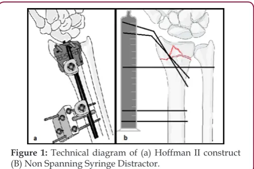
A total number of 32 cases of distal radius fracture treated by Non-Spanning Syringe Distractor from 2015 to 2016 admitted in Primary Level Hospital, India. Ethical committee approval was obtained prior to initiation of the study. All fractures were classified as per the AO classification [5]. Presence of distal ulnar fracture was recorded separately. Our study group was compared with a historical control group of 30 (mean age 61) patients who were operated for Hoffman II type of non-bridging external fixator (Figure 1) between 2015 to 2016 in India and followed up for 1 year by the same author but at the Tertiary Level center.
Inclusion criteria
a. Displaced unstable comminuted fracture of the distal
radius, which was defined as any distal radial fracture with more than 20° of dorsal angulation, metaphyseal comminution with or without intra-articular extension, and more than 10
mm loss of radial height
b. Fresh fracture (reported within 7 days of injury) c. Age >50 years (elder population)
d. Informed consent for operative care.
Exclusion criteria
a. Open fracture b. Pathological fracture
c. Fractures where adequate reduction was not achieved on
operative table
d. Injury severity score (ISS) of >17
e. Ipsilateral upper limb pathology which would affect the
functional outcome e.g. arthritis, scaphoid fracture.
Non Spanning Syringe Distractor Technique
All surgeries were performed under regional anesthesia. Once reduction was achieved by ligament taxis, usually 2-3 K-wires (size 2 or 2.5 mm) were used for fixation. Then maintaining the stability at fracture, K-wires were bended carefully by wire bender up to an angle perpendicular to radial shaft. Proximally 1 or 2 threaded K-wires (size 3mm) inserted in distal radial shaft in dorso-lateral plane 4-5 cm proximal to fracture level depending upon fracture pattern and quality of bone. Distal ulno radial trans-fixation k wire if used for distal radio ulnar joint instability can also be used for distraction purpose if provides enough length. Length of K-wires were cut approximately 4-5 cm away from the skin making them blunt for fixation inside syringe. Under the image intensifier, the required distraction was applied across the wires, and approximate length judged by scale for hole making in syringe. A 5 or 10 cc plastic syringe used as distracter after drilling holes in one plane by similar sized K-wire over T handle. This makes opposite surface of holes as shield covering blunt cut ends of K wires. All the patients were given below elbow plaster (slab) applied meticulously by cotton bandage and soft paddings (Figure 1).
Figure 1: Technical diagram of (a) Hoffman II construct (B) Non Spanning Syringe Distractor.
Figure 2: Case 1 of Non Spanning Syringe Distractor.
Figure 3: Case 2 of Non Spanning Syringe Distractor.
A sample size calculation showed that in order to show a difference with a 5% significance level and 80% power, 30 individuals in each group would be needed for radial tilt as outcome, while more than 8,000 individuals would be needed with
inclination as the outcome. The mean differences, 95% confidence
intervals and p-values for comparisons of the mean values at pre
month, 6 month and 1 year follow-ups when evaluating functional measures. We considered p-values less than 0.05 to be statistically significant.
Results
Table 1: AO classification of fractures.
AO Fracture
Type Hoffman II (n = 30 control) Distractor (n=30 study)Non Spanning Syringe
A3 7 5
C1 9 9
C2 8 10
C3 6 6
Out of 32 one patient died and one lost follow up reducing sample size to 30 same as comparison group. The mean age of patients treated was 62 years as compared to 61 years of control group. The patients were predominantly males (60%) in both groups. The dominant hand was injured in 21 (70%). Domestic falls followed by road traffic accidents were the predominant (>80%) modes of injury. Fractures belong to A3 or C1-3 class according to
AO classification (Table 1).All the fractures were united within 3 months. 4 patients had pain mainly due to prominent ulnar styloid secondary to malunion or DRUJ instability. Wrist and finger pain and stiffness significantly improved after physiotherapy except in one due to Reflex Sympathetic Neuropathy (RSN). One out of two diabetic patients had developed pin track infection, which was healed subsequently. No one developed radial neuritis.
Anatomical assessment: Preoperatively, the median radial tilt was 29 degrees of dorsal angulation in the group and 32 in the Non Spanning Syringe Distractor group. Postoperatively, the median tilt was 8 degrees of volar angulation in the Hoffman II group and 2 degrees volar in the Non Spanning Syringe Distractor group (p = 0.002). At the time of removal of the fixators, there was still a statistically significant difference in radial tilt: 9 degrees of volar angulation in the Hoffman II group and 4 degrees in the. Non Spanning Syringe Distractor group (p= 0.04). At 1 year, the difference was no longer statistically significant. For the other anatomical variables, no statistically significant differences were found (Table 2).
Table 2: Comparison of radiological assessments, mean (95% CI).
Hoffman II (n = 30 control) Non Spanning Syringe Distractor (n=30 study) Mean Difference Radial Tilt (degrees)
Preoperatively 29 (25-34) dorsal 32 (27-36) dorsal
Post Operatively 8 (6-10) volar 2 (0-5) volar 6 (1-9) At time of Removal (6 week) 9 (7-11) volar 4 (2-6) volar 5 (1-8) At 1 year 8 (6-10) volar 4 (2-6) volar 4 (0-8)
Ulnar variance (mm)
Preoperatively 4 (3-4) 4 (3-5)
Post Operatively 0 (0-1) 0 (-1-0) 0 (0-2) At time of Removal (6 week) 1 (0-2) 0 (-1-0) 1 (0-2)
At 1 year 1 (0-2) 0 (0-1) 1 (0-2)
Radial inclination (degrees)
Preoperatively 17 (15-19) 18 (16-20)
Post Operatively 23 (22-24) 23 (22-24) 0 (-1-2) At time of Removal (6 week) 23 (22-24) 24 (23-24) -1 (-2-2) At 1 year 23 (21-24) 23 (21-24) 0 (-2-3)
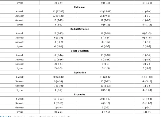
Functional assessment: At 6 weeks, the mean loss of flexion was 24 degrees in the Hoffman II group and 34 degrees in the Non Spanning Syringe Distractor group (p = 0.001). At the other times, the differences between the groups were not statistically significant. There were no statistically significant differences between the groups concerning loss of extension, radial and ulnar deviation, supination, or pronation at the different times (Table 3). There were no statistically significant differences in mean values of
the VAS score between the groups at any time (data not shown). At 1 year, the mean (CI 95%) DASH score was 9 (3–14) in the Hoffman II group and 13 (8–20) in the Non Spanning Syringe Distractor group. According to Gartland and Werley score, 24(80%) excellent to good and 6 (20%) fair to poor results were achieved by Non Spanning Syringe Distractor. These results are also compared with different other landmark studies (Table 4).
Table 3: Comparison of functional assessments by mean (95% CI) loss of movement (in degrees) in the injured wrist compared to the uninjured wrist.
Hoffman II (n = 30 control) Non Spanning Syringe Distractor (n=30 study) Mean Difference Flexion
6 week 24 (20-28) 35 (28-40) -10 (5-16)
6 month 8 (4-12) 9 (7-12) -2 (-8-3)
1 year 3 (-1-8) 8 (5-10) -5 (-11-6)
Extension
6 week 42 (37-47) 43 (35-49) -1 (-5-6)
3 month 23 (14-31) 25 (19-29) -1 (-8-7)
6 month 10 (7-13) 11 (7-15) -1 (-4-7)
1 year 4 (1-6) 9 (6-12) -5 (-1-11)
Radial Deviation
6 week 12 (8-15) 12 (7-18) 0 (-5 - 5)
3 month 6 (1-10) 6 (-3-14) 0 (-4 - 8)
6 month -1 (-4-2) 0 (-4-5) -1 (-3-7)
1 year -1 (-3-1) -1 (-2-5) 0 (-3-7)
Ulnar Deviation
6 week 12 (8-16) 13 (9-18) -1 (-3-6)
3 month 10 (4-16) 7 (-2-16) -3 (-7-6)
6 month 2 (-1-5) 5 (1-9) -3 (-2-8)
1 year 2 (-1-5) 2 (-1-5) 0 (-5-5)
Supination
6 week 30 (23-37) 31 (22-42) -1 (-5 - 10)
3 month 9 (4-14) 13 (3-22) -4 (-5-13)
6 month 7 (3-10) 10 (6-12) -1 (-9-6)
1 year 4 (2-7) 8 (5-11) -4 (-11-4)
Pronation
6 week 15 (9-23) 20 (14-27) -5 (-10-1)
3 month 4 (-2-10) 6 (1-12) -2 (-10-5)
6 month 1 (-1-4) 2 (0-5) -1 (-2-1)
1 year 0 (-2-2) -1 (-7-5) 1 (5-7)
Table 4: Comparison of various study results (functional).
Sr. No. Modality of Treatment Name of series No. of Cases Results
Excellent/Good Fair/Poor
1 Pins Dowling & Sawyer [16] 51 84% 16%
2 Pins And Plaster Cole & Obletz [17] 33 94% 06%
3 Ra Frame Cooney et al. [18] 60 87% 13%
4 Hoffman D’ Anca et al. [19] 54 94% 06%
5 Ao Delta GS Edwards et al. [20] 30 96% 04%
6 Jess Rajeev shukla et al. [21] 72 78% 22%
7 Non Spanning Syringe Distractor Our series(Present study) 30 80% 20%
Discussion
The fracture of distal end radius is the most common fracture
we treat. Management of fracture distal end of radius is still a
challenge for orthopedic surgeon and pose therapeutic problem in term of reduction of fracture, maintenance of reduction till the
fracture unites mobility of the joint after fracture union. Moreover outcome of these fractures is not uniformly good regardless of treatment instituted. We agree with D.L. Fernandez et al that a good functional result usually accompanies a good anatomical
reduction [7]. Collapse, loss of palmar tilt, radial shortening, and
articular incongruity is frequent after closed treatment of unstable
and comminuted intra-articular fractures of the distal radius
describe bridging constructs that cross and necessarily immobilize the wrist during the bony healing process [11]. The ligamentotaxis is the basic principle used by external fixation [12]. Prolonged rigid immobilization of the wrist in spanning external fixator leads to decreased blood supply to bone and soft tissues and causes periarticular fibrosis. This leads to osteoporosis, poor motion, and
compromised functional outcome.
The early mobilization of the wrist leads to normalization of blood supply, hastened functional recovery, earlier resolution of wrist swelling, and decreased joint stiffness [2,4,8]. The dynamic external fixators have also been developed to provide mobilization of the wrist while reduction and fixation are maintained [13]. One such fixator was first designed by Penning (1990) [14]. The device allows wrist flexion by a hinge joint, with the center of motion being at the capito-lunate joint. This is based on several anatomic and biomechanical studies by Short WH et al. [15]. Our case series attributes to 24(80%) excellent to good and 6(20%) fair to poor results, which are quite comparable with all other landmark studies. No statistically significant difference found in radiological and clinical variables or DASH scores between control Hoffman II fixator group and study group of Non Spanning Syringe Distractor. Thus we believe that Non-Spanning Syringe Distractor is an alternative mechanical device especially for those who are prompted to not to use external fixator because abovementioned reasons. It combines the advantages of pins only and wrist spanning external fixator
preventing their disadvantages at the same time.
Conclusion
Finally we would like to conclude that Non-Spanning Syringe Distractor is an easy, cost effective and reliable treatment in treating intra articular and unstable extra articular distal end radial fractures where limited resources in rural part of developing country prompt the orthopedic surgeons to opt for more conservative modality of plaster +/-pins and more frequently complicated by collapse and mal-union. We recommend this device as an alternative method to the currently used modalities and not as a superior substitute .We cannot pretend to present well-objectified functional result unless controlled multicenter trial in larger patient group, but can confirm the feasibility of the Non Spanning Syringe Distractor technique.
References
1. Colles A (1814) On the fracture of the carpal extremity of the radius. Edinb Med Surg J 10:182-186.
2. Collert S, Isacson J (1987) Management of redislocated Colles’ fractures. Clin Orthop Relat Res 135: 183-186.
3. Ruch DS (2006) Fractures of the distal radius and ulna. In: Bucholz RW, Heckman JD, Court-Brown C. (Eds,) Rockwood and Green’s Fractures in
Adults (6 edn.). Philadelphia: Lippincott Williams & Wilkins, USA, 909-964.
4. Vaughan PA, Lui SM, Harringtom JJ, Maistrelli GL (1985) Treatment of unstable fractures of distal radius by external fixation. JBJS 67: 385. 5. CE Plant, C Hickson, H Hedley, NR Parsons, ML Costa (2015) Is it time to
revisit the AO classification of fractures of the distal radius? Bone Joint J Jun 97B(6): 818-823.
6. Manish Changulani, Ugochuku Okonkwo, Tulsi, Yegappan Kalairajah (2008) Outcome evaluation measures for wrist and hand - which one to choose? Int Orthop 32(1): 1-6.
7. DL Fernandez (2000) Should anatomic reduction be pursued in distal radial fractures? The Journal of Hand Surgery: British & European 25(6): 523-527.
8. Robert W, Bocholz James, D Hackman. Rockwood and Greens fracture in adults (5th edn.). 1: 829-880.
9. Agee JM (1993) External fixation: Technical advances based upon multiplanar ligamentotaxis. Orthop Clin North Am 24(2): 265-274. 10. Edward GS (1991) Intra articular fractures of the distal radius treated
with the small AO external fixator. J Bone Joint Surg 73(A) : 1241-1250. 11. Ombrédanne L (1929) L’ostéosythése temporaire chez les enfants.
Presse Med 52: 845-848.
12. Vidal J, Buscacyret C, Paran M, Mulka J (1983) Ligamentotaxis. In Mears DC (Eds), External skeletal Fixation. Baltimore: Williams & Wilkins, USA, 493-96.
13. Clyburn TA (1987) Dynamic external fixation for comminuted intra-articular fractures of the distal end of the radius. J Bone Joint Surg Am 69(2): 348-354.
14. Pennig D (1993) Dynamic external external fixation of distal radius fractures. Hand Cli 9(4): 587-602.
15. Short WH, Palmer AK, Werner FW, Murphy DJ (1987) A biomechanical study of distal radial fractures. J Hand Surg Am 12(4): 529-534. 16. Dowling JJ & Sawyer (1961) Blackwell: Comminuted Colles fractures
evaluation of method of treatment J Bone Joint Surg 43-A: 657-668. 17. Cole JM, Obletz BE (1966) Comminuted fractures of the distal end of the
radius treated by skeletal transfixion in plaster cast. An end-result study of thirty-three cases. J Bone Joint Surg Am 48(5): 931-945.
18. Cooney WP, Agee JM, Hastings HI, Melone CPJ, Rayhack JM (1990) Management of Intraarticular Fractures of the Distal Radius. Contemporary Orthopedics 21: 71-104.
19. D Anca AF, Byron TW, Feinstein PA (1984) External fixator management of unstable Colles fractures: an alternative method. Orthopedics 7(5): 853-859.
20. G S Edwards (1991) Intra-articular fractures of the distal part of the radius treated with the small AO external fixator.J Bone Joint Surg Am 73(8): 1241-1250.
Assets of Publishing with us
• Global archiving of articles
• Immediate, unrestricted online access
• Rigorous Peer Review Process
• Authors Retain Copyrights
• Unique DOI for all articles