Ruby laser-assisted dépilation: the mode of
action and potential ways of improving
outcome
Adam Topping FRCS
2000
A thesis submitted to the University of London
for the degree of
Doctor of Medicine (M.D.)
The RAFT Institute of Plastic Surgery
Mount Vernon Hospital
Northwood
Middlesex
&
The North wick Park Institute for Medical Research
Northwick Park Hospital
Harrow
All rights reserved
INFORMATION TO ALL USERS
The quality of this reproduction is dependent upon the quality of the copy submitted.
In the unlikely event that the author did not send a complete manuscript and there are missing pages, these will be noted. Also, if material had to be removed,
a note will indicate the deletion.
uest.
ProQuest U643687
Published by ProQuest LLC(2016). Copyright of the Dissertation is held by the Author.
All rights reserved.
This work is protected against unauthorized copying under Title 17, United States Code. Microform Edition © ProQuest LLC.
ProQuest LLC
789 East Eisenhower Parkway P.O. Box 1346
DESCRIPTION OF THESIS
Name o f candidate Adam Partington Topping
Title o f thesis Ruby laser-assisted dépilation: The mode o f action and potential ways o f improving outcome.
TEXT
The technique of ruby laser-assisted dépilation has become commonly used in the fields o f reconstructive and cosmetic plastic surgery. Nevertheless it is clear that this technique, as presently practised, produces sub-optimal results in the majority of patients. One o f the contributing factors towards the inefficiency of current treatment regimes is the reluctance to produce the side effect o f epidermal damage.
This thesis examines the clinical technique of ruby laser-assisted dépilation. Attempts are made to define this technique’s mode of action and then to devise new methods of protecting the skin fi*om the associated side effect o f epidermal damage. The work presented uses cell biology and biochemical techniques, specifically: human cell and tissue explant culture, animal models, histology, immunohistochemistry, protein gel analysis (SDS PAGE and western blotting), thermal imaging.
The work shows that laser dépilation treatment causes heat production within the confines o f the hair follicle which then dissipates to the surrounding dermal stroma. The maximum temperature induced in each follicle differs between follicles within the same treatment area and also between different individuals. Laser light penetration through the skin’s depth was found to be deeper than previously theorised. In
addition, an acceptable animal model was established and characterised. Two differing methods of potentially protecting the epidermis from side effect damage were assessed. The first relied on physical blocking of the laser light from reaching the viable cells o f the epidermis, whilst the second involved the induction o f the cells’ own intrinsic stress protection mechanisms (specifically heat shock proteins). Both potential techniques were assessed using cell and/or tissue explant culture and
This thesis is dedicated to my wife, Lucy and my son Sam.
I wish to thank my supervisors Dr C Linge and Professor C Green for their invaluable
supervision and constant enthusiasm, Mr D T Gault for his support and supply of
material, Mr A O Grobbelaar for his advice and help and Professor R Sanders for
making it all possible. I also thank Dr A Cambrey for her help and advice during the
progress o f my work.
I also would like to thank Mr J Shelton for teaching me tissue culture techniques, Mrs
F Daley for immunohistochemical assistance and Mrs S Barnet, Ms C Noel and Mrs E
Clayton for the preparation of all the histological sections. I would furthermore like to
thank Mr K Ladhani for all his help and assistance during my two years of research.
My thanks go to Dr Paul Sibbons for his help in obtaining the Animal Licence and for
being the Project Licence holder during the work performed at Northwick Park. I am
also grateful for the assistance of the theatre staff at Northwick Park during the
experimental work performed at the Institute, in particular to Mr D Shepherd and Ms
C Grey.
I would finally like to thank everyone at RAFT, both trustees and employees, for
finding the necessary funds to continue my work and for making my research time
ABSTRACT
Aim - To improve efficacy and lessen side effects resulting from normal mode ruby
laser (NMRL)-assisted dépilation via a greater understanding o f its mode o f action
and the development o f novel methods of reducing associated epidermal damage.
Employing a thermal imaging camera and ex vivo hair-bearing skin, the targets for the
NMRL (pulse duration 900 psec and spot size 7 mm) were defined, the temperatures
reached and the heat dissipation rates determined. Production of heat was confined to
the hair follicles, with the peak temperatures reached varying considerably between
hairs within the same treatment area and also between individuals. Histological
assessment for a known indicator of cellular damage (p53 expression) identified the
sites and extent o f damage, which correlated with the peak temperatures measured.
An energy meter was used to detect the penetration o f NMRL light through ex vivo
skin, which was found to be deeper than previously theorised. The black-haired
mouse (C57B1/10) was assessed both macroscopically and histologically and found to
be an acceptable animal model of NMRL dépilation and associated epidermal
damage. Attempts to reduce the epidermal damage by simply stopping the light
reaching the epidermis using a chromophore block were assessed. Chromophore did
indeed reduce the amount o f epidermal damage detected in laser-irradiated ex vivo
human skin, whereas in contrast it increased the wounding seen in the much thinner
skin of the mouse. Nevertheless the mouse model showed that this technique did not
affect the dépilation efficacy. An alternative method o f reducing epidermal damage
using induction o f the cells’ intrinsic protective mechanisms (heat shock proteins,
HSP) was assessed using cultured kératinocytes and the mouse model. Primarily, the
sub-lethal temperature optimum for HSP expression in human kératinocytes was
determined, then an in vitro model o f NMRL-associated epidermal damage was
established and the heat pre-treatment assessed. The temperature to precondition
mouse skin was then determined and its protective capacity and effect on depilatory
CONTENTS
CHAPTER 1 - GENERAL INTRODUCTION
1.1 HAIR 34
1.2 ANATOMY OF HAIR 36
1.3 PHYSIOLOGY OF HAIR 42
1.4 PATHOLOGY OF HAIR 47
1.4.1 Excess Hair 47
Hypertrichosis 50
Hirsutism 51
1.4.2 Treatment of Excess Hair 52
1.5 LASERS 56
1.5.1 Terminology 56
1.5.2 The Ruby Laser 58
1.5.3 Present Knowledge Regarding Laser/Skin Interaction 58
1.5.4 Which Laser is Best Suited for Dépilation? 64
1.5.5 Present Depilatory Results 68
1.5.6 Present Skin Side Effects 71
1.6 REDUCTION OF SIDE EFFECTS 75
1.7 HEAT SHOCK PROTEINS AND THE STRESS RESPONSE 76
1.8 HYPOTHESES 78
CHAPTER 2 - MATERIALS AND METHODS
2.1 MATERIALS 80
2.1.1 Animals 80
2.1.2 Anaesthesia 80
Histology 82
Sodium dodecylsulphate-polyacrylamide gel electrophoresis
(SDS-PAGE) and Western Blotting 82
Immunohistochemistry 82
2.1.6 Specialised Equipment 83
Ruby Laser 83
Thermal Imaging Camera 85
2.1.7 Imaging 86
2.1.8 Photography 86
2.2 TISSUE CULTURE METHODS 86
2.2.1 General Procedures 86
Media 86
Trypsinisation/passaging 88
Cell Counting 88
Trypan Blue Viability Test 89
Feeder Cells 89
2.2.2 Initiation of a Keratinocyte Population 90
2.2.3 Initiation of a Fibroblast Population 90
2.2.4 Cryopreservation of Cells 92
2.2.5 Organ Culture 93
2.3 STAINING METHODS 93
2.3.1 Histochemistry 93
Haematoxylin and Eosin 93
Modified SACPIC Technique 94
Massons Fontana 94
Massons Trichrome 94
2.3.2 Immunohistochemistry 95
Detection of p53 and Heat Shock Protein 70 (HSP 70) Expression
in Human Tissue 95
2.4 METHODS FOR SDS PAGE AND WESTERN BLOT ANALYSIS 97
Cellular Protein Extraction 97
Preparation of Gels 97
Protein Assay 97
Electrophoresis 98
Western Blotting 98
2.5 METHODS - THE BLACK-HAIRED MOUSE MODEL 101
2.5.1 Husbandry 101
2.5.2 Anaesthesia 102
2.5.3 Laser Procedure 102
2.5.4 Analysis of Results 103
Clinical 103
Histological 103
2.6 STATISTICAL METHODS 105
CHAPTER 3 - THE INTERACTION BETWEEN A LASER PULSE
AND HUMAN SKIN
3.1 INTRODUCTION 106
3.2 AIMS 108
3.2.1 Thermal Imaging Experiment 108
3.2.2 Laser/Skin Penetration Experiment 108
3.3 METHOD 109
3.3.1 Thermal Imaging Experiment 109
3.3.2 Laser/Skin Penetration Experiment 111
3.4 STATISTICS 112
3.5 RESULTS 112
3.5.1 Thermal Imaging Experiment 112
Thermal Imaging 112
3.6.1 Thermal Imaging Experiment 129
3.6.2 Laser/Skin Penetration Experiment 133
3.7 CONCLUSIONS 136
CHAPTER 4 - AN ANIMAL MODEL ASSESSING RUBY
LASER-ASSISTED DEPILATION
4.1 INTRODUCTION 139
4.2 AIMS 143
4.3 METHODS 143
4.4 STATISTICS 145
4.5 RESULTS 145
4.5.1 Dépilation 145
Resting Hair Sites 145
Growing Hair Sites 146
Comparison of Resting and Growing Hair Site Data 150
4.5.2 Histology of Hair Damage 152
4.5.3 Epidermal Damage 152
Daily Comparison of Epidermal Damage Between Resting and
Growing Hair Sites 152
Time Taken to Fully Heal in Resting and Growing Hair Sites 157
4.5.4 Histology of Epidermal Damage 158
4.6 DISCUSSION 166
CHAPTER 5 - THE EFFECT ADDITIONAL CHROMOPHORE
HAS UPON RUBY LASER/SKIN INTERACTION
5.1 INTRODUCTION 172
5.2 AIMS 174
5.3 METHODS 175
5.3.1 The Colour Most Suited to Reflect or Absorb Ruby Laser Light 175
5.3.2 The Effect the Addition of Black Ink as a Chromophore to the
Surface of Ex vivo Human Skin Prior to Ruby Laser Irradiation has upon
Histological Damage Noted 175
5.3.3 The Addition of Black Ink to the Surface of Ex vivo Human Skin and
its Effect Upon Laser Light Transfer through Skin 176
5.3.4 The Addition of Black Ink to the Skin of the Black-haired Mouse
Prior to Ruby Laser Irradiation and its Effect Upon Depilatory Efficacy
and Skin Side Effects 177
5.4 STATISTICS 178
5.5 RESULTS 178
5.5.1 The Colour Most Suited to Reflect or Absorb Ruby Laser Light 178
5.5.2 The Effect the Addition of Black Ink as a Chromophore to the
Surface of Ex vivo Human Skin Prior to Ruby Laser Irradiation has upon
Histological Damage Noted 179
5.5.3 The Addition of Black Ink to the Surface of Ex vivo Human Skin and
its Effect Upon Laser Light Transfer through Skin 181
5.5.4 The Addition of Black Ink to the Skin of the Black-haired Mouse
Prior to Ruby Laser Irradiation and its Effect Upon Depilatory Efficacy
and Skin Side Effects 185
Dépilation 185
Growing Hair Sites 185
Resting Hair Sites 187
Skin Damage 187
Histology 196
Dépilation 196
Skin Damage 199
5.6 DISCUSSION 204
5.6.1 The Colour Most Suited to Reflect or Absorb Ruby Laser Light 204
5.6.2 The Effect the Addition of Black Ink as a Chromophore to the
Surface of Ex vivo Human Skin Prior to Ruby Laser Irradiation has upon
Histological Damage Noted 204
5.6.3 The Addition of Black Ink to the Surface of Ex vivo Human Skin and
its Effect Upon Laser Light Transfer through Skin 205
5.6.4 The Addition of Black Ink to the Skin of the Black-haired Mouse
Prior to Ruby Laser Irradiation and its Effect Upon Depilatory Efficacy
and Skin Side Effects 206
5.7 CONCLUSIONS 209
CHAPTER 6 - POTENTIAL PROTECTION FROM EPIDERMAL
SIDE EFFECTS BY PRETREATMENT -
IN VITRO
PROOF OF
PRINCIPLE
6.1 INTRODUCTION 210
6.2 AIMS 213
6.3 METHODS 214
6.3.1 Determining Heat-induced Cell Death of Seven Paired Fibroblast
and Keratinocyte Cell Lines Exposed to a Range of Temperatures 214
6.3.2 Determining the Effect of Temperature Upon Heat Shock Protein 70
6.3.3 Determining the Effect of Heat Preconditioning of Kératinocytes in
Culture Upon Cell Survival after Subsequent Exposure to Ruby Laser
Irradiation 216
6.4 STATISTICS 218
6.5 RESULTS 219
6.5.1 Determining Heat-induced Cell Death of Seven Paired Fibroblast
and Keratinocyte Cell Lines Exposed to a Range of Temperatures 219
6.5.2 Determining the Effect of Temperature Upon Heat Shock Protein 70
Synthesis within Seven Keratinocyte Cell Lines 222
6.5.3 Determining the Effect of Heat Preconditioning of Kératinocytes in
Culture Upon Cell Survival after Subsequent Exposure to Ruby Laser
Irradiation 225
6.6 DISCUSSION 229
6.6.1 Determining Heat-induced Cell Death of Seven Paired Fibroblast
and Keratinocyte Cell Lines Exposed to a Range of Temperatures 229
6.6.2 Determining the Effect of Temperature Upon Heat Shock Protein 70
Synthesis within Seven Keratinocyte Cell Lines 231
6.6.3 Determining the Effect of Heat Preconditioning of Kératinocytes in
Culture Upon Cell Survival after Subsequent Exposure to Ruby Laser
Irradiation 232
6.7 GENERAL DISCUSSION 233
6.8 CONCLUSIONS 237
6.9 THE ABILITY OF E X VIVO NORMAL HUMAN SKIN TO BE HEAT
PRECONDITIONED BY APPLICATION OF A HEATING APPARATUS 237
6.9.1 Aims 238
6.9.2 Method 238
6.9.3 Results 240
6.9.4 Discussion 241
PRINCIPLE
7.1 INTRODUCTION 249
7.2 AIMS 251
7.3 METHODS 251
7.3.1 Determining the Ideal Temperature at which to Precondition the
Skin of the Black-haired Mouse whilst producing Minimal Side
Effects 251
7.3.2 Determining the Consequences of Heat Preconditioning Upon the
Effects of Ruby Laser Irradiation of the Skin of the Black-haired
Mouse 253
7.4 STATISTICS 255
7.5 RESULTS
7.5.1 Determining the Ideal Temperature at which to Precondition the
Skin of the Black-haired Mouse whilst producing Minimal Side
Effects 256
Macroscopic Analysis 256
Histological Analysis 256
7.5.2 Determining the Consequences of Heat Preconditioning Upon the
Effects of Ruby Laser Irradiation of the Skin of the Black-haired
Mouse 260
Macroscopic Analysis 260
Skin Damage 260
Dépilation 266
Histological Analysis 269
7.6 DISCUSSION 273
7.6.1 Determining the Ideal Temperature at which to Precondition the
Skin of the Black-haired Mouse whilst producing Minimal Side
7.6.2 Determining the Consequences of Heat Preconditioning Upon the
Effects of Ruby Laser Irradiation of the Skin of the Black-haired
Mouse 275
Skin Damage 275
Dépilation 276
7.7 CONCLUSIONS 277
CHAPTER 8 - DISCUSSION
8.1 RELATING THE SCIENTIFIC EXAMINATION OF LASER-SKIN
INTERACTIONS TO THE RESULTS ACHIEVED CLINICALLY 278
8.2 CHOICE OF MODELS TO INVESTIGATE RUBY LASER-ASSISTED
DEPILATION 280
8.3 CHROMOPHORE BLOCKS AND THEIR ROLE IN SKIN
PROTECTION 281
8.4 HEAT PRETREATMENT AND ITS ROLE IN SKIN PROTECTION 284
8.5 THE FUTURE OF LASER-ASSISTED DEPILATION 286
APPENDICES
APPENDIX A 287
Establishing the Correct Seeding Density of Kératinocytes Required for the In
vitro Model of Epidermal Damage
Results and Discussion
APPENDIX B 289
Does Laser Treatment of the In vitro Model Cause Similar Levels of Cell Damage
and Death to That Seen In vivo?
Results
Results
Discussion
REFERENCES
299LIST OF FIGURES
CHAPTER 1
Figure 1.1: Anatomical representation of a human hair follicle. The solid black
region represents the keratogeneous zone, the straight-line regions the hard
keratin and the speckled regions the soft keratin. 35
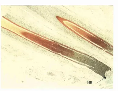
Figure 1.2: Modified SACPIC stain of a transverse section taken from human
hair-bearing skin showing the different keratinised regions of a hair shaft. The
viable hair kératinocytes are stained green, whilst those in the keratogenous zone
are stained red. The fully keratinised, mature cells of the hair shaft are stained
yellow. HB is the hair bulb (xlOO). 37
Figure 1.3: The three stages of the hair cycle. Note that during the telogen phase
the position of the resorbed dermal papilla of the old hair bulb lies adjacent to
the bulge region. 43
Figure 1.4: How a laser beam is produced. 57
Figure 1.5: A diagrammatic representation of the electromagnetic spectrum
Figure 1.6: Absorption spectra of the major skin pigments according to
wavelength at the concentrations they usually occur in vivo. Note the relative
difference in absorbance of melanin compared with oxyhaemoglobin at around
700 nm. 67
Figure 1.7: Photograph showing superficial skin burning 2 days after exposure to
normal mode ruby laser irradiation. 72
Figure 1.8a: Hypopigmentation of the skin occurring 2 weeks after exposure to
normal mode ruby laser irradiation. 73
Figure 1.8b: Hyperpigmentation of the skin occurring 2 weeks after exposure to
normal mode ruby laser irradiation. 73
CHAPTER 2
Figure 2.1 : The “Chromos 694 nm Dépilation” normal mode ruby laser. 84
Figure 2.2: The FLIR Systems “Thermovision 900” thermal imaging camera. 84
Figure 2.3: Keratinocyte cells grown in culture and viewed through an inverted
phase contrast microscope. The larger cells of the colony, seen lower right,
represent differentiating, suprabasal kératinocytes. 91
Figure 2.4: Fibroblast cells grown in culture and viewed through an inverted
Figure 3.1:
a: Dermal aspect of a specimen from patient 3 after microdissection revealing the
hair bulbs and lower hair shafts (xlO). The outlined areas have identified the
groups of hairs shown in the thermal image in Figure b. 113
b: The first thermal image of the specimen in Figure a taken during ruby laser
exposure at 15 J/cm^ and showing the peak temperatures in Centigrade obtained
by the identified hairs in Figure a (x8.5). 113
c: A thermal image recorded at approximately 0.75 seconds after ruby laser
exposure showing a reduction in temperature at the sites of the hair follicles but
an increase in temperature within the intervening skin (x8.5). 113
Figure 3.2a: The first thermal image as shown in Figure 3.1b but with an arrow
across three hairs (A l, A2 and A3) depicting the pixels along which the
temperature changes were recorded over time. 115
3.2b: Graphical representation of the changes in temperature over time
recorded by each of the pixels along the arrow shown in (a). Hairs A l, A2 and A3
have been identified and the increase in the pixel number follows the direction of
the arrow. 115
Figure 3.3: Graph showing the distribution of maximum temperature rise
recorded for each hair (N=80) measured from all patients. The commonest
temperature rise occurred between 5.1 and 10®C. 116
Figure 3.4: Graph showing the distribution of maximum temperature rises
according to patient of all hairs measured. The range (hollow dots), the mean
Figure 3.5: Graph showing the change in temperature over time of the hair A3
(at pixel 59) taken from Figure 3.2a and the intervening skin (at pixel 50). 117
Figure 3.6: Graph showing the rate of heat loss from all hairs measured from a
specimen taken from patient 3. 117
Figure 3.7: Graph showing the best fit curves for the rate of heat loss from all
follicles measured according to patient. 118
Figure 3.8: Modified SACPIC staining of a tangential section of hair-bearing
skin showing a hair follicle in the anagen phase (A?) and a hair follicle in the
telogen phase (TP) characterised by the brush border (BB)( xlOO). 120
Figure 3.9: Modified SACPIC staining of a section of hair bearing skin having
undergone ruby laser exposure showing a follicle containing an undamaged
vellus hair (VH) and a damaged terminal hair (TH). The outer root sheath
(ORS) has been disrupted (x200). 120
Figure 3.10: p53 immunostaining of a section of a specimen having undergone
UV irradiation. Positive nuclei are stained brown and negative nuclei
counterstained blue with haematoxylin. Positive nuclei are seen within cells
throughout the layers of the epidermis and into the dermis (x200). 122
Figure 3.11: p53 immunostaining of a section of a specimen having undergone
ruby laser irradiation. p53 expression can be seen to have occurred throughout
the epidermal layers within the majority o f cells (x200). 122
Figure 3.12: p53 immunostaining of a tangential section of a specimen from
patient 3 at the level of the sebaceous gland (SG). Damage has occurred to the
outer root sheath (ORS) shown by the asterisk and the cells lying adjacent to this
area in both the follicle and the sebaceous gland are expressing p53 protein
Figure 3.14: Graph showing an extrapolation of the plots in Figure 3.13 above to
the X axis revealing the probable maximum depth of penetration of the ruby
laser beam in ex vivo skin. 127
CHAPTER 4
Figure 4.1: Transverse section through mouse skin showing a hair in anagen
phase. Melanin can be seen throughout the length of the hair shaft (xlOO). 141
Figure 4.2: Photograph of the trimmed back of a black haired mouse illustrating
the black patches synonymous with hairs in the anagen phase and pink patches
synonymous with hairs in telogen phase. 141
Figure 4.3: Transverse section of mouse skin stained with Massons fontana
showing that for hairs in telogen phase, the melanin is restricted to that portion
of the hair shaft which protrudes from the skin surface. 142
Figure 4.4: Photograph of a mouse 1 day after exposure to ruby laser irradiation
with the fluences starting at top left and rotating anti clockwise being 5, 6, 7 and
8 J/cm^ 142
Figure 4.5: Graph representing the hair regrowth scores achieved over time in
resting hair regions irradiated with different fluences. 147
Figure 4.6: Graph representing the hair regrowth scores achieved over time in
Figure 4.7: Photograph of a mouse 8 weeks after exposure to ruby laser
irradiation at 8 J/cm^ upon a growing hair region. An area of decreased hair
regrowth is visible with depigmentation of the hairs regrowing in that site. 151
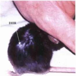
Figure 4.8: Photograph of a mouse 8 weeks after exposure to ruby laser
irradiation at 8 J/cm^ upon a growing hair region. Decreased hair regrowth
(DHR) is evident in the area identified. 151
Figure 4.9: Transverse section through a resting hair region stained with the
modified SACPIC technique showing a telogen hair. Ruby laser irradiation has
occurred at 5 J/cm^ and shaft damage (SD) can be seen only in the external
aspect of the shaft above the level of the skin. This would coincide with the limit
of the melanin pigment. The section is representative of the higher fluences too
(x200). 153
Figure 4.10: Transverse sections stained with the modified SACPIC technique
having been exposed to a) 5 J/cm^ and b) 8 J/cm^ fluence showing disruption of
the melanin (DM) within the hair shafts which is greater at 8 J/cm^ (HB = hair
bulb) x200. 154
Figure 4.11: Graph representing the wound scores achieved over time in resting
hair regions irradiated with different fluences. 155
Figure 4.12: Graph representing the wound scores achieved over time in the
growing hair regions irradiated with different fluences. 156
Figure 4.13: Graph representing the time taken to complete wound healing in
hoth resting and growing hair regions as a result of ruby laser exposure at the
gland (SG), the probable bulge region (BR) and the hair bulb (HB) have been
identified (x200). 161
Figure 4.15a: Transverse section of a resting hair follicle within mouse skin
exposed to a ruby laser fluence of 5 J/cm^ and stained with H&E. The epidermis
shows signs of damage with areas of complete loss (EL), but the cells lining the
hair canal look relatively undamaged in comparison (x200). 162
Figure 4.15b: Transverse section of mouse skin taken from a resting hair region
exposed to a ruby laser fluence of 5 J/cm^ and stained with H&E. Note that
epidermal damage is present with areas of cell loss (CL), nuclear pyknosis (NP)
and cell vacuolisation (CV) x400. 162
Figure 4.16a: Transverse section of mouse skin in a resting hair region after
exposure to ruby laser irradiation at 8 J/cm^. Note almost complete epidermal
loss but the cells lining the hair canal appear undamaged (x200). 163
Figure 4.16b: Transverse section of mouse skin in a resting hair region after
exposure to ruby laser irradiation at 8 J/cm^. Note that islands of epidermal cells
are still present within the treatment field although their viability cannot be
ascertained (x200). 163
Figure 4.17: Transverse section stained with H&E of a growing hair region
exposed to ruby laser irradiation at 5 J/cm^ Note the epithelial damage, for
instance cellular vacuolisation (CV) and nuclear pyknosis (NP) x200. 164
Figure 4.18: Transverse section stained with H&E of a growing hair region
exposed to ruby laser irradiation at 8 J/cm^. Note epithelial damage manifesting
as cellular vacuolisation and nuclear pyknosis with areas of almost complete
Figure 4.19: Transverse section stained with Massons Fontana of a) a resting
hair region and b) a growing hair region of the skin of the black-haired mouse
showing no melanin within the epithelial or follicular cells. 166
Figure 4.20: Transverse sections stained with Massons Fontana of a) a resting
hair region exposed to 5 J/cm^ and b) a growing hair region exposed to 8 J/cm^
Melanin granules can be seen scattered over the epithelial surface and disrupted
(DM) within the hair follicle identified (x200). 167
Figure 4.21: Transverse section stained with Massons trichrome of a resting hair
region biopsied 8 weeks after laser exposure. No scarring or disruption is evident
within the collagen of the dermis (stained blue) suggesting that the skin damage
incurred as a result of laser exposure is not permanent (xlOO). 168
CHAPTER 5
Figure 5.1: Graph representing the laser light penetration profiles through
differing thicknesses of ex vivo skin whose surfaces have either been painted or
not with black ink prior to laser exposure at a fluence of 9.24 J/cm^. 180
Figure 5.2: Graph showing an extrapolation of the plots in Figure 5.1 to the x
axis revealing the probable maximum depth of penetration of the ruby laser
beam in ex vivo skin whose surfaces had either been painted or not with
chromophore prior to laser exposure at a fluence of 9.24 J/cm^. 180
Figure 5.3: Transverse section of a negative control of human ex vivo skin having
undergone immunohistochemical staining for expression of p53. Virtually all the
nuclei have taken up the counterstain (haematoxylin) and so appear blue
Positive nuclei (brown) are seen throughout the layers of the epidermis and into
the dermis (x200). 183
Figure 5.5: Transverse section of a specimen of ex vivo human skin having
undergone ruby laser irradiation at 15 J/cm^ and stained for p53 protein
expression. The nuclei in the basal layer and above have stained positive for the
protein (brown) showing damage has occurred to the cells within this region
(x200). 184
Figure 5.6: Transverse section of a specimen of ex vivo human skin having had
chromophore added to the surface prior to ruby laser exposure at 15 J/cm^ and
stained for p53 protein expression. No increase in p53 protein expression is
apparent in the basal layers as compared to the section above (x200). 184
Figure 5.7: Graphs representing the weekly hair regrowth scores in growing hair
regions irradiated at a) 6 J/cm^ and b) 8 J/cm^. 186
Figure 5.8: Photograph showing hair dépilation in a growing hair region 8 weeks
after ruby laser exposure at 8 J/cm^ Chromophore had been added to this site
prior to exposure. 188
Figure 5.9: Graphs representing the weekly hair regrowth scores in resting hair
regions irradiated at a) 6 J/cm^ and b) 8 J/cm^. 189
Figure 5.10: Graphs representing the daily wound scores achieved upon growing
hair regions where chromophore had either been added or not prior to laser
Figure 5.11: Photograph showing the skin damage (SD) in a growing hair site 7
days after ruby laser exposure at 8 J/cm^ which did not have chromophore
added prior to irradiation. 193
Figure 5.12: Photograph showing the skin damage (SD) in a growing hair site 7
days after ruby laser exposure at 8 J/cm^ which had chromophore added to the
site prior to irradiation. 193
Figure 5.13: Graph showing the mean times to full healing of the skin wounds in
growing hair sites which had either chromophore added or not to the skin
surface prior to ruby laser exposure at 6 and 8 J/cm^. 194
Figure 5.14: Graphs representing the daily wound scores achieved upon resting
hair regions where chromophore had either been added or not prior to laser
exposure at a) 6 J/cm^ and b) 8 J/cm^ fluence. 195
Figure 5.15: Photograph of the back of a mouse 2 days after ruby laser exposure
upon resting hair regions showing increased skin damage in the two upper sites
where chromophore had been added prior to irradiation. The two cephalad sites
have been exposed to a fluence of 6 J/cm^ whilst the caudal sites have been
exposed to 8 J/cm^. 197
Figure 5.16: Graph showing the mean times to full healing of the skin wounds in
resting hair sites which had either chromophore added or not to the skin surface
prior to ruby laser exposure at 6 and 8 J/cm^. 198
Figure 5.17a: Transverse section stained using the modified SACPIC technique
showing the extent of damage occurring to the shaft of a resting hair irradiated
chromophore applied to the surface prior to irradiation at 6 J/cm^. Little
difference is apparent in the extent of shaft damage occurring within the two
sections (x400). 200
Figure 5.18a: Transverse section stained with H&E of a resting hair specimen
irradiated at 6 J/cm^ showing epithelial damage by the presence of cell
vacuolisation (CV) and nuclear pyknosis (NP) x200. 201
Figure 5.18b: Transverse section stained with H&E of a resting hair specimen
that had chromophore added to the skin surface prior to irradiation at 6 J/cm \
Greater damage to the skin surface has occurred than in Figure 6.6a with
epithelial cell loss evident (CL) x200. 201
Figure 5.19a: Transverse section of a specimen of mouse skin irradiated at 8
J/cm^ showing epithelial damage. Cell vacuolisation (CV) and nuclear pyknosis
(NP) is evident (x200). 202
Figure 5.19b: Transverse section from a specimen of mouse skin having had
chromophore added prior to irradiation at 8 J/cm \ Virtually the whole of the
epidermis has been damaged or removed as a result of laser exposure (x200). 202
Figure 5.20a: Transverse section stained with H&E of a specimen from a
growing hair region exposed to laser irradiation at 6 J/cm^. Signs of epidermal
damage are present as described before (x200). 203
Figure 5.20b: Transverse section stained with H&E of a specimen from a
growing hair region where chromophore was applied prior to laser irradiation at
6 J/cm^. Skin damage is evident with areas of epithelial loss (EL), but the damage
CHAPTER 6
Figure 6.1: Graph showing the percentage of dead cells against temperature for
each of the seven fibroblast cell lines. 220
Figure 6.2: Graph showing the percentage dead cells against temperature for
each of the seven keratinocyte cell lines. 221
Figure 6.3: Graph showing the mean plot with standard deviation of the
percentage of dead cells against temperature for all seven keratinocyte and
fibroblast cell lines. 223
Figure 6.4: Representative Western Blot with a positive staining for HSP 70 5
hours after heat stressing at the range of temperatures shown (M.M. is molecular
marker, 37°C I is 37°C in the incubator, 37°C W is 37°C in the waterbath). The
product of the optical density and the area for each temperature was established
by the Seescan computer and are shown beneath the relevant well. 224
Figure 6.5: Second representative Western Blot with a positive staining for HSP
70 5 hours after heat stressing at the range of temperatures shown ( M.M. is
molecular marker, 37°C I is 37°C in the incubator, 37°C W is 37°C in the
waterbath). The product of the optical density and the area for each temperature
was established by the Seescan computer and are shown beneath the relevant
well. 224
Figure 6.6: Representative Western Blot with a positive staining for HSP 70 24
hours after heat stressing at the range of temperatures shown (M.M. is molecular
marker, 37°C I is 37°C in the incubator, 37°C W is 37°C in the waterbath). The
product of the optical density and the area for each temperature was established
the optical density and the area shown beneath the relevant well. 226
Figure 6.8: Graph showing the relationship between the total number of viable
cells and increasing fluence of exposure from a normal mode ruby laser.
Irradiation occurred 5 hours after heating and the wells contained 100 pg/ml
melanin. 227
Figure 6.9: Graph showing the relationship between the total number of viable
cells and increasing fluence of exposure from a normal mode ruby laser.
Irradiation occurred 24 hours after heating and the wells contained 100 pg/ml
melanin. 227
Figure 6.10: Graph showing the relationship between the total number of viable
cells and increasing fluence of exposure from a normal mode ruby laser.
Irradiation occurred 5 hours after heating and the wells contained 200 pg/ml
melanin. 228
Figure 6.11: Graph showing the relationship between the total number of viable
cells and increasing fluence of exposure from a normal mode ruby laser.
Irradiation occurred 24 hours after heating and the wells contained 200 pg/ml
melanin. 228
Figure 6.12: Diagrammatic representation of the experiment described in section
6.6 which was performed in a Class II hood maintaining tissue sterility. 239
Figure 6.13: Transverse section of ex vivo human skin acting as a negative
control and stained for HSP 70 expression. Minimal protein expression is evident
within the nuclei of all epidermal cells which appear blue from the haematoxylin
Figure 6.14: Transverse section of ex vivo human skin exposed to the heating
probe at 37°C and stained for HSP 70 protein. Minimal expression is evident
within the nuclei of the epidermis which are blue from the haematoxylin
counterstain (x200). 243
Figure 6.15: Transverse section of ex vivo human skin exposed to 42°C from the
heating probe and stained for HSP 70 protein. A few of the cells of the basal
layer are expressing the protein with the nuclei appearing brown (x200). 244
Figure 6.16: Transverse section of ex vivo human skin exposed to 46°C from the
heating probe and stained for HSP 70 protein. Positive expression has occurred
in all nuclei denoted by the brown stain and is particularly strong at the basal
layer (x200). 245
Figure 6.17: Transverse section of ex vivo human skin exposed to 50°C from the
heating probe and stained for HSP 70 protein. All the nuclei have stained
positive for expression of the protein but cellular damage is now becoming
evident in terms of vacuolisation (CV) x200. 246
Figure 6.18: Transverse section of ex vivo human skin exposed to 54°C from the
heating probe and stained for HSP 70 protein. The nuclei of the cells of the
epidermis are expressing the protein but increased damage is now evident in
terms of the epidermis becoming removed from the dermis in places (x200). 247
CHAPTER 7
Figure 7.1: Photograph of an anaesthetised mouse being exposed to the heating
expression has occurred in the nuclei of the cells within the epidermis (x200). 254
Figure 7.3: Transverse section of a specimen stained for HSP 70 having been
exposed to 45°C from the heating apparatus for 15 minutes. Increased expression
has occurred within the nuclei of the cells of the epidermis and also the cells
lining the hair follicles to the extent of the sebaceous gland (x200). 257
Figure 7.4: Transverse section of a specimen stained with H&E having been
exposed to 45°C from the heating apparatus for 15 minutes. No evidence of
cellular damage is apparent within the epidermis (x200). 257
Figure 7.5: Transverse section of a specimen stained for HSP 70 having been
exposed to 47°C from the heating apparatus for 15 minutes. Increased expression
is present within the nuclei of the epidermis and throughout the cells of the
follicle to the hair bulb (x200). 258
Figure 7.6a: Transverse section through mouse epidermis exposed to 47C for 15
minutes showing damage to the epidermal cells (CV=cell vacuolisation) x400.
b: Transverse section through mouse skin in a hair resting region
exposed to the same temperature as a) showing damage to the cells lining the
follicular canal (x200). 258
Figure 7.7: Graphs showing the wound scores achieved in preconditioned and
non-preconditioned sites in resting hair regions at a) 5 J/cm^ and b) 6 J/cm^. 261
Figure 7.8: Photograph showing a non-preconditioned (NPC) and
preconditioned (PC) laser exposed site in a resting hair region 2 days after
Figure 7.9: A photograph also showing a non-preconditioned (NPC) and
preconditioned (PC) laser exposed site in a resting hair region 2 days after
irradiation at 6 J/cm^. 263
Figure 7.10: Graphs showing the wound scores achieved in preconditioned and
non-preconditioned sites in resting hair regions at a) 5 J/cm^ and b) 6 J/cm^. 264
Figure 7.11: Mean time in days for the laser induced wounds to heal in
preconditioned and non-preconditioned sites a) in resting hair regions and b) in
growing hair regions. 265
Figure 7.12: Graphs representing the hair regrowth over time in resting hair
regions that were either preconditioned or not prior to laser exposure at a) 5
J/cm^ and b) 6 J/cm^. 267
Figure 7.13: Graphs representing the hair regrowth over time in growing hair
regions that were either preconditioned or not prior to laser exposure at a) 5
J/cm^ and b) 6 J/cm^. 268
Figure 7.14a: Transverse section through a resting hair region stained with H&E
having been exposed to ruby laser irradiation at 5 J/cm^. Note the extent of the
epidermal damage in terms of cell vacuolisation (CV) xlOO. 270
Figure 7.14b: Transverse section through a resting hair region stained with H&E
having been preconditioned 5 hours before exposure to ruby laser irradiation at
5 J/cm2. Note decreased epidermal cell damage compared to Figure 8.11a above
(xlOO). 270
Figure 7.15a: Transverse section through a growing hair region stained with
H&E having been exposed to ruby laser irradiation at 5 J/cm^. Note the
increased extent of epidermal damage in terms of cell vacuolisation (CV)
the decreased extent of epidermal damage in terms of cell vacuolisation (CV)
compared to the irradiated growing hair region above (Figure 8.12a) xlOO. 271
Figure 7.16a: Transverse section through a resting hair region stained with H&E
having been exposed to ruby laser irradiation at 6 J/cm^. Note the extent of the
epidermal damage in terms of cell vacuolisation (CV) xlOO. 272
Figure 7.16b: Transverse section through a resting hair region stained with H&E
having been preconditioned prior to exposure to ruby laser irradiation at 6
J/cm^. Note minimal epithelial damage has occurred (xlOO). 272
APPENDICES
Figure B .l: Diagrammatic representation of the second pilot study (see Appendix
B) to determine the ruby laser fluence required to induce keratinocyte cell
death. 291
Figure B.2: Graph representing the cell death of cultured human kératinocytes
after exposure to ruby laser irradiation at a range of fluences
(see Appendix B). 292
Figure C .l: Diagrammatic representation of the third pilot study (see Appendix
C) to discover the effect of varying concentrations of melanin and ruby laser
irradiation upon cultured human kératinocytes. 295
Figure C.2: Graph representing the cell death of cultured human kératinocytes
with the addition of two concentrations of synthetic melanin after exposure to
LIST OF TABLES
CHAPTER 1
Table 1.1: Fitzpatrick sun-reactive skin typing. The categorising of individuals is
based upon verbal response regarding first, moderate (three minimal erythema
doses-MED) unprotected sun exposure for a period of 45 to 60 minutes. 1 MED
is equivalent to 15 to 30 minutes of noon exposure in northern (20® to 45®)
latitudes or 30 mJ/cm^. 66
Table 1.2: A comparison of recent depilatory studies using the ruby laser. 69
Table 1.3: A comparison of recent depilatory studies using lasers other than the
ruby laser. 70
CHAPTER 2
Table 2.1 : Constituents of tissue culture media. 87
a. Fibroblast culture medium (FCM).
b. Keratinocyte culture medium (KCM).
Table 2.2: Constituents of the 7.5% (running) gel and the 4% (stacking) gel used
for the Western Blot analysis. 99
Table 2.3: Constituents of buffer solutions used in the Western Blot analysis. 99
a. Sample buffer.
b. Running buffer (a stock of 2 litres).
Table 2.5: Modification of the wound scoring system shown in Table 2.4. 104
Table 2.6: Classification and scoring of hair regrowth. 104
CHAPTERS
Table 3.1: Table showing the mean percentage of hair follicles from both patient
specimens exhibiting histological damage to the hair shaft, with the total number
of hairs examined within both patient specimens also shown. The mean and
range of depth to which damage was seen to occur to the hair shafts, according to
patient, is shown (* depicts those patient specimens in which damage to the
follicular hair shafts was noted to extend to the hair bulb) along with the mean
percentage of hairs from both patient specimens noted to be in telogen phase. 121
Table 3.2: Table showing the percentage of follicles containing histologically
damaged hair shafts whose follicular cells expressed p53 protein. The mean and
range of depth to which p53 expression was found within those damaged follicles
is also tabulated. 121
Table 3.3: Table showing that no change in fluence was recorded by repetitive
firing of the same fluence value upon the same thickness of skin sample. 126
Table 3.4: Table showing the maximum depth of penetration according to
CHAPTER 5
Table 5.1: Table showing the maximum depth of penetration according to
CHAPTER 1 - GENERAL INTRODUCTION
Recent advances in the field o f laser technology have led to the development and
production o f a variety of lasers with the ability to target and destroy hairs within the
skin. The capacity to permanently depilate skin could have important consequences in
the fields of both reconstructive and cosmetic surgery but as yet, studies performed
have only reported a moderate and mainly temporary success rate. Skin side effects
have been noted including superficial burning, hypopigmentation and
hyperpigmentation which can limit the strength o f treatment. This thesis aims to
improve laser-assisted dépilation by firstly understanding the interaction between the
laser and skin and then determining methods to lessen the side effects whilst either not
affecting or even improving depilatory efficacy. To enable this requires an intimate
knowledge o f both lasers and skin before discussing the present data available on how
lasers interact with skin.
1.1 HAIR
The precise role of hair on the human body is not known. In primates and ancestors it
would have been primarily that of heat retention for the warm-blooded mammals.
Since then the human has experienced loss of total body hair through evolution stated
to be a selective advantage on descending from trees and hunting in a warm climate so
allowing greater temperature control (Ehling, et al., 1991). Presently its surmised role
is predominantly that o f sociosexual interaction (eyebrows, axillae and pubis). This
occurs through visual signalling and olfactory means by scent dispersal from
neighbouring apocrine and sebaceous glands. The greater psychological than physical
effect of abnormalities of hair on the individual concerned would serve to reinforce
this belief, as does the change of hair type and distribution that occurs upon sexual
maturity. Its protective role is minimal except possibly against irradiation and heat
loss, particularly upon the scalp region. Hair also supplies a sensory function that
Chapter 1 - General Introduction
Sebaceous gland
Arrector pili muscle
Internal root sheath
Bulge region
;
Matrixz ^ i r a t u m corneum
External root sheath JTpidermis
Hair cuticle
Cortex
Medulla
(3)
i
Connective tissue sheath
Connective tissue papilla
Figure 1.1: Anatomical representation of a human hair follicle. The solid black region
represents the keratogeneous zone, the straight-line regions the hard keratin and the
1.2 ANATOMY OF HAIR
Hair is a filamentous, keratinised structure forming part of the Pilosebaceous Unit,
which in addition, consists o f the follicle, the arrector pilli muscle, sebaceous gland
and depending on the site, an apocrine gland (see Figure 1.1). Keratins are a group of
insoluble proteins o f which there are two types, hard and soft. Hair contains hard
keratin whilst desquamating tissues such as skin contain soft keratin. Keratins form
long fibres which are bound tightly via di-sulphide bonds and cross-linking with other
proteins. Consequently, they show great resistance to changes in pH, temperature and
enzymatic digestion (Rushmer, et al., 1966). Hair is essentially dead except at its base
where the growing region, or hair bulb, is situated. Hair tracts have been mapped in
man showing that a general “flow” o f hair exists particularly noticeable on the
forearm and fingers in the ulnar direction. There are parts o f the body where hair is
absent. These are the palms of the hands, soles o f the feet, the umbilicus, nipples,
glans penis, clitoris and labia minora where it is probable that their presence would be
a hindrance to function.
Hair is contained within a follicle whose depth can vary from between 1 and 3 mm
below the dermis so the lower end protrudes into subcutaneous fat. The hair emerges
from the skin surface at an oblique angle which can be changed by action of the
arrector pilli muscle. This muscle originates from the papillary dermis and attaches to
the follicle at approximately l/3rd the way dovm the hair shaft. Action of the muscle,
which is influenced by cold and the sympathetic nervous system, makes the angle
between skin and hair more acute and so the hair “stands on end”. The diameter of
hair can vary between 15 to 120 pm and is dependent on the type and the site the hair
originates from (see Figure 1.2).
Microscopically, the mature hair shaft has been described as usually having 3
concentric zones differing in the type of keratin present. From inside out the zones are
described as the medulla, the cortex and the cuticle. The medulla may be absent in
thinner hairs. It is formed of disintegrating cells producing columns which contain air
Chapter 1 - General Introduction
Figure 1.2: Modified SACPIC stain of a transverse section taken from human
hair-bearing skin showing the different keratinised regions of a hair shaft. The viable hair
kératinocytes are stained green, whilst those in the keratogenous zone are stained red.
The fully keratinised, mature cells of the hair shaft are stained yellow. HB is the hair
hair-shafl and is composed of many tightly packed keratinised cells set in a dense
matrix. The cells contain melanosomes and nuclear remnants. The cuticle, which
forms the surface o f the hair, consists of several layers of keratinised squamous
epithelial cells directed in an upward and slightly outward direction similar to the tiles
on a roof. They are fused with the cells of the inner root sheath at the level of the
isthmus (just below the sebaceous gland) so helping to fix the hair-shaft in its
containing piliary canal. The outer surface o f the cuticle cells is rich in high sulphur
protein which increases their environmental resistance. Nevertheless, as the hair shaft
emerges from the skin so wear and tear causes it to become jagged as the cells are
removed until ultimately it gains a brush-like appearance.
The inner root sheath is a layer which lies adjacent to the hair shaft almost up to the
level o f the duct of the sebaceous gland (isthmus) but then degenerates just before
reaching it. Its role has been described as that of a temporary filling material for the
growing hair shaft. It consists of 3 layers; an inner cuticle, a middle Huxley’s layer
and an outer Rente’s layer. The cuticle layer undergoes kératinisation as the cells
ascend the canal. Huxley’s layer keratinises after those o f the cuticle at the level o f the
middle o f the follicle and Rente’s at the level o f the upper bulb. All layers then
degenerate at the level of the isthmus.
The outer root sheath extends from the level o f the upper bulb as a single or double
layer o f undifferentiated cells becoming multilayered higher up the follicle and then
continuous with the superficial epithelium of the skin. Melanocytes and Langerhans
cells are contained within it. At the level o f the sebaceous duct the outer root sheath
forms the wall of the piliary canal after the breakdown o f the inner root sheath. Two
types of cells containing different keratin and antigen expressions during
differentiation have been described recently within the outer root sheath (Ito, et al.,
1986). Further to this, a bulge region situated at and below the insertion of the arrector
pilli muscle has been described. The cells within this region have features consistent
with those of stem cells (Cotsarelis, et al., 1990), (Rochat, et al., 1994) and could play
a fundamental part in hair cycling to be discussed later (see physiology section).
Chapter 1 - General Introduction
kératinocytes which have occasionally been seen to contain melanin under electron
microscopy (Narisawa, et a l, 1995). Narisawa looked at scalp and eyebrow hair with
reference to the bulge and found that melanisation was independent o f hair cycling
(Narisawa, et al., 1997). The bulge of eyebrow hair follicles always contained melanin
in varying amounts whilst the bulge of scalp hair was free o f melanin, it only being
seen in the bulb and during specific growth phases. Thus the site and distribution of
melanocytes and the melanin they produce would appear to vary throughout the body,
particularly in the two regions most associated with hair growth, which could have an
important influence on ruby laser-assisted dépilation described later. Surrounding the
outer root sheath is the glassy membrane which is a non-cellular layer equivalent to
the basal layer of the skin. The whole follicle is invested in a connective tissue sheath
called the outer dermal sheath into which is inserted the arrector pilli muscle.
The base of the pilosebaceous unit is called the hair bulb being the lowermost part of
the follicular epithelium. It encloses the dermal papilla, which during hair growth
consists predominantly o f highly cellular connective tissue continuous with the outer
dermal sheath, but during development induces formation of the hair germ. The hair
bulb generates the hair and its inner root sheath and can be divided into two sections.
The lower section, called the germinal matrix, consists of close-packed, mitotically
active pluripotential kératinocytes interspersed with melanocytes and Langerhans
cells. The upper section, or the upper bulb, consists o f cells derived from the lower
germinal matrix which move apically, differentiating along several lines. Those at the
centre form the hair medulla whilst successive, radial cells give rise to the cortex and
cuticle o f the hair and finally the layers of the inner root sheath respectively.
Hair pigmentation derives from melanocytes present in the bulb adjacent to the apex
of the dermal papilla. Melanocytes are also present in the outer root sheath and other
parts o f the follicle as described earlier. Within the bulb the melanocytes donate
melanosomes to the medulla and cortex of the hair mainly and are active during hair
growth alone producing both eumelanin (dark brown) and pheomelanin (red/yellow)
melanin (eu or phaeo) becomes lighter in colour. The melanin granules present in the
hair cortex are at a stronger concentration at the periphery.
Hair colouration appears to have no protective role against environmental irradiation.
The colour may change, usually during adolescence, as a result o f the change in the
dominant melanin. This is primarily under genetic control via the probable production
o f melanocyte stimulating hormone. However, other factors can affect hair colour
such as drugs (Chloroquine), nutritional deficiencies and metabolic disorders
(Phenylketonuria). Hair greying results from a reduction in the number and activity of
melanocytes. Albinism occurs when the melanocytes are present at the normal
quantity but are inactive. Both circumstances appear to be genetically determined.
The blood supply o f the hair follicle is via collateral vessels from the reticular
arteriolar plexus to the dermal papilla and from ascending branches to networks
around the bulb and the inferior segment of the follicle. These latter vessels travel
with the sensory nerve endings within the outer dermal sheath.
Embryologically, the hair follicle is composed of epidermal and dermal tissue. The
latter, namely the dermal papilla and dermal sheath, are derived from mesenchymal
tissue. This is situated just below the epidermis and instructs the epidermis to form a
downward projection which progresses and envelopes the mesenchymal grouping so
becoming the dermal papilla and sheath. This is accomplished by a series of messages
sent between the two embryological cell lines during the 2nd and 5th month of fetal
life. Approximately equal numbers of such groupings occur in all regions o f the body
initially taking the form o f focal crowding of basal cell nuclei in the epidermis of the
eyebrow, upper-lip and chin (Pinkus, 1958). These are the primary germs and
subsequently more primary, and then secondary germs form between existing follicles
so forming groups of three. Production o f hair follicles is complete by 6 months
gestation and is equal in amount in both males and females (Szabo, 1958). The total
number has been estimated at 5 million. After birth, new follicles are not thought to
Chapter 1 - General Introduction
uneven regional growth o f the skin causes the differing densities noted over the body.
After its development, a certain follicle may produce several different types of hair.
Three types of hair are recognised on the human; lanugo (lana = wool), vellus (Fleece)
and terminal (Rook, 1965). Lanugo covers the fetus by the fifth or sixth month in-
utero and is shed before birth except in the region of the eyelids, eyebrows and scalp
where it becomes initially stronger hut then replaced several months post-partum.
New hair growth then occurs over the body giving a downy appearance. These are the
vellus hairs which persist until puberty. They are soft, rarely exceed a length o f 2cm,
are occasionally pigmented and do not have a medulla. At puberty, in certain regions
o f the body and under hormonal control, coarse hairs develop. This occurs most
notably in the axilla and groin in both sexes and the back and chest to a greater or
lesser extent in the male. They are termed, along with the hairs of the scalp and
eyebrows, the terminal hairs. These hairs are longer and coarser than vellus hairs and
are often medullated and pigmented, although there are gradations between the two.
The process from vellus to terminal may reverse with ageing.
O f the estimated 5 million hair follicles on the body, approximately 1 million are
present on the scalp. 100 000 of these have been stated to be terminal but this number
appears to vary according to the hairs pigmentation; light hairs 140 000, dark 102 000
and red 88 000 (Friedenthal, 1908). Ageing seems to produce a reduction in follicular
count on the scalp firom 615 per cm^ between 20 and 30 years to 435 per cm^ between
80-90 years and baldness reduces this figure still further (305 per cm^ on average
between ages 45 and 85) (Giacommetti, 1965). Distribution o f hair follicles in other