FUNCTIONAL AND STRUCTURAL
CHANGES IN THE ANORECTUM
FOLLOWING RADIOTHERAPY FOR
UROLOGICAL MALIGNANCY
A thesis submitted to the University of London for the
Degree of Doctor of Medicine
By
Mr Dickon Hayne MBBS FRCS
Department of Surgery,
The Royal Free and University College Medical School, London
ProQuest Number: U643075
All rights reserved
INFORMATION TO ALL USERS
The quality of this reproduction is dependent upon the quality of the copy submitted.
In the unlikely event that the author did not send a complete manuscript and there are missing pages, these will be noted. Also, if material had to be removed,
a note will indicate the deletion.
uest.
ProQuest U643075
Published by ProQuest LLC(2016). Copyright of the Dissertation is held by the Author.
All rights reserved.
This work is protected against unauthorized copying under Title 17, United States Code. Microform Edition © ProQuest LLC.
ProQuest LLC
789 East Eisenhower Parkway P.O. Box 1346
ABSTRACT
FUNCTIONAL AND STRUCTURAL CHANGES IN THE ANORECTUM FOLLOW ING RADIOTHERAPY FOR UROLOGICAL MALIGNANCY
Pelvic radiotherapy (RT) is a common treatment modality for bladder and prostate
cancer. Current understanding o f the effects o f radiation on the anorectum is based on
a limited number o f studies with a paucity o f prospective studies. The aetiology and
extent o f functional and structural anorectal injury remains unclear.
The first aim o f this thesis was to use in-vivo techniques and computerised RT
planning predictions to determine the dose o f RT received by the anorectum. The
second aim o f this thesis was to prospectively measure the acute effects o f RT on
function and structure o f the anorectum using a combination o f interview, anorectal
physiological investigations (ARP), endoanal ultrasonography and dynamic contrast
enhanced MR! and to relate dose to the changes that occurred.
Thirty-two patients were recruited and 29 underwent investigations before and six
weeks after radiotherapy. 18 patients underwent in-vivo dosimetry and ten patients
were re-investigated six months after RT. Faecal urgency, frequency and incontinence
were seen in 17(59%), 15(52%) and 9(31%) patients six weeks after RT and ARP
demonstrated significantly decreased rectal sensation and rectal capacity suggesting a
causative association. After RT, a reduction o f 0.2mm in the thickness o f the sub-
epithelial layer o f the anal canal on endoanal ultrasound and increased degree and rate
o f anal enhancement on dynamic contrast MRI (40%, 50% respectively) were seen.
No functional anal canal disturbance was detected on ARP six weeks after RT.
External sphincter ftinction improved six months after RT demonstrating a degree o f
training o f the external sphincter. Radiation doses to the anorectum were highly
variable but could not be correlated with any o f the ftinctional or structural changes
that were demonstrated in this study.
Conclusion
The functional disturbance experienced at this stage results mainly from rectal injury,
though evidence for acute anal canal injury exits. Efforts to reduce symptoms should
ACKNOWLEDGEMENTS______________________________
This thesis is dedicated to my wife Anna.
I am most grateful to Professor P.B. Boulos, Professor o f Surgery for giving me the
opportunity to undertake this research, for his supervision throughout the work and
for his advice and patience during the writing up o f this thesis. I would like to thank
Dr Heather Payne, Consultant uro-oncologist for her help and enthusiasm in recruiting
to the study, for her guidance and constant support throughout the study. I am very
grateful to Dr Chris Hare, Consultant Radiologist for performing all the endoanal
ultrasounds and for his invaluable input with the MRI work. I was fortunate to have
the reliable assistance o f Miss Elisa Wrightham, Clinical Scientist in the GI Clinical
Measurement Unit and for the invaluable help o f Miss Ursula Johnson and Dr Andy
Priest, Medical Physicists. I would also like to thank Miss Carolynne Vaizey
Consultant Surgeon for her guidance and support and Dr Joseph Eliahoo, Medical
LIST OF ABBREVIATIONS
AAA = Abdominal aortic aneurysm
AES = Anal electrical sensitivity
AF = Atrial fibrillation
ARP = Anorectal physiology
CT = Computerised tomography
CTV = Clinical target volume
CRF = Chronic renal failure
DNA = Deoxyribose nucleic acid
DVH = Dose volume histogram
HAS = External anal sphincter
EORTC = European Organisation for Research and Treatment o f Cancer
3DCRT =Three-dimensional conformai radiation therapy
GTV = Gross Tumour Volume
Gy = Gray
HT = Hypertension
ICRU = International committee o f radiation units and measurements
IMRT = Intensity-modulated radiation therapy
IAS = Internal anal sphincter
LENT = Late effects normal tissue task force
LINAC = Linear accelerator
LM = Longitudinal muscle
MI = Myocardial Infarction
MRC = Medical Research Council
MRI = Magnetic Resonance Imaging
NIDDM = Non insulin dependant diabetes mellitus
PTV = Planning target volume
PR = per rectum
PSA = Prostatic specific antigen
PSI = Pounds per square inch
RF = Radio-frequency
ROI = Region o f interest
RTOG = Radiation Therapy Oncology Group
SOMA = Subjective objective management analytic
TLD - Thermo-luminescent dose-meter
TRUS = Trans rectal ultrasound
TURBT = Trans urethral resection o f bladder tumour
TURP = Trans urethral resection o f prostate
LIST OF FIGURES
Page
Fig. 1.1 Central Planning CT Slice (Prostatic Carcinoma) 27
Fig. 1.2 Patient receiving pelvic radiotherapy 36
Fig. 1.3 Radiation proctitis 55
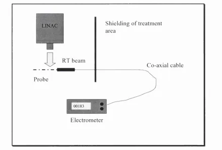
Fig. 3.1 Scanditronix rectal probe with restricting collar 79
Fig. 3.2 Set-up for dosimetry experiments 80
Fig. 3.3 Schematic representation o f anorectal probe 85
Fig. 3.4 Diodes position in the anorectum on CT scannogram 86
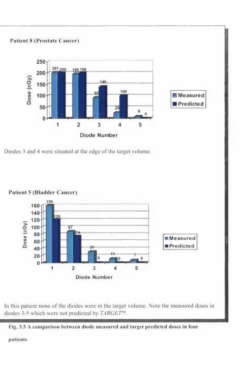
Fig. 3.5 Comparison between diode measured and TARGET predicted
doses in four patients (GRAPHS A,B,C,D) 90
Fig. 4.1 Changes In Proctitis and Incontinence Scores After RT 112
Fig. 4.2 GI Clinical Measurement Unit 113
Fig. 4.3 Change In Maximum Tolerable Volume After RT 122
Fig. 4.4 Change In Anal and Rectal Electrical Sensitivity After RT 123
Fig. 4.5 Scatter plot o f change in Incontinence Score before and 6
weeks after RT vs. rectal dose 127
Fig. 4.6 Scatter plot o f change in Proctitis Score before and 6 weeks
after RT vs. rectal dose 128
Fig. 4.7 Scatter plot o f change in Incontinence Score before and 6
weeks after RT vs. anal dose 129
Fig. 4.8 Scatter plot o f change in anal canal resting pressure before
and 6 months after RT vs. anal dose 130
Fig. 4.9 Scatter plot o f change in rectal electrical sensitivity before
and 6 weeks after RT vs. rectal dose 131
Fig. 4.10 Scatter plot o f change in maximum tolerable volume before
and 6 weeks after RT vs. rectal dose 132
Fig. 5.1 Endoanal ultrasound image 136
Fig. 5.2 Endoanal ultrasound probe 138
Fig. 5.3 Open magnet MRI scanner 143
Fig. 5.5 Anal enhancement curves after RT in one patient 147
Fig. 5.6 Mean anal canal enhancement in the lower and upper anal
canal after RT 148
Fig. 5.7 Mean gradient o f enhancement curves (1*^ 55 seconds)
after RT 149
Fig. 5.8 Scatter plot for change in rate o f anal canal enhancement
pre and 6/52 post RT and anal dose 152
Fig. 5.9 Scatter plot for change in degree o f anal canal enhancement
LIST OF TABLES
Table 1.1 Scoring System For Symptoms, Endoscopic and
Histological Results
Table 1.2 RTOG Acute Intestinal Toxicity Score
Table 1.3 RTOG Late Intestinal Toxicity Score
Table 1.4 Effects o f radiation on anorectal physiology
Table 2.1 Study Protocol
Table 3.1 Correction Factors For Individual Diodes
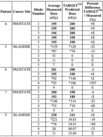
Table 3.2 Targef^ Predicted/Diode Measured Dosimetry Comparison
-Readings On 5 Fractions
Table 3.3 T argef^ Predicted/Diode Measured Dosimetry Comparison
-Readings On 2 Fractions
Table 3.4 HELA)C^ Predicted and Average Measured Dose with
Percentage Difference
Table 3.5 Summary o f anal and rectal doses
Table 4.1 Proctitis Score from Talley et al
Table 4.2 ‘Vaizey modification’ o f Wexner Incontinence Score
Table 4.3 Incontinence and Proctitis Score Results
Table 4.4 Prevalence o f Individual Proctitis Symptoms
Table 4.5 Prevalence o f Individual Incontinence Symptoms
Table 4.6 Manometry Results
Table 4.7 Rectal Volume Results
Table 4.8 Anal and Rectal Electrical Sensitivity Results
Table 5.1 Changes In Endosonographic Thickness O f Anal Canal Layers
After RT
Table 5.2 Degree o f anal canal enhancement before and after RT
Table 5.3 Rate O f Anal Canal Enhancement Over The 55 Seconds
Before and After RT
Table 5.4 Comparison o f upper and lower anal canal enhancement
STATEMENT OF ORIGINALITY
The studies described and the analysis o f the data in this thesis is the original work o f
the author. All studies took place at The Middlesex Hospital and were performed by
the author with the following exceptions:
1. Some dosimetric data were collected in conjunction with Miss Ursula Johnson
and Dr Derek D ’Sousa, Medical Physicists, Department O f Medical Physics.
2. All anorectal physiological studies were performed in conjunction with Miss
Elisa Wrightham, Clinical Scientist, Clinical Measurement Unit,
3. All endoanal ultrasound images were taken by Dr Chris Hare, Consultant
Radiologist, Department O f Radiology
4. Some MRI data were analysed in conjunction with Dr Chis Hare, Consultant
Radiologist, Department O f Radiology and Dr Andy Priest, MRI Medical
Physicist, Department O f Medical Physics.
5. The statistical tests used in this thesis have been discussed at length with the
statisticians o f the Research & Development Department o f the University
College Hospital NHS Trust, and in particular with Dr Joseph Eliahoo. All
statistical analysis was performed by the author using a combination o f
SPSS™ statistical software package and Microsoft Excel™
No part o f this work has been submitted to any other university for consideration
CONTENTS
Title page
1
Abstract
2
Acknowledgements
3
List o f abbreviations
4
List o f figures
6
List of tables
8
Statement o f originality
9
CHAPTER 1:
INTRODUCTION AND HISTORICAL REVIEW
1.1 BACKGROUND 20
1.2 RADIATION PHYSICS AND MECHANISMS
OF RADIATION INJURY 22
1.3 RADIOTHERAPY TREATMENT PLANNING 25
1.3.1 Patient Immobilization
1.3.2 Definition O f Treatment Volumes
1.3.3 Localization O f The Target Volume
1.3.4 Calculation O f Dose Distribution
1.3.5 Dosimetry
1.4 THERAPIES FOR UROLOGICAL MALIGNANCY 31
1.4.1 Prostate cancer
1.4.2 Radiotherapy for prostate cancer
1.4.3 Bladder cancer
1.4.4 Radiotherapy for bladder cancer
1.5 FUNCTIONAL DISTURBANCE AFTER
PELVIC RADIOTHERAPY 42
1.5.1 Clinical Features O f Rectal Complications
1.5.2 Clinical Features O f Anal Complications
1.5.3 Anorectal Injury And Incontinence
1.5.4 Anorectal Physiological Testing
1.6 STRUCTURAL CHANGES IN THE ANORECTUM AFTER
PELVIC RADIOTHERAPY 53
1.6.1 Histopathological Features In The Rectum
1.6.3 Imaging The Radiation Injured Anorectum
1.7 THERAPEUTIC OPTIONS AFTER RADIATION INJURY
TO THE ANORECTUM 59
1.7.1 Haemorrhagic Proctitis
1.7.2 Rectal Stricture
CHAPTER 2:
AIMS OF STUDY AND STUDY PLAN
2.1 INTRODUCTION 68
2.2 AIMS OF THE STUDY 73
2.3 PATIENTS AND STUDY PROTOCOL 74
2.3.1 Patient Selection
2.3.2 Study Protocol
CHAPTER 3:
ASSESSMENT OF ANORECTAL DOSE
3.1 INTRODUCTION 78
3.2 DEVELOPMENT OF DOSIMETRY TECHNIQUES 79
3.2.1 The anorectal probe in a simulated model
3.2.2 Calibrating the diodes
3.2.3 A problem with the dosimetry
3.3 COMPARISON OF TARGET™ PREDICTED AND DIODE
MEASURED DOSES IN PELVIC RADIOTHERAPY 84
3.3.1 Patients and methods
3.3.2 Results
3.3.3 Conclusions
3.4 COMPARISON OF HELAX^^ PREDICTED AND DIODE
MEASURED DOSES IN RADIOTHERAPY FOR PROSTATE
CANCER 93
3.4.1 Introduction
3.4.2 Patients and methods
3.4.3 Results
3.4.4 Conclusions
3.5 DETERMINATION OF ANORECTAL DOSE IN INDIVIDUAL
PATIENTS 97
3.5.1 Introduction
3.5.2 Methods
3.5.3 Results
3.5.4 Conclusion
3.6 DISCUSSION OF DOSIMETRY EXPERIMENTS 101
3.6.1 Probe Movement
CHAPTER 4:
FUNCTIONAL CHANGES IN THE ANORECTUM AFTER
RADIOTHERAPY
4.1 INTRODUCTION 105
4.2 ASSESSMENT OF ANORECTAL SYMPTOMS 107
4.2.1 Methods
4.2.2 Results
4.2.3 Conclusions
4.3 ANORECTAL PHYSIOLOGICAL STUDIES 113
4.3.1 Patients & Methods
4.3.2 Results
4.3.3 Conclusions
4.4 RELATIONSHIP OF ANORECTAL FUNCTION TO DOSES
RECEIVED 126
4.4.1 Methods
4.4.2 Results
CHAPTER 5:
STRUCTURAL CHANGES AFTER RADIOTHERAPY
5.1 INTRODUCTION 135
5.2 ENDOANAL ULTRASOUND OF THE ANAL CANAL
BEFORE AND AFTER RT 136
5.2.1 Background
5.2.2 Methods
5.2.3 Results
5.2.4 Conclusions
5.3 CONTRAST ENHANCED DYNAMIC MRI OF THE ANAL
CANAL BEFORE AND AFTER RT 141
5.3.1 Introduction
5.3.2 Methods
5.3.3 Results
CHAPTER 6:
DISCUSSION
6.1 FUNCTIONAL CHANGES AFTER RT 157
6.2 STRUCTURAL CHANGES AFTER RT 159
6.3 AETIOLOGY OF ANORECTAL DISTURBANCE AFTER RT 161
6.3.1 Neurological Injury
6.3.2 Reduced Rectal Capacity After Radiation Injury
6.3.3 Failure O f Correlation Between Dose And Post RT Changes
6.4 CONCLUSION OF THESIS 166
APPENDICES
Appendix 1. Patient Information Sheet
Appendix 2. Consent Form
Appendix 3. Patient Demographics
Appendix 4. Patient Completion
Appendix 5.a Comparison between TLD measured and diode measured doses in 3 patients
Appendix 5.b TARGET Predicted/Diode Measured Comparison -Readings On 5 Fractions
Appendix 5.c TARGET Predicted/Diode Measured Comparison -Readings On 2 Fractions
Appendix 5.d HELAX Predicted/Diode Measured Dosimetry Comparison
Appendix 5.e Histograms Showing Predicted Vs Measured Dose in 10 patients
Appendix 5.f Patient Dose Data Set
Appendix 6. Incontinence and Proctitis Score Results
Appendix 7. Manometry Results
Appendix 8. Rectal Volume Data
Appendix 9. Anal & Rectal Electrical Sensitivity Data
Appendix 10. Endoanal Ultrasound Results a) Summary
b) 3 o’clock
c) 9 o ’clock
176 177 178 179 180 181 182 183 185 189 190 191 192 193 194 195 197
Appendix 11. MRI Results a) Lower anal canal 199
b) Upper anal canal 200
CHAPTER 1:
INTRODUCTION
AND
1.1 BACKGROUND
Urological malignancies, particularly prostate and bladder cancer have shown a
dramatic increase in incidence over the last two decades [Majeed and Burgess, 1994].
Pelvic radiotherapy (RT) is a common treatment modality for these malignancies,
which is being increasingly adopted. Current knowledge o f the effects o f radiation on
the anorectum is based on a limited number o f studies. Variability in delivery
techniques both currently and historically, combined with a paucity o f prospective and
randomised studies makes interpretation o f the reported results difficult. This
introduction aims to present the existing evidence and to identify those areas that
require further work, some o f which will be addressed in this thesis.
The use o f radiation as a medical therapy is a relatively recent development. Wilhelm
Rontgen discovered x-rays in 1895. The first therapeutic use for x-rays was reported
in 1897 by a German surgeon called Wilhelm Freund. He used x-rays to induce
regression o f a hairy mole and presented his work to the Vienna Medical Society. In
1901 Pierre Curie deliberately used a radium tube to produce an ulcer on his arm and
charted its progress and ultimate healing [Hall, 1993]. From these early beginnings
the study o f radiobiology and radio-therapeutics began. Modem external beam RT
was bom in the 1950’s with the development o f mega-voltage linear accelerators.
Radiation therapy now has a major role in the treatment o f a number o f malignancies
arising in the pelvis. Carcinomas o f the prostate, bladder, rectum and gynaecological
Rectal injury after pelvic RT is well documented [Mathes and Alexander, 1996] and
minimizing the rectal dose is an important issue for the radiotherapist [Brizel, 1998].
Anal canal injury and subsequent dysfunction receives scant attention in the literature.
1.2 RADIATION PHYSICS AND MECHANISMS OF RADIATION INJURY
Therapeutic radiation is known as ionising radiation due to its effects on cellular
processes through the ionisation o f intracellular molecules. This ionisation o f
intracellular molecules, in turn affects the processes o f cell division. Ionising radiation
may be classified as particulate radiation or electromagnetic radiation. Particulate
radiation consists o f subatomic particles including electrons, neutrons, protons and
alpha particles. At present only electrons are used in radiotherapy and only for limited
indications, usually skin lesions due to their poor penetration. Electromagnetic
radiation refers to x-rays or gamma rays. These are physically identical but the two
names are used to distinguish their means o f production. Both can be described in
terms o f ‘rays’, or as individual packets o f energy (photons). Gamma rays are
produced from the decay o f radioactive isotopes, whereas x-rays are usually produced
artificially by accelerating electrons to a high energy and stopping them abruptly with
a heavy metal target. Part or all o f this kinetic energy is converted into x-rays. The
energies required to produce x-rays capable o f penetrating tissue are in the
megavoltage (MV) range and are produced by machines known as a linear
accelerators (LINACs). The higher the energy o f the x-rays, the greater the degree o f
penetration into the body, and the less they interact with superficial tissues. In the
treatment o f urological tumours energies o f greater than 8MV are required. This
process is called teletherapy, but is most commonly referred to as extemal beam
radiotherapy.
When x-rays collide with body tissues they produce fast electrons that ultimately
different absorption mechanisms that are largely dependent on the energy o f the
incident x-rays. Low energy x-rays (diagnostic x-rays) are absorbed by a process
known as the photoelectric effect. The incident photon interacts with an inner orbital
electron shell o f an atom. The absorbed energy causes an electron (known as a fast
electron) to be ejected from its orbit shell, which then goes on to ionise other atoms,
breaking chemical bonds in cellular DNA. The x-rays most commonly used for
radiotherapy are in the 1-lOMV range. This energy o f photon generally produces an
absorption interaction known as the Compton effect. The effect is similar to the
photoelectric effect, in that energy is given up resulting in release o f a fast electron.
However, the photon is not absorbed, but deflected from its original path and
proceeds through the tissues resulting in further interactions and fast electron
productions. At treatment energies in excess o f lOMV pair production predominates.
The incident photon interacts with the nucleus o f the atom, giving up all its energy in
the process. This results in production o f a positron and a fast electron. The fast
electron results in ionisation as before. The positron collides with adjacent electrons
and is rapidly annihilated with the creation o f two new photons both capable o f
causing subsequent ionisations.
The resultant biological damage produced by fast electrons occurs directly by
ionisation and breaking o f chemical bonds in cellular DNA and indirectly by
interaction with biological molecules to produce free radicals. These free radicals, in
particular hydroxyl, are highly reactive molecules, which can diffuse far enough to
reach and damage cellular DNA. A single strand DNA break may be recognised and
repaired using the complimentary strand as a template. When several ionisations
irreparable lesion and the damage caused prevents normal mitosis from occurring,
resulting in cell death during attempted division. Despite DNA damage most cells
continue to carry out their other normal physiological functions until mitosis
occurs[Tubiana M, 1999;Hall et al., 1988]. In early responding tissues such as skin,
mucosal epithelium and bone marrow, radiotherapy damage is rapidly apparent,
usually within the course o f RT itself. Late responding tissues (e.g. neurological
tissue) may not show the effects o f RT for months or even years after treatment. This
explains delayed RT damage in some tissues.
In the clinical setting radiation is usually delivered in ‘fractions’ o f the total treatment
dose over several weeks. This fractionation has a number o f advantages, namely
repopulation, repair, reassortment and reoxygenation. Fractionation allows rapidly
proliferating normal tissues to recover cell mass between fractions (repopulation).
Normal tissues that are dividing slowly are able to repair DNA damage more
effectively than tumour (repair). Fractionation also allows more tumour cells to move
out o f the relatively protected S phase o f the cell cycle into the G2 and M phases o f
the cell cycle making them vulnerable to radiation (reassortment). Finally,
revascularisation o f hypoxic areas in a tumour mass between fractions, results in their
reoxygenation, enhancing their radiosensitivity [Mould RF, I98I];[H uddart RA,
1.3 RADIOTHERAPY TREATMENT PLANNING
The initial phase o f radiotherapy is treatment planning. This involves patient position
and immobilization, definition o f treatment volumes, choice o f technique and
calculation o f dose distribution. Dosimetry is the assessment o f dose distribution.
1.3.1 Patient Position And Immobilization
Accurate patient set-up on the planning machine is achieved by aligning bony
landmarks using wall-mounted lasers and then tattooing the patient at three sites.
These tattoos are then aligned with wall-mounted lasers on the treatment machines to
ensure patients are in an identical position for each fraction o f RT once treatment
begins. The patient is always planned and treated supine and with the bladder full for
prostate cancer patients and empty for bladder cancer patients.
1.3.2 Definition O f Treatment Volumes
When delivering RT, parameters such as volume and dose have to be specified for the
purposes o f prescription, recording and reporting. This has been standardised by the
International Committee o f Radiation Units and Measurements -IC R U (1993)
allowing international comparisons o f various RT treatments. The report defines the
Gross Tumour Volume (GTV), Clinical Target Volume (CTV) and Planning Target
Volume (PTV). The GTV is the demonstrable macroscopic extent o f the tumour. To
encompass sub clinical or microscopic disease a margin is added to the GTV; this is
the CTV. The PTV is a further margin added to the CTV to allow for internal organ
1.3.3 Localization O f The Target Volume
The target volume may be localized using plain x-rays on a machine called a
simulator (conventional planning). Alternatively a dedicated spiral CT scanner with
an integrated computer planning system (CT Planning) can be used. The area to be
treated is scanned, in the case o f prostate cancer from the sacro-iliac joints to the
below the inferior pubic rami. The radiation oncologist then outlines the GTV and
may also outline other areas o f interest such as the rectum using dedicated planning
software. The clinical target volume and planning target volume are then added. The
increase in margin to allow for microscopic disease and internal movement are then
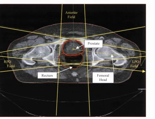
added. An example o f a planning CT (central slice) with the CTV and rectum outlined
Prostate
Rectum Femoral
Head
Fig. 1.1 Central P lanning C T Slice (Prostatic C a rcinom a )
K E Y :
RPO = Right Posterior O blique Field
LPO = Left Posterior O blique Field
— = Planning Target V olu m e
1.3.4 Calculation O f Dose Distribution
The calculation o f dose distribution is performed by medical physicists. The
arrangement o f the radiation fields and their relative contribution to the total dose,
facilitated by a Treatment Planning System are determined and then the planning
images are processed. A Treatment Planning System is a computer program to help
calculate the expected doses at any given point in the pelvis accounting for changes in
the energy o f the radiation field as it passes through different structures such as bone
and soft tissue as well as adjusting for complex photon interactions and scatter. Two
Treatment Planning systems are referred to in this thesis. TARGET™ , which is an
older treatment planning system, utilises a two-dimensional calculation o f dose based
on measured beam data, and HELAX™, calculates dose using a sophisticated three-
dimensional photon interaction model, which fully includes radiation scatter.
1.3.5 Dosimetry
Dosimetry is the quantitative measurement o f radiation dose. This usually involves
physical measurements although computerised simulations and dose predictions are
frequently referred to as dosimetry. Dosimetry can be used as a form o f quality
control, which is usually performed by medical physicists. The LINACs themselves
are frequently evaluated to ensure that they are accurately producing the expected
doses o f radiation. There are a number o f devices used for assessing radiation dose.
An ionisation chamber is considered the gold standard although a number o f other
electronic devices exist. Devices called thermo-luminescent dose-meters (TLDs),
which are pieces o f lithium fluoride, are frequently used. TLDs are exposed to
radiation and then heated, which results in light being produced. The degree o f
amount o f radiation received. Other devices such as radiation detecting diodes can
also provide dosimetric data. Silicon diodes contain a sandwich o f a second metal that
acts as an electron donor or recipient. The disparity in electron number provides a
potential difference across the diode. P type diodes are positive (less electrons) and N
type are negative (more electrons). As photons o f radiation collide with the diodes
they result in ionisation and liberated electrons flow as current driven by the potential
difference described above. This current flows to an electrometer and is registered as
a count. The count is directly proportional to the number o f photons hitting the diodes
and hence the radiation dose. Diodes are in many ways superior to TLDs because,
once calibrated, they can provide an immediate and continuous reading o f dose.
Radiation doses to specific organs can be estimated from radiotherapy planning data.
Presenting a dose to a specific organ (such as the rectum) in a clinically meaningful
way can be achieved by using a dose volume histogram (DVH). The DVH plots the
percentage volume o f a specific organ against the percentage o f the prescribed
radiation dose received. The DVH has become accepted amongst radiotherapy
oncologists as useful tool for treatment plan evaluation [Cheng and Das, 1999] and has
more clinical relevance than a single dose at any given point, because the volume o f
an organ that is irradiated to a certain degree is displayed graphically.
Under some circumstances dosimetry is carried out in-vivo. This means that radiation
doses are assessed during the actual treatment o f a patient using TLDs or diodes. Most
commonly a skin dose is measured, although dosimetry within body cavities is
The SI unit o f radiation dose is the Gray. One Gray is equal to one joule o f energy
absorbed per kilogram o f mass. The magnitude o f a Gray in clinical terms is realised
from the following dose examples. A total body CT is a dose o f about 0.01 Gray.
Abortion would be considered if a foetus received a radiation dose o f greater than 0.2
Gray. The tolerance dose o f the rectum (rectal tolerance dose TD is 45-50Gy,
which is defined as the dose expected to result in a serious rectal complication in 5%
1.4 THERAPIES FOR UROLOGICAL CANCER
1.4.1 Prostate Cancer
Prostate cancer is the second commonest cause o f death from cancer in men and in
1998 was responsible for 8570 deaths in England and Wales [Majeed et al, 2000]. The
directly age-standardized incidence (i.e. accounting for an increasing elderly
population) has more than doubled in the last ten years with a corresponding increase
in prostate cancer deaths. There has been no improvement in 5-year survival for cases
diagnosed since 1985 when compared with preceding decades [Majeed et al., 2000]. It
has been estimated that 30% o f American men aged over 50 have histological
evidence o f prostate cancer [Whitmore et al.,1973 ]. However, the lifetime risk o f
developing clinically apparent disease is 10% with a 3% lifetime risk o f mortality
from prostate cancer.
Prostate cancer is an adenocarcinoma in more than 90% o f cases and arises in the
peripheral zone o f the prostate in 75% o f cases. In the early stages patients may be
asymptomatic or have symptoms o f urinary outflow obstruction as in benign prostatic
hypertrophy. Asymptomatic patients can present either as an incidental finding
following abnormal digital rectal examination or after prostatic specific antigen (PSA)
measurement. Those patients with lower urinary tract symptoms may also have PSA
estimation or prostatic cancer may be an incidental finding in the histological
examination o f the specimen following trans urethral resection o f the prostate
(TURP). In the late stages skeletal pain from bony métastasés, nodal disease and
Localised prostate cancer refers to disease confined within the capsule o f the prostate
without lymph node or metastatic spread. This equates to a TNM classification o f T2
NO MO or less.
TNM Classification of Prostate Cancer
Primary tumour (T)
TX: Primary tumour cannot be assessed TO: No evidence o f primary tumour
T 1 : Clinically unapparent tumour not palpable nor visible by imaging T la: Tumour incidental histological finding in 5% or less o f tissue resected T ib : Tumour incidental histological finding in more than 5% o f tissue
resected
T ic: Tumour identified by needle biopsy T2: Tumour confined within prostate T2a: Tumour involves 1 lobe
T2b: Tumour involves both lobes
T3: Tumour extends through the prostatic capsule T3a: Extra capsular extension
T3b: Tumour invades seminal vesicle(s)
T4: Tumour is fixed or invades adjacent structures other than seminal
vesicles: bladder neck, external sphincter, rectum, levator muscles, and/or pelvic wall
Regional lymph nodes (N)
Regional lymph nodes are the nodes o f the true pelvis, are the pelvic nodes below the bifurcation o f the common iliac arteries.
Distant lymph nodes are outside the confines o f the true pelvis and their involvement constitutes distant metastasis.
NX: Regional lymph nodes cannot be assessed NO: No regional lymph node metastasis
N 1 : Metastasis in regional lymph node or nodes
Distant metastasis (M)
MX: Distant metastasis cannot be assessed MO: No distant metastasis
M 1 : Distant metastasis
M ia: Non regional lymph node(s) M lb: Bone(s)
M lc: Other site(s)
H istopathological g rad e (G)
GX: Grade cannot be assessed
G 1 : Well differentiated (slight anaplasia)
G2: Moderately differentiated (moderate anaplasia)
G3-4: Poorly differentiated or undifferentiated (marked anaplasia)
However, a different histopathological grading system is commonly adopted that has
been shown to provide prognostic information. The Gleason system [Gleason DF et
al., 1974] relies on the low-power microscopic appearance o f the glandular
architecture o f the prostate. A primary grade (1-5) is assigned to the most commonly
observed glandular architecture and a secondary grade (1-5) to the second most
commonly occurring glandular architecture in the specimen. The Gleason score or
Gleason sum is obtained by adding the primary and secondary grades thus Gleason
sums range from 2 to 10. Well-differentiated tumours score 2-4, moderately
differentiated score 5-6 and poorly differentiated score 8-10. Gleason score 7 have
been grouped as both poorly and moderately differentiated. As the primary grade is
the more important in terms o f prognosis Gleason score 7 tends to be graded as 4+3
(worse prognosis) or 3+4 (better prognosis).
The treatment options are controversial as the natural history o f the disease is not fully
understood. Post mortem evidence shows early prostatic cancer is far more common
than clinical prostatic cancer thus many men develop localised prostate cancer which
never presents in their lifetime [Whitmore et al., 1973]. Localised prostate cancer is
amenable to cure both with surgery or radical RT. However, both cause significant
morbidity and which modality o f treatment is the more appropriate in any given
advocates. Based on the current evidence there is little to choose between these
treatment options.
Radical prostatectomy has a ten-year survival o f 90% and a disease-free survival o f
75%, although long-term morbidity is significant [Wilt TJ et al., 1998]. Urinary
incontinence rates after radical prostatectomy are acceptable; total urinary
incontinence is rare (<3%) but stress incontinence may occur in up to 20% o f patients.
The return o f urinary continence is gradual over the first year after surgery [Steiner
MS et al., 1991]. Impotence rates may reach 50% [Moffat L., 2000], although nerve
sparing prostatectomy when feasible has made the operation more acceptable [Wilt TJ
et al., 1998]. Preservation o f potency varies as a function o f age. In men under the age
o f 60 reported rates o f potency are 40-82% when both neurovascular bundles are
preserved dropping to 20-60% if only one nerve is preserved. Recovery o f sexual
function also occurs in the first year [Walsh et al., 1994].
External beam RT offers a disease-specific survival o f 76% at 10 years in T1 and T2
prostate cancer [Leibel SA et al., 1996 ]. In locally advanced disease local control and
probably survival after radiotherapy is further improved with adjuvant androgen
blockade [Mason MD et al., 2000 ]. Significant urinary disturbance following external
beam RT for prostate cancer is uncommon [Hamilton et al., 2001]. Gastrointestinal
complications following radiotherapy are common and are discussed in detail (1.5).
The watch and wait policy was reported in two studies involving untreated organ-
confined prostate cancer. The 10-year survival was described as 85%, but patients
To determine the optimal approach for these patients a prospective randomised trial is
required. The MRC randomised trial o f radiotherapy versus surgery versus no
immediate treatment in early prostate cancer (PR06) was discontinued in 1996 due to
recruitment difficulties. There are two similar ongoing studies; The Prostate Cancer
Intervention Versus Observation Trial (PIVOT) in North America and a smaller
Swedish study, the results o f which are keenly awaited [Wilt TJ et al., 1998]. With a
lack o f scientific evidence treatments tend to be offered based on the patient or the
clinician’s preference. Radiotherapy is traditionally offered to an older patient group,
or those who are unwilling or deemed unfit to undergo major pelvic surgery.
However, until more information becomes available the uncertainty about the best and
most appropriate treatment modality remains.
1.4.2 Radiotherapy Techniques For Localised Prostate Cancer
RT for localised prostate cancer is most commonly delivered by external beam using
a three-field technique, usually with an anterior and two lateral (or posterior oblique)
beams. This provides the maximum dose to the target volume whilst allowing
minimal doses to adjacent radiosensitive areas such as the rectum and the femoral
heads [Dobbs J, 1985]. 64Gy given as 32 fractions o f 2Gy over a six-week period is a
Fig. 1.2 P atien t receivin g pelvic rad ioth erap y
T hree-dim ensional conform a! radiation therapy (3D C R T ) and intensity-m odulated
radiation therapy (IM R T) are new er techniques w hich are b eing adopted, although
these are currently o n ly being used at a few centres in the UK. T hree-dim ensional
conform ai radiation therapy and IMRT allow d ose distributions that conform c lo sely
to the three-dim ensional shape o f the target volu m e w h ile m in im izin g d ose to adjacent
structures [V erhey, 1999]. H ow ever, a benefit o f 3D C R T and IMRT is to a llow dose
escalation to the target volu m e but this m ay result in surrounding areas receivin g
sim ilar d oses to traditional field set-ups.
R adioactive prostatic im plants are an attractive alternative to external beam RT.
Pasteau im planted the first radium needles in 1910. In the I 9 7 0 ’s open retro-pubic
im plantation o f iodine seeds w as carried out. D ue to operative m orbidity and poor
introduction o f Trans Rectal Ultrasound (TRUS) and template guidance allowed
trans-perineal insertion o f needles and much more accurate positioning o f the seeds
[Holm et al., 1983]. The current options are iodine or palladium seeds that have the
advantage o f rapid dose fall o ff according to the inverse square law effect and short
range o f emission, in theory providing high doses to the target volume without
affecting the surrounding structures. The procedure has the advantage that it can be
performed as a single outpatient procedure and is reported to cause less morbidity
than external beam RT. Seed implantation does however require live radioactive
sources and is only carried out in specialist centres. Control o f disease as defined by
PSA level, has been reported in 98% o f patients at 2 years and 76% at 5 years.
Irritative urinary symptoms are common but the risk o f urinary incontinence is less
than 0-1% unless the patient had recently undergone TURP, when this rises to 50%.
Recent TURP is therefore now considered a contraindication for this treatment
modality [Holm, 1997].
High-dose rate brachytherapy is another method o f dose escalation. A brachytherapy
boost (e.g. 16.5 Gy in 3x 5.5Gy fractions) is received in addition to a shorter course o f
fractionated external beam RT (e.g. 46Gy in 23fractions over 4V2 weeks). Hollow
rods are positioned in the prostate via a trans-perineal route under trans-rectal
ultrasound guidance. The brachytherapy source can then be introduced via the hollow
rods into the prostate. Remote after-loading systems allow post-implant manipulation
o f source position and treatment duration to optimise the dose distribution within the
target volume. The isotope used is iridium-192 which has higher emission energies
than iodine or palladium resulting in higher rectal and bladder doses than seed
suitable for bulkier tumours. High dose rate monotherapy involving giving the whole
RT dose via high emission energy brachytherapy is being investigated at a few centres
although results are not yet available [Duchesne GM, 2001].
1.4.3 Bladder Cancer
Bladder cancer is the fourth commonest malignancy in men and the ninth commonest
in women. Between 1971 and 1997 the total number o f cases o f bladder cancers in
England and Wales has increased by 66.7% from 7245 to 12080 [Arya M, Hayne D,
et al., 2001]. The 5-year survival in the UK has been improving although this may
largely be related to improved diagnosis and treatment o f early lesions. Bladder
cancer typically presents with painless haematuria, irritative urinary symptoms,
abdominal pain or with metastatic disease. The standard investigations for any patient
with haematuria, microscopic or overt, are cystoscopy, urine cytology and either
intravenous urography or a plain abdominal x-ray with a renal ultrasound.
Transitional cell carcinoma accounts for more than 90% o f all cases o f bladder cancer,
80% o f which are superficial with a low rate o f progression to invasive disease. The
full TNM staging o f transitional cell carcinoma o f the bladder is described below.
TNM Staging For Bladder Cancer
Primary tumour (T)
TX: Primary tumour cannot be assessed TO: No evidence o f primary tumour Ta: Non-invasive papillary carcinoma Tis: Carcinoma in situ: "flat tumour"
T1 : Tumour invades subepithelial connective tissue
T2: Tumour invades muscle T2a: Tumour invades superficial muscle (inner half)
T2b: Tumour invades deep muscle (outer half) T3: Tumour invades perivesical tissue
T3b: macroscopically (extravesical mass)
T4: Tumour invades any o f the following: prostate, uterus, vagina, pelvic wall, or abdominal wall.
T4a: Tumour invades the prostate, uterus, vagina T4b: Tumour invades the pelvic wall, abdominal wall
Regional lymph nodes (N)
Regional lymph nodes are those within the true pelvis; all others are distant lymph nodes.
NX: Regional lymph nodes cannot be assessed NO
N1 N2
No regional lymph node metastasis
Metastasis in a single lymph node, 2 cm or less in greatest dimension Metastasis in a single lymph node, >2 cm but <5 cm; or multiple lymph nodes
N3: Metastasis in a lymph node more than 5 cm in greatest dimension
Distant metastasis (M)
MX: Distant metastasis cannot be assessed MO: No distant metastasis
M 1 : Distant metastasis
Superficial bladder cancer (Ta, T1 and Tis) is managed differently from muscle
invasive bladder cancer (T2-T4). Superficial bladder cancer is initially treated with
transurethral resection. In patients with low-grade Ta tumours, cystoscopy at regular
intervals and repeat resection or cystodiathermy as necessary is usually adequate. For
multiple or frequent recurrences and those with high grade or T1 tumours at
presentation, intravesical immunotherapy (e.g. BCG) or chemotherapy (e.g.
mitomicin or epirubicin) is generally employed. The 5-year survival for superficial
disease is around 80%. Muscle invasive bladder cancer (T2-T4) requires radical
cystectomy with urinary diversion or radiotherapy. Radical cystectomy involves
bilateral pelvic lymphadenectomy and wide excision o f the bladder and prostate
including the urachal remnant, the overlying peritoneum, and the vascular pedicles. In
posterior vesical pedicles is indicated and does not compromise cancer control. In
women radical cystectomy may also involve anterior pelvic exenteration including
uterus, fallopian tubes, ovaries, anterior vaginal wall, complete excision o f the urethra
and urinary diversion. In a long term follow up study o f 1054 patients, the 10-year
recurrence-free survival in patients with muscle invasive lymph node-negative
tumours was 78% in T2 and 76% in T3a, 61% for T3b and 45% in T4 tumours [Stein
et al., 2001]. Unfortunately, a significant proportion o f patients (24% in the above
series) have lymph node involvement at the time o f surgery.
Multi-modality bladder preservation therapies involving transurethral resection
followed by planned combination chemotherapy and radiotherapy are employed in
selected patients. The outcomes o f such bladder preservation therapies may be similar
to those reported in a like population treated with radical cystectomy. The major
benefit in conservatively treated patients is a functioning bladder in 50% o f
cases.[Petrovich et al., 2001].
1.4.4 Radiotherapy For Bladder Cancer
External beam RT for bladder cancer is administered using a similar regime to
prostate cancer, although the patients are planned with the bladder empty. Higher
stage tumours unsurprisingly have a worse prognosis. Higher-grade tumours have a
better initial response to RT but the chance o f distant métastasés is greater resulting in
poorer survival. Shelly et al reviewed the randomised controlled trials assessing
surgery versus radiotherapy for muscle invasive bladder cancer. Three randomised
trials comparing pre-operative radiotherapy followed by radical cystectomy (surgery)
eligible for assessment. These trials represented a total o f 439 patients, 221
randomised to surgery and 218 to radical radiotherapy. The mean overall survival
(intention-to-treat analysis) at 3 and 5 years were 45% and 36% for surgery, and 28%
and 20% for radiotherapy, respectively. The results were also significantly in favour
o f surgery at 3 years and at 5 years when analysed on a treatment received basis
suggesting an overall survival benefit with surgery. However, only three trials were
included for analysis and many patients did not receive the treatment they were
randomised to. It must also be noted that many improvements in both radiotherapy
1.5 FUNCTIONAL DISTURBANCE AFTER PELVIC RADIOTHERAPY
1.5.1 Clinical Features O f Rectal Complications
More than 75% o f patients [Counter et al., 1999;Sedgwick et al., 1994] undergoing
pelvic RT will experience some rectal symptoms during the treatment period [Perez et
al., 1999] and these can be so severe as to interrupt treatment [Sandeman, 1980]. This
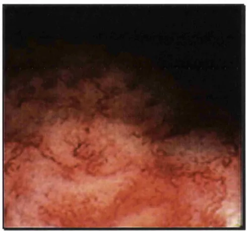
is acute phase radiation proctitis; late phase radiation proctitis describes proctitis
presenting or persisting 3 months after completion o f RT. Late phase radiation
proctitis is also described as chronic radiation proctitis. Radiation-induced proctitis
causes significant rectal bleeding in 6-8% o f patients [Silva et al., 1999] as a result o f
mucosal friability and neovascular telangiectasias [Taylor et al., 1993]. Proctitis can
present with a number o f other distressing symptoms including rectal pain, diarrhoea,
tenesmus, faecal frequency and urgency.
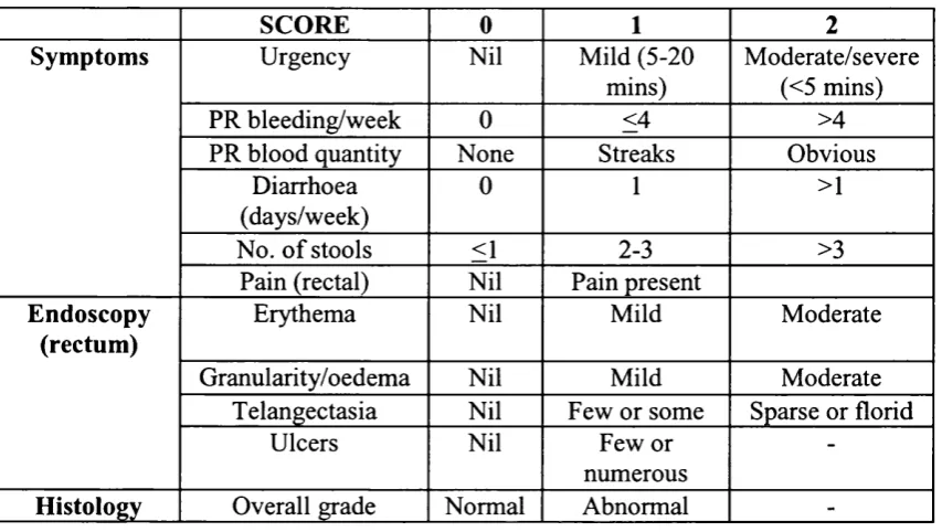
Proctitis can be graded on reported symptoms, endoscopic appearance and histology.
Talley et al (1997) described a scoring system that provided an objective
measurement o f proctitis. (Table 1.1)
Wachter et al (2000) described a more detailed endoscopic scoring system for post
radiation changes seen in the rectum. Neither o f these systems has been widely
adopted. The Radiation Therapy Oncology Group(RTOG) scoring system for acute
Table 1.1 Scoring System For Symptoms, Endoscopic and Histological Results
SCORE 0 1 2
Symptoms Urgency Nil Mild (5-20
mins)
Moderate/severe (<5 mins)
PR bleeding/week 0 <4 >4
PR blood quantity None Streaks Obvious
Diarrhoea (days/week)
0 1 >1
No. o f stools <1 2-3 >3
Pain (rectal) Nil Pain present
Endoscopy (rectum)
Erythema Nil Mild Moderate
Granularity/oedema Nil Mild Moderate
Telangectasia Nil Few or some Sparse or florid
Ulcers Nil Few or
numerous
-Histology Overall grade Normal Abnormal
-PR= per rectum
Table 1.2 RTOG Acute Intestinal Toxicity Score ORGAN
TISSUE 0 GRADE 1 GRADE 2 GRADE 3 GRADE 4 G RADES
LOWER G.I INCLUDING PELVIS No change Increased frequency or change in quality o f bowel habits not requiring medication/ rectal discomfort not requiring analgesics Diarrhoea Diarrhoea requiring parenteral requiring support/ severe parasympatholytic mucous or
drugs (e.g.. blood
Lomotil)/ mucous discharge discharge not necessitating necessitating sanitary sanitary pads/ pads/abdominal
rectal or distension (flat
abdominal pain plate
requiring radiograph
analgesics demonstrates distended bowel loops)
Acute or sub acute obstruction, fistula or perforation; GI bleeding requiring transfusion; abdominal pain or tenesmus requiring tube decompression or bowel diversion Death directly related to RT
The long-term sequlae following pelvic RT can be disabling. It has been shown that
pelvic irradiation causes widespread persistent disturbance o f gastrointestinal function
fat, bile acids and vitamins [Yeoh et al., 1993a]. Other permanent effects may take
months or even years to become apparent[Anseline et al., 1981], the peak incidence
for late complications occurring between two and five years after RT [Perez et al.,
1994]. For example, patients with radiation induced rectal stricture often present with
obstruction a number o f years after RT. Obstructive symptoms, therefore, should not
be assumed to be secondary to incurable malignancy as these patients may be
amenable to surgery [Schofield et al., 1983]. In a series reported by Lucarotti et al
(1991), a patient presented with late rectal complication 30 years after receiving
pelvic RT. Irradiated tissue has vastly reduced regenerative properties and relatively
trivial traumatic or infective insults can result in severe tissue breakdown, leading to
problems when operating in an irradiated field [Gilinsky et al., 1983].
The precise incidence o f late radiation-induced injury depends on the delivery
technique, combination with chemotherapy and patient factors including co-existing
diseases such as diabetes[Greven et al., 1991];[Herold et al., 1999]. In a series o f 738
patients treated with external beam RT for prostate cancer, there was a cumulative 10-
year incidence o f 8% o f moderate intestinal injury such as proctitis, enteritis and
anorectal stricture and a 3% incidence o f severe intestinal injury such as severe
proctitis, small bowel obstruction and fistula formation [Perez et al., 1994]. Another
large retrospective study reported long-term toxicity in 199 men treated with radical
RT for localized prostate cancer. In the rectum, toxicity o f grade 2 or above according
to the RTOG score was seen in 10 (5%) patients [Maartense et al., 2000]. In a further
study by [Gerard et al., 1999] o f 231 patients treated with curative intent external
beam RT for carcinoma o f the prostate, nine (4%) patients developed severe acute
colostomy and thirty-three (14.3%) patients had rectal bleeding, although only 7 (3%)
required local treatment. Anorectal function after RT, according to the Memorial
Sloan Kettering Cancer Centre scoring system was excellent in 90% o f patients with
a median follow up o f 5 years. In a prospective long-term follow up (mean follow up
time o f 46 months) study after RT for prostate cancer Borghede et al (1997) reported
mild gastrointestinal complications in 42% o f patients. Only 16 (9%) patients had
moderate or severe complications. Interestingly, the risk o f complications strongly
correlated with the presence o f intestinal symptoms prior to treatment. Boersma et al
(1998) investigated late gastrointestinal (GI) complications after conformai
radiotherapy for localized prostate cancer in 130 patients. Intestinal complications
were classified using the RTOG/European Organisation for Research and Treatment
o f Cancer (EORTC) and the Subjective Objective Management Analysis / Late
Effects Normal Tissue Taskforce (SOMA/LENT) scoring systems. The incidence at 2
years for GI complications o f Grade 2 or greater was 14% and 20% for the
(RTOG/EORTC) and (SOMA/LENT) scores respectively. Dose volume histogram
(DVH) parameters did not identify risk groups for late complications. No significant
correlation was found between any o f the DVH parameters and the actuarial incidence
o f complications. However, a trend was observed that a total radiation dose above 74
Gy resulted in a higher incidence o f severe rectal bleeding.
An increased incidence o f rectal cancer is reported in the long term following pelvic
RT[Kleinerman et al., 1995]. MacMahon and Rowe, (1971) showed that early and late
radiation proctitis, rectal stenosis and induration o f the rectovaginal septum
constituted risk factors for development o f secondary cancer. Jao et al (1987) reported
histological evidence o f a radiation reaction around the cancer but only 17% had
presented with symptoms o f proctitis. Denman et al., (1978) suggested high radiation
dose and severe radiation damage are not essential for radiation-associated colorectal
cancer. This was supported by animal studies that involved irradiating the descending
colon o f rats with a range o f doses from 25-65Gy. The dose producing the maximum
number o f tumours was 45Gy. Due to these risks Cohen and Winawer (2000)
recommended surveillance for rectal cancer, beginning five years after pelvic RT even
in the absence o f clinical symptoms o f proctitis.
1.5.2 Clinical Features O f Anal Complications
Anal discomfort can occur in the acute phase after RT and may be compounded by
radiation-associated diarrhoea. The dose received by the anal canal depends on its
proximity to the target volume. Injury to the anal sphincter complex after pelvic RT
has been reported although evidence is indirect and is mainly based on observed
functional disturbances[Varma et al., 1986].
1.5.3 Anorectal Injury And Incontinence
Anal continence is dependant on stool consistency, bowel activity, sphincter function
[Engel et al., 1995] rectal compliance and rectal capacity [Cherry and Rothenberger,
1988]. Pelvic RT can affect all these factors [Yeoh et al., 1993a] thus impairing
continence. The reported incidence o f faecal incontinence following pelvic RT ranges
from 0-27% [Iwamoto et al., 1997]. The wide variation may be explained by
differences in target site, patient mix, delivery techniques, dose received and the
vigilance in symptomatic assessment. Furthermore, much o f the RT morbidity data
anal/rectal pain, faecal urgency and faecal incontinence (Table 1.3). Patients are
reluctant to report these symptoms and their omission from the scoring systems may
be a major cause o f under-reporting.
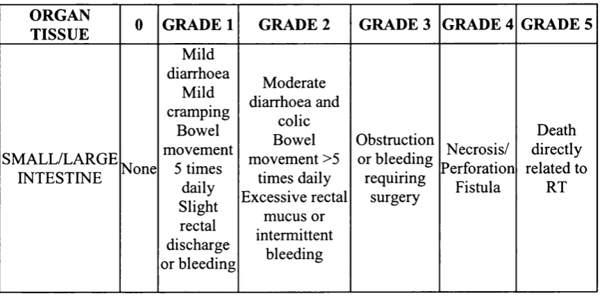
Table 1.3 RTOG Late Intestinal Toxicity Score Grade 1-5
ORGAN
TISSUE GRADE 1 GRADE 2 GRADE 3 GRADE 4 G RADES
SMALL/LARGE INTESTINE None Mild diarrhoea Mild cramping Bowel movement
5 times daily Slight rectal discharge or bleeding Moderate diarrhoea and colic Bowel movement >5 times daily Excessive rectal mucus or intermittent bleeding Obstruction or bleeding requiring surgery Necrosis/ Perforation Fistula Death directly related to RT
Hanlon et al drew further attention to the failings o f the RTOG Late Intestinal
Toxicity Score and stressed the importance o f the inclusion o f late chronic rectal
bleeding requiring multiple fulgurations [Hanlon et al., 1997]. This group reported
late rectal toxicity in 352 patients treated with external beam RT for non-metastatic
prostate cancer using three different morbidity scales. The median dose was 74 Gy
(range 63-81) and the median follow-up for these patients was 36 months (range 2-
76). The morbidity scales compared were: the RTOG, the Late Effects Normal Tissue
Task Force (LENT), and Hanlon et al’s own modification o f the LENT (FC-LENT)
complications by each scale was 0.7%, 2%, and 6% respectively. This clearly
demonstrated how widely different morbidity scales can vary and the importance o f a
meaningful uniformly agreed criteria.
The reported rate o f incontinence following external beam RT for anal carcinoma
varies widely from 0 to 57% [Papillon et al., 1987];[Doggett, Green, et al. 1988];
[Cummings, 1990];[Miller et al., 1991];[Tanum et al., 1991];[Martenson and
Gunderson, 1993];[Touboul et al., 1994];[Touboul et al., 1995]with an average
incidence o f about 25%. These patients receive 45-60 Gray directly to the anus, a
greater anal dose than that received in any o f the pelvic RT regimes for urological
malignancy. Were direct sphincter injury the main aetiology o f incontinence
following pelvic RT, one would expect a higher incidence o f incontinence in patients
receiving RT for anal carcinoma, than that which is reported.
1.5.4 Anorectal Physiology Studies
The results o f anorectal physiological studies after pelvic RT have been inconsistent
(Table 1. 4). The reported studies comprise retrospective and prospective data on
different groups o f patients undergoing different radio-therapeutic regimens
[Varma et al., 1986];[Bimbaum et al., 1992 ];[Bimbaum., et al. 1994];[Iwamoto et
Table 1.4 Effects of radiation on anorectal physiology
Author DXT Dose Target Organ Maximum anal canal resting pressure Squeeze increment Rectal threshold volume Maximum tolerated rectal volume
Varma et al
1985, 1986 50Gy Prostate V ► •
Birnbaum et al
1994 45Gy Rectum ► ► • •
Birnbaum et al
1996 45Gy Rectum ► ► • #
Yeoh et al
1996 44-50Gy Cervix 'W V V
Iwamoto et al
1997 8.5-9.5Gy Cervix Jk. ► • V
Kim et al
1998 44-54Gy Cervix ► V • V
Yeoh et al
1998 55-64Gy Prostate 'W yr V #
Yeoh et al
2000 55-64Gy Prostate ► 'W ► e
KEY:
►
#
Varma et al (1985,1986) studied a group o f 10 patients with chronic radiation proctitis
and incontinence. This group had significantly lower resting anal canal pressures and
a markedly reduced maximum tolerable rectal volumes when compared to
asymptomatic matched controls. Kim et al (1998) studied 24 patients with late
radiation proctitis following treatment for carcinoma o f the cervix. Again in
comparison to aged matched female controls the rectal threshold, urge and maximum
tolerable volumes were reduced, perhaps explained by reduced rectal compliance
related to radiation induced inflammation and fibrosis. The maximal squeeze pressure
was significantly reduced but the resting pressure was unchanged.
Bimbaum et al investigated the acute (1992) and chronic (1994) effects o f
preoperative RT for rectal cancer. No significant change in anal canal resting or
squeeze pressures was demonstrable four weeks after treatment in the 20 patients
initially investigated. In 10 o f these patients who were followed up for 14 to 42 (mean
35.5) months, the sphincter pressures remained unaltered. However due to variations
in the site o f the tumours within the rectum, patients would have received very
different doses to the anal canal, indeed the authors described the anal canal as being
in the target volume in only three o f the patients. Iwamoto et al (1997) published a
study investigating manometric changes during and six months after RT for cervical
cancer. A significant reduction in maximum tolerated rectal volume and in rectal
compliance was demonstrated. While there was no effect on anal squeeze pressures
the resting pressure was increased. However, these patients were mainly treated with
intracavitary RT and according to the authors, the dose to the anal canal was
negligible. They hypothesised that the increase in anal canal resting pressure may
the dose to the anal canal dose was negligible, anal canal oedema seems an unlikely
explanation. A response to diarrhoea stool might be a better explanation o f the finding
o f an increased anal canal resting pressure. However, anal canal resting pressure
should be a measure the involuntary tone o f the internal anal sphincter. An increase in
the measured value suggests that some external sphincter component was being
measured and a true resting pressure was not recorded.
Yeoh et al (1996) examined the prevalence o f anorectal dysfunction in a randomly
selected group o f 15 women who had received pelvic RT five to ten years previously.
When compared with controls there was a reduction in anal resting and squeeze
pressures, rectal compliance and maximum tolerable rectal volume. Fourteen o f the
15 patients had at least one physiological parameter o f anorectal function outside the
control range. Yeoh and his colleagues then undertook a prospective evaluation o f the
effects o f pelvic RT in 34 patients with prostatic carcinoma. Anorectal symptoms and
anorectal physiology were assessed before, four to six weeks (1998) and one year
(2000) after RT. After six weeks 19 (56%) had faecal frequency, 16 (47%) faecal
urgency and 8 (23%) patients had faecal incontinence. The basal and squeeze sleeve
recorded pressures were significantly reduced (54 v 49 mm Hg and 111 v 102 mm
Hg) before and after RT respectively. The rectal compliance was also reduced (1.2 v
1.4 mm Hg/ml). Those patients, who experienced urgency, were found to have a
lower threshold volume to rectal distension after RT.
One year after RT the anorectal disturbance persisted with 19 (56%), 17 (50%) and
incontinence, respectively. The threshold volume to rectal distension remained lower,