In schizophrenia, deficits in natural IgM isotype antibodies including those directed to
malondialdehyde and azelaic acid strongly predict negative symptoms, neurocognitive
impairments and the deficit syndrome.
Michael Maes, M.D., Ph.D. (a,b,c), Buranee Kanchanatawan, M.D. (a), Sunee Sirivichayakul, Ph.D.
(d), André F. Carvalho, M.D., Ph.D. (e,f)
a Department of Psychiatry, Faculty of Medicine, Chulalongkorn University, Bangkok, Thailand
b Department of Psychiatry, Medical University of Plovdiv, Plovdiv, Bulgaria
c IMPACT Strategic Research Center, Barwon Health, Deakin University, Geelong, Australia
d Faculty of Medicine, Chulalongkorn University, Bangkok, Thailand
e Department of Psychiatry, University of Toronto, Toronto, ON, Canada
f Centre for Addiction and Mental Health (CAMH), Toronto, ON, Canada
Corresponding author:
Prof. Dr. Michael Maes, M.D., Ph.D.
Department of Psychiatry
Faculty of Medicine
Chulalongkorn University
Bangkok
Thailand
https://scholar.google.co.th/citations?user=1wzMZ7UAAAAJ&hl=th&oi=ao
Buranee Kanchanatawan: [email protected]
Sunee Sirivichayakul: [email protected]
Abstract
Schizophrenia is characterized by an interrelated activation of the immune-inflammatory response
system (IRS) and the compensatory immune-regulatory reflex system (CIRS), which
downregulates the IRS. Deficit schizophrenia is characterized by a deficit in natural regulatory
autoimmune responses to tryptophan catabolites. The presence and correlates of IgM isotype
antibodies to oxidative specific epitopes (OSEs), nitroso (NO) and nitro (NO2) adducts in
schizophrenia remain unknown.
This study measured IgM antibodies to malondialdehyde (MDA), azelaic acid,
phosphatidylinositol, oleic acid, NO-tryptophan, NO-albumin, NO-cysteinyl and NO2-tyrosine in
a sample of 80 schizophrenia patients, divided into those with and those without deficit
schizophrenia, and 38 healthy controls.
Deficit schizophrenia was characterized by significantly lower IgM antibody levels to all OSEs as
compared with non-deficit schizophrenia and controls. Lowered IgM antibodies to MDA coupled
with increased IgM levels to NO-cysteinyl and NO2-tyrosine strongly predict deficit schizophrenia
versus non-deficit schizophrenia with an area under the ROC curve of 0.913. A large part of the
variance (21.2 – 42.2 %) in the negative symptoms of schizophrenia and excitation is explained
by IgM antibody titers to MDA (inversely) and NO-cysteinyl and/or NO2-tyrosine (both
positively). Lower IgM antibodies to MDA are significantly associated with impairments in
episodic memory including direct and delayed recall.
These findings further indicate that deficit schizophrenia is a distinct phenotype of schizophrenia,
which is characterized by lower natural IgM antibody levels to OSEs and relative increments in
antibodies of the IgM isotype attenuate CIRS functions and that this impairment may drive
negative symptoms and impairments in episodic memory and thus deficit schizophrenia.
Key words: immune, inflammation, natural IgM autoimmune, oxidative stress, kynurenine,
Introduction
In 1995, Smith and Maes [1] proposed the monocyte-T lymphocyte theory of
schizophrenia, which considered the role of activated immune-inflammatory pathways in the
neurodevelopmental pathology of schizophrenia through effects of prenatal infections, causing
increased oxidative and nitrosative stress (O&NS), cytokine-induced stimulation of the tryptophan
catabolite (TRYCAT) pathway, modulation of glutamate production and microglial activation.
Now, more than 2 decades later, there is abundant evidence that the different schizophrenia
phenotypes are characterized by activated immune-inflammatory processes in the peripheral blood
[2] and brain [3,4].
Thus, the acute (first episode psychosis and acute relapses) and more chronic phases of
schizophrenia (chronic, treatment resistant and stable-phase schizophrenia) are accompanied by
activation of the immune-inflammatory response system (IRS) as indicated by increased plasma
concentrations of acute phase proteins (APPs), complement factors and pro-inflammatory
cytokines and chemokines [2]. Those different schizophrenia phenotypes are characterized by
elevated levels of interleukin (IL-1)-1β, IL-6, IL-2, IL-12, IL-17, tumor necrosis factor (TNF)-α
and interferon (IFN)-γ, indicating activation of M1 macrophagic, T helper (Th)-1 and Th-17
immune cell phenotypes [2]. In accordance with M1 macrophagic activation, there are also data
that schizophrenia is accompanied by increased production of nitric oxide (NO) and nitro-tyrosine
coupled with lipid peroxidation and consequent aldehyde production, including malondialdehyde
(MDA) [5].
Nevertheless, the same patients also show increased levels of IL-4, IL-5, IL-13, IL-10 and
transforming growth factor (TGF)-β1, indicating activated Th-2 and T regulatory (Treg) immune
increased levels of soluble IL-2 receptor (sIL-2R), sIL-1R antagonist (sIL-1RA), sTNF-R1 and
sTNF-R2, findings which not only indicate IRS activation but also concomitant
immune-regulatory effects on IL-1, IL-2 and TNF-α pro-inflammatory signaling [2]. Those Th-2 and Treg
cytokines, soluble cytokine receptors and APPs (including haptoglobin) exert multiple negative
feedback signals on the IRS thereby attenuating the primary IRS response [2]. This system
therefore was named the compensatory immune-regulatory system (CIRS) [2,6,7]. Interestingly,
in schizophrenia both activation of the IRS and CIRS are strongly interrelated phenomena, while
first-episode psychosis is accompanied by a significantly increased IRS / CIRS ratio [2,8]. Immune
mediators produced by M1 cells (e.g. IL-1, IL-6 and TNF-α), and 1 (e.g. IL-2 and IFN-γ),
Th-17 (e.g. IL-Th-17) and Th-2 (e.g. IL-4, IL-5, IL-13 and CCL3 or eotaxin) cells coupled with activated
oxidative and nitrosative stress (O&NS) pathways, may exert neurotoxic effects and hence cause
neuroprogressive processes [2,8,9,10,11].
Most importantly, deficits in the CIRS were observed in different schizophrenia subtypes.
For example, FEP is accompanied by a relative lack of plasma sIL-2R, sTNF-R1, sTNF-R2 and
sIL-1RA responses, which may increase the vulnerability to develop more prominent IRS
responses after immune injuries [8]. Plasma levels of CC16 or uteroglobulin, an endogenous
anti-cytokine, are significantly lowered in patients with schizophrenia versus healthy controls [12,13].
Deficit schizophrenia is characterized by a highly significant deficit in IgM antibody levels
directed against tryptophan catabolites (TRYCATs) [14]. Since natural IgM antibodies directed
against endogenous antigens are generally immune-regulatory, such findings may point towards a
deficit in the CIRS functions in deficit schizophrenia [14]. Importantly, the deficit in IgM isotype
antibody responses to TRYCATs was highly significantly associated with the negative symptoms
Another component of the CIRS consists of IgM antibodies directed against oxidative
specific epitopes (OSEs), including MDA and azelaic acid [15]. For example, in women with
perinatal depression, IgM isotype-mediated responses directed to MDA are inversely associated
with multiple signs of nitro-oxidative stress and depressive symptoms as well [15]. IgM antibodies
to MDA protect against cardio-vascular disorder, are a first line defense against micro-organisms,
have anti-inflammatory activities and eliminate apoptotic cells thereby promoting tissue-
homeostasis [16,17]. Nevertheless, no studies have examined IgM antibodies to OSEs (including
MDA and azelaic acid) in deficit and non-deficit schizophrenia. Since nitric oxide (NO) production
may be enhanced in schizophrenia [18,19], it is plausible that increased nitrosylation (with
consequent formation of nitroso-adducts) and nitration (with consequent formation of NO2
-adducts) is present in schizophrenia. Nevertheless, no studies have examined IgM responses to
NO- and NO2-adducts in schizophrenia.
Hence, the current study was carried out to examine 1) whether deficit schizophrenia is
accompanied by a deficit in IgM antibody levels to OSEs as compared with non-deficit
schizophrenia and healthy controls; and whether these antibodies are inversely associated with
negative symptoms and neurocognitive deficits; and 2) whether IgM isotype antibody levels to
NO- and NO2-adducts are increased in schizophrenia.
Methods
Participants
This study recruited 118 participants, including 38 healthy controls and 80 participants
with schizophrenia who attended the Policlinic of the Department of Psychiatry at the King
schizophrenia without any acute episodes for at least one year. They all fulfilled the diagnostic
criteria for schizophrenia according to DSM-IV-TR criteria. Moreover, patients were divided into
those with and without deficit schizophrenia according to the Schedule for Deficit syndrome SDS
[20]. Healthy controls were recruited by word of mouth from the same catchment area as the
patients, namely Bangkok, Thailand. Controls were excluded when they had suffered from lifetime
or current diagnoses of axis I diagnoses according to DSM-IV-TR criteria and when they had a
positive family history of schizophrenia. We employed the following exclusion criteria for
schizophrenia patients: a) acute psychotic episodes the year prior to inclusion; b) axis-1
DSM-IV-TR disorders other than schizophrenia, including bipolar disorder, major depression,
schizoaffective disorder, psycho-organic disorders, and substance use disorders; c) neurological
disorders including Parkinson’s disease, stroke, Alzheimer’s disease and multiple sclerosis; d) use
of any medication that could interfere with immune functions, including immunomodulatory
drugs, antioxidant supplements and supplements with ω3-polyunsaturated fatty acids; and e)
medical illness including rheumatoid arthritis, psoriasis, diabetes (type 1 and 2), COPD, and
inflammatory bowel disease.
All controls and patients as well as the guardians of patients, namely parents or other close
family members, provided written informed consent prior to participation in this study. The study
was conducted according to International and Thai ethics and privacy laws. Approval for the study
(298/57) was obtained from the Institutional Review Board of the Faculty of Medicine,
Chulalongkorn University, Bangkok, Thailand, which is in compliance with the International
Guidelines for Human Research protection as required by the Declaration of Helsinki, The
Belmont Report, CIOMS Guideline and International Conference on Harmonization in Good
Measurements
Clinical assessments
All socio-demographic and clinical data in all subjects were assessed using a
semi-structured interview by one and the same senior psychiatrist, specialized in the treatment of
schizophrenia (BK). The latter also scored the SDS [20] and the Scale for the Assessments of
Negative Symptoms [21]. We also used the Positive and Negative Syndrome Scale (PANSS) to
assess negative (PANSSneg) and positive (PANSSpos) symptoms [22]. The DSM-IV-TR
diagnostic criteria of schizophrenia were made using the Mini-International Neuropsychiatric
Interview (M.I.N.I.) in a validated Thai translation [23]. Based on the items of the PANSS and the
Brief Psychiatric Rating Scale [24] we computed four z-unit weighted composite scores reflecting
four different symptom dimension scores, namely psychotic symptoms, hostility, excitation and
mannerism [25]. Psychotic symptoms were assessed as the sum of the z score of PANSS P1
(delusion) (zP1) + zP3 (hallucinations) + zP6 (suspiciousness) + zBPRS11 (suspiciousness) +
zBPRS12 (hallucinatory behavior) + BPRS15 (unusual thought content). Hostility was computed
as the sum of zP7 (hostility) + zPANSS general14 (zG14, poor impulse control) + zBPRS10
(hostility) + zBPRS14 (uncooperativeness). The excitement subscore was computed as zP14
(excitement) + zP5 (grandiosity) + zBPRS8 (grandiosity) + zBPRS17 (excitement); and
mannerism was computed as zG5 + zBPRS7 (both mannerism and posturing). The diagnosis of
Tobacco Use Disorder (TUD) was made using DSM-IV-TR criteria. Body mass index (BMI) was
assessed the same day as the clinical interview and rating scale scoring as body weight (kg) / length
In addition, a well-trained research assistant, master in mental health and blinded to the
clinical diagnosis, measured four CERAD (Consortium to Establish a Registry for Alzheimer’s
disease)-Neuropsychological [26] and three CANTAB (Cambridge Neuropsychological Test Automated Battery) tests [27], which were performed the same day the semistructured interview and clinical scoring were completed. The four CERAD tests are: a) the Mini-Mental State
Examination (MMSE), which probes different functions including orientation, naming,
concentration, constructional praxis and memory; b) Verbal Fluency Test (VFT) to probe semantic
memory and fluency; c) Word List Memory (WLM) to assess verbal episodic memory and learning
ability; and d) Word List Recall, true recall (True Recall) to assess be verbal episodic memory
recall. In addition, we used an Episodic Memory principal component (PC) extracted from
CERAD episodic memory tests [28]. We have also used a latent vector extracted from three
CANTAB tests reflecting severity of executive functions [29], namely a) Spatial working memory
between errors (SWM_BE) and Strategy (SWM_STR) to probe executive working memory ability
and task strategy used by the central executive; and b) One touch stockings of Cambridge,
probability solved on first choice (OTS_PSOFC), to probe spatial planning.
Assays
In patients and controls, fasting blood was sampled at 8.00 a.m. for the assay of
IgM-mediated autoimmune responses directed against OSEs, NO-adducts and NO2-tyrosine. An
enzyme-linked immunosorbent assay (ELISA) was used to measure IgM levels directed against
conjugated azelaic acid, MDA, phosphatidylinositol (Pi) and oleic acid [30-33]. Azelaic acid,
MDA, PI and oleic acid were linked to fatty acid free-BSA according to previously described
before [32]. In order to mimic nitrosylation and nitration processes, tryptophan (NOW),
NO-cysteinyl and NO2-tyrosine were synthesized by linking haptens to BSA (Sigma-Aldrich) using
glutaraldehyde [31,35,36]. The synthesis of these conjugates has been described previously [37].
Each hapten conjugate was nitrosylated using sodium nitrite (NaNO2) dissolved in 2 ml of each
conjugate, in 0.5 M HCl at 37°C for 2 h, while shaking in the dark. Conjugates were then dialyzed
at 4°C for 24 h against a Phosphate Buffered Saline (PBS: 10-2 M NaH
2PO4, 12H2O; 0.15M NaCl;
pH 7.4) solution. nitrosothiol bond formation was determined by spectrophotometry. The
S-nitrosothiol compounds possess two absorbance maxima, at 336 and 550 nm, respectively: e336
nm= 900 M-1cm-1 for the conjugates, e
550nm = 4000 M-1cm-1 for BSA. Absorbance was evaluated
in order to determine NO concentrations linked to the compounds. The detection of IgM autoantibodies to the conjugates was performed by an indirect ELISA tests [33,37]. Briefly, polystyrene 96-well plates (NUNC) were coated with 200 µl solution containing the conjugates or
BSA in 0.05 M carbonate buffer at pH 9.6. Well plates were incubated at 4°C for 16 h under
agitation. Then, a 200 µl of blocking solution (PBS, 2.5 g/l BSA) was added for 1 h and placed at
37°C. Following three washes with PBS, plates were filled up with 100 µl of sera diluted at 1:1000
in the blocking buffer A (PBS, 0.05% Tween 20, 10% Glycerol, 2.5 g/l BSA, 1 g/l BSA-G) and
incubated at 37°C for 2 h. After three washes with PBS-0.05% Tween 20, plates were incubated
at 37°C for 1 h with peroxidase-labeled anti-human IgM secondary antibodies diluted respectively
at 1: 15,000, in the blocking buffer (PBS, 0.05% Tween 20, 2.5 g/l BSA). They were then washed
three times with PBS-0.05% Tween 20, and incubated with the detection solution for 10 min in
the dark. Chromogen detection solution was used for the peroxidase assay at 8% in 0.1 M acetate
µl 2-N HCl. ODs were measured at 492 nm using a multiscan spectrophotometer. All assays were
carried out in duplicate. The intra-assay coefficients of variation (CV) were < 6%.
Statistical analysis
Analysis of variance (ANOVAs) was employed to assess differences in scale variables
between groups, while analysis of contingency tables (Χ2-test) was used to check associations
between nominal variables. Multinomial regression analysis was used to assess the most important
IgM responses predicting deficit versus non-deficit schizophrenia versus normal controls, while
binary regression analysis was used to delineate the most significant predictors of deficit
schizophrenia versus non-deficit schizophrenia + normal controls. Odds ratios (OR) and 95%
confidence intervals were computed in both multinomial and binary regression analysis. Multiple
regression analysis was employed to check the most significant IgM responses that predict
dependent variables including negative symptoms and cognitive test results. Correlation matrices
were assessed employing Pearson’s product moment and Spearman’s rank order correlation
coefficients. We used multivariate general linear model (GLM) analysis to check whether the IgM
responses to OSEs and NO/NO2 adducts are predicted by extraneous variables including age, sex,
BMI, and drug state of the patients. Consequently, tests for between-subject effects were used to
examine the effects of the significant explanatory variables on the IgM responses. Receiver
Operating Characteristics (ROC) analysis was used to compute the area under the ROC curve. We
used Ln transformations of the OD values in order to normalize the data distribution of the
IgM-responses. All Ln OD data were consequently processed in z transformations and we computed a
z-unit weighted composite score reflecting total IgM responses to OSEs (sum zOSEs), as zIgM
index we divided our study group (n=118) into two subsamples (n=59 each) using the median-split
method. All results of regression analyses were checked for multicollinearity. In addition, we also
interpreted the bootstrapped (n=1000) results and report when there are differences between results
with and without bootstrapping. Statistical analyses were performed using IBM SPSS windows
version 22. Tests were 2-tailed and a p-value of 0.05 was used for statistical significance.
Results.
1. Socio-demographic data
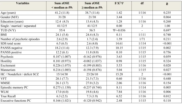
Table 1 shows the socio-demographic and clinical data in two subgroups divided according
to the sum zOSE values using the median split method (median = 0.1186). Doing so we have two
study groups, one with increased and a second with lowered IgM levels to sum zOSEs. We did not
correct the p-values in this table for multiple testing as these data together with the intercorrelation
matrices were only used to delineate the variables to be used as explanatory variables in the
ultimate regression analysis. Table 1 shows that there were no differences in age, sex, education,
marital status, smoking behavior, BMI, number of psychotic episodes, PANSS positive score,
psychosis, hostility, VFT, MMSE and a first PC extracted from the three executive tests between
both study groups. Subjects allocated to the low sum zOSE group showed significantly higher
scores on SDS, PANSS negative, excitement, mannerism, and episodic memory PC as compared
with subjects with higher OSE values. Subjects allocated to the low sum zOSE group showed
significantly lower values of WLM and true recall as compared with subjects belonging to the
group with higher sum zOSE values.
Figure 1 shows the measurements of the IgM antibodies to OSEs, NO and NO2-adducts
(all in z transformations of the Ln transformed values). In order to examine the associations
between the IgM responses to OSEs and NO/NO2-adducts we have performed multinomial logistic
regression analysis with diagnosis (deficit versus non-deficit schizophrenia versus control) as
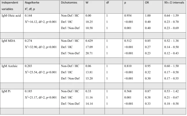
dependent variables and the 8 IgM responses as explanatory variables. Table 2 shows the outcome
of these multinomial regression analyses. IgM responses to oleic acid, MDA, azelaic acid and Pi
were significantly associated with deficit versus non-deficit schizophrenia and versus normal
controls, whilst these IgM levels did not differ between non-deficit schizophrenia and controls.
P-correction for FDR (8 IgM measurements) showed that the differences in IgM responses to oleic
acid, MDA, Pi and azelaic acid remained significant at the p=0.002 level. The largest impact size
was observed for IgM responses to MDA, followed by azelaic acid and Pi. Entering age, sex and
education as additional variables showed that sex had a significant effect on diagnosis, but did not
change the associations between the IgM antibody levels and diagnosis.
For example, IgM antibodies to MDA (Χ2=32.17, df=2, p<0.001) and sex (Χ2=9.18, df=2,
p=0.010) were significant in the multinomial regression analysis with diagnosis as dependent
variable, whilst sex was significant in the differentiation of non-deficit schizophrenia versus
controls (Wald=8.33, df=1, p=0.004) but not the two other differentiations. The IgM antibodies to
NO/NO2 adducts did not significantly predict diagnosis when entered alone in the analysis.
Nevertheless, entering IgM levels to OSEs and NO / NO2-adducts in the same regression showed
that also the IgM responses to NO/NO2-adducts were significant. Table 2 (last regression) shows
that IgM antibodies to MDA (inversely) and NO-cysteinyl (positively) strongly predict deficit
versus non-deficit schizophrenia and normal controls, while there are no significant effects
Table 3 examines the best predictors of deficit schizophrenia as dependent variable (versus
the combined group of subjects with non-deficit schizophrenia and controls as reference group)
and IgM responses, age, sex and education as explanatory variables. Binary logistic regression
analysis shows that IgM antibodies directed against MDA (inversely) and NO-albumin (positively)
significantly predict deficit schizophrenia; 79.9% of all subjects were correctly classified with a
sensitivity of 67.5% and a specificity of 85.9%. The area under the ROC curve was 0.870
(SE=0.033, 95% confidence intervals: 0.805-0.935). The second logistic regression analysis in
Table 3 shows that deficit schizophrenia (versus all other subjects) was also significantly
associated with IgM responses directed against MDA (inversely) and to NO-cysteinyl (positively)
whereby 81.4% of all subjects are correctly classified with a sensitivity of 70.0% and a specificity
of 87.2%. The area under the ROC curve was 0.873 (SE=0.034, 95% confidence intervals:
0.806-0.940). Regression #3 shows that IgM responses to MDA (inversely) and NO-cysteinyl and NO2
-tyrosine (both positively) significantly predicted deficit versus non-deficit schizophrenia; 85.0%
of the subjects were correctly classified with a sensitivity of 82.5% and a specificity of 87.5%. The
area under the ROC curve was 0.913 (±0.35; 95% CI intervals: 0.843-0.982). Age, sex and
education were not significant in these regression analysis.
Effects of extraneous variables on the IgM responses
In order to examine possible effects of age, sex, BMI, and education on the IgM levels we
performed multivariate GLM analysis with the IgM responses to 4 OSEs and 4 NO/NO2-adducts
as dependent variables. There were no significant effects of sex (F=1.62, df=8/99, p=0.128), BMI
(F=0.98, df=8/99, p=0.468), and education (F=1.14, df=8/99, p=0.344) on the IgM antibodies.
p<0.001), although none of the tests for between-subjects effects was significant. In any case, the
regression analyses used in this study were adjusted for possible effects of age, sex and education
by entering those variables as additional explanatory variables in the regression analyses. In
addition, there were no significant effect of smoking (yes or no) on the 8 IgM levels (F=1.04,
df=8/98, p=0.415). We have also examined possible effects of the drug state, namely use of
risperidone (n=33), clozapine (n=10), haloperidol (n=8), perphenazine (n=20), antidepressants
(n=26), mood stabilizers (n=12) and anxiolytics/hypnotics (n=27). Multivariate GLM analysis
showed no significant effects of risperidone (F=1.47, df=8/89, p=0.179), clozapine (F=1.89,
df=8/89, p=0.072), perphenazine (F=0.31, df=8/89, p=0.960), antidepressants (F=1.74, df=8/89,
p0.101), mood stabilizers (F=0.59, df=8/89, p=0.783) and anxiolytics/hypnotics (F=0.99, df=8/89,
p=0.446). Without p-correction there was a significant effect of haloperidol on the IgM values
(F=2.15, df=8/89, p=0.039). After p-correction for FDR these differences were no longer
significant (p=0.236). Tests for between-subject effects showed significant effects (without
p-correction) of haloperidol on IgM to Pi (p=0.042) and NO-albumin (p=0.011). Nevertheless, these
differences were no longer significant after p-correction for FDR, namely IgM against Pi (p=0.168)
and NO-albumin (p=0.088).
Associations between IgM antibodies and schizophrenia phenomenology
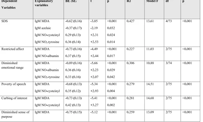
In order to examine which IgM values predict schizophrenia symptomatology we have
carried out multiple regression analysis in schizophrenia patients with symptoms as dependent
variables and the IgM antibodies as explanatory variables. Table 4 shows that 42.7% of the
variance in the SDS score was explained by the regression on IgM levels to MDA and azelaic acid
restricted affect was explained by IgM responses to MDA (inversely) and NO-albumin
(positively). 30.6% of the variance in diminished emotional range was explained by IgM responses
to MDA (inversely) and NO-albumin and NO2-tyrosine (both positively). 27.9% and 28.1% of the
variances in poverty of speech and curbing of interest, respectively, were explained by the
regressions on IgM responses directed against MDA (inversely) and NO-cysteinyl (positively).
25.9% of the variance in diminished sense of purpose was explained by the regression on IgM
levels to MDA and NO2-tyrosine. 42.2% of the variance in diminished social drive was explained
by the regression on IgM levels to MDA and azelaic acid (both inversely) and NO-albumin and
NO2-tyrosine (both positively).
Further analysis showed that positive symptoms as measured with the PANSS were only
very moderately (5.8% of the variance) associated with IgM antibodies to NO2-tyrosine
(positively), while a large part (25.8% of the variance) of the negative subscale score of the PANSS
was associated with IgM to MDA (inversely) and NO2-tyrosine (positively). A large part of the
variances in excitation (21.2%) was explained by the regression on IgM antibodies to MDA and
NO-albumin. There were no significant associations between the IgM responses and either
psychotic symptoms, hostility or mannerism.
All above-mentioned analyses were rerun using age, sex and education as additional
explanatory variables. Age and sex were not significant in these analyses whereas education was
a significant predictor variable that however did not change the association between the
schizophrenia symptoms and IgM levels except in the case of excitation. Table 4 shows a second
regression with excitation as dependent variable including education as explanatory variable. We
found that 28.8% of the variance in excitation was explained by the regression on education and
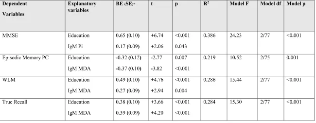
Associations among IgM antibodies and cognitive probes
We have also examined associations between the IgM antibodies to OSEs and NO/NO2-
adducts and neurocognitive tests (see Table 5) using multiple regression analysis with the
cognitive tests as dependent variables and the IgM responses, age, sex and education as
explanatory variables. There were no significant associations between VFT and the IgM levels.
Education and IgM antibodies directed against Pi explained 38.6% of the variance in MMSE,
whereby IgM responses to Pi had a weak albeit significant effect (3.7% of the variance) on MMSE
scores. Education and IgM antibodies to MDA explained 21.9% of the variance in the episodic
memory PC with MDA alone explaining around 12.7%. Education and IgM levels to MDA
explained 28.6% of the variance in WLM with the IgM responses to MDA explaining around
8.8%. Education and IgM responses to MDA explained 28.4% of the variance in true recall with
IgM responses explaining around 12.9% of the variance.
Discussion
The first major finding of this study is that the IgM isotype antibodies to different OSEs
were significantly lower in patients with deficit schizophrenia as compared with non-deficit
schizophrenia and controls. The decrease in these IgM levels to OSEs is highly sensitive and
specific for deficit schizophrenia versus non-deficit schizophrenia. MDA and azelaic acid are both
products of oxidative damage to membrane polyunsaturated fatty acids (PUFAs), which make up
a large part of membrane phospholipids and are highly susceptible to oxidative damage by reactive
oxygen species (ROS). The latter may induce lipid peroxidation resulting in higher levels of
and causing the production of reactive aldehydes including MDA [40,41]. Aldehydes such as
MDA may in turn react with DNA thereby forming mutagenic DNA-adducts and promote toxic
stress in cells which ultimately may lead to cell death [40,41]. Following aldehyde formation these
neoepitopes such as MDA may be expressed on the surface of dying and apoptotic cells and
oxidized LDL cholesterol particles as well as on circulating microparticles [42,43]. Expressed on
these surfaces, the neoepitopes are recognized by immunocytes and consequently autoimmune,
including adaptive IgM responses may be generated directed against these neoepitopes [44-46]. In
addition, natural IgM antibodies, which have specificity for many OSEs, including MDA, are
present without antigenic contact and in fact are part of the innate first-line defense against
microorganisms [16,17]. Increased MDA levels are frequently, but not always, observed in
schizophrenia [47,48], while mood disorders including major depression and bipolar depression
are characterized by increased MDA levels or increased IgM responses to MDA [49-51, in
preparation]. Thus, the findings in depression and bipolar disorder type 1 reporting increased IgM
isotype-mediated responses to MDA, Pi and azelaic acid contrast the findings in deficit
schizophrenia which is characterized by a highly significant decrease in IgM isotype antibodies to
MDA. To the best of our knowledge there are no reports on MDA in the deficit phenotype of
schizophrenia while we were unable to find significant associations between chronic schizophrenia
and oxidative stress measurements including lipid peroxides and advanced oxidation protein
products (AOPPs) [52].
Most importantly, IgM-mediated antibodies to MDA regulate immune-inflammatory
responses by clearing inflammatory debris, including apoptotic and dying cells [42,44]. These IgM
antibodies participate in first line defense through early recognition and elimination of invading
reviewed in the introduction, in prenatal depression there are inverse associations between IgM
isotype-mediated autoimmune responses directed to MDA and indicants of oxidative stress and
depressive symptoms, findings which indicate that these IgM antibodies have protective functions
[15]. Therefore, we proposed that this type of IgM antibodies are part of the CIRS [7,15].
Azelaic acid (or nonanedioic acid) is produced following oxidative damage to linoleic acid
through formation of oxo-nonanoic acid or alternatively by oxidation of oleic acid at the 9 carbon
with consequent degradation to azelaic acid [54]. Interestingly, azelaic acid has anti-inflammatory
and anti-oxidant effects and therefore should be regarded as another component of the CIRS. For
example, besides its anti-inflammatory functions, azelaic acid is a strong antioxidant which may
inhibit the production of O2-, OH and H2O2 and the peroxidation of arachidonic acid by reactive
hydroxyl ions [55-57]. Moreover, azelaic acid may inhibit neutrophil functions including the
production of ROS produced by neutrophils [58]. Here we observed that deficit schizophrenia is
accompanied by highly significant decreases in IgM antibodies to azelaic acid, which are not
detected in non-deficit schizophrenia, while depression and bipolar depression are accompanied
by increased IgM responses to azelaic acid [49, in preparation]. IgM antibodies to OSEs are, in
general, immune-regulatory and play a protective role against immune-inflammatory disorders
including cardio-vascular disorder [59-61]. All in all, our results indicate that lower IgM levels to
azelaic acid increase risk against deficit schizophrenia probably via lowered immune-regulation.
Also the IgM isotype antibodies directed against two other membrane components, namely
oleic acid and Pi, were significantly lowered in deficit schizophrenia. Oleic acid is a
monounsaturated omega 9 fatty acid, which plays a key role in membrane fluidity and additionally
acts as a neurotrophic factor inducing neuronal differentiation [62]. Pi is another important
regulate cell survival, proliferation, calcium levels and polarization [63]. After oxidative disruption
of lipid membranes and oxidative modifications both oleic acid and Pi may be recognized by the
immune system, which consequently mounts an IgM-mediate autoimmune response [45,64].
Depression, for example, is accompanied by increased IgM responses to both oleic acid and Pi
indicating oxidative damage to lipid biomembranes [45,50]. Increased circulating levels of IgM
antibody titers to Pi are observed in other inflammatory disorders including multiple sclerosis in
association with the inflammatory responses during acute relapses [65]. Our findings show that
lowered IgM antibody titers directed against both oleic acid and Pi are specific for the deficit
phenotype as compared with the nondeficit phenotype and therefore that these natural IgM
antibody titers confer protection against the deficit phenotype.
It is important to note that deficit schizophrenia is also accompanied by lowered IgM
antibody titers to TRYCATs, while the IgA responses to TRYCATs are increased [14,28]. Thus,
our studies show that the deficit phenotype is characterized by more general deficits in natural IgM
isotype antibody titers to OSEs and TRYCATs and that such deficits do not occur in nondeficit
schizophrenia. This deficit in CIRS functions may result in attenuated regulation of neuro-immune
responses and increased responsivity of immune-inflammatory and TRYCAT pathways.
The second major finding of this study is that after considering the effects of IgM titers to
OSEs, the IgM responses to NO and NO2-adducts significantly and positively predicted deficit
schizophrenia. Increased IgM responses to NO-adducts, including NOW, albumin and cysteinyl,
and NO2-adducts (NO2-tyrosine) indicate increased nitrosylation and nitration of proteins,
respectively. Interestingly, the IgM antibody titers to OSEs and NO-adducts were significantly
correlated (r=0.763, p<0.001, n=118) and, therefore, the relative increments in IgM isotype
namely a more general decrease in natural IgM isotype levels as well as a relative increase in
nitrosylation and nitration. Interestingly, schizophrenia is accompanied by increased nitro-tyrosine
production although the production of nitric oxide is not always increased [5,18,19], while
hypernitrosylation is a hallmark of mood disorders, either depression or bipolar disorder [66; Maes
et al., in preparation].
Recently, we have reviewed that mild nitrosylation when occurring in physiologic levels
has immune-regulatory and neuroprotective effects [67,68]. For example, mild nitrosylation
regulates cellular processes, has DNA repairing properties, mediates synaptic plasticity and
neuronal survival [67,68]. Nevertheless, hypernitrosylation as a consequence of chronically
activated nitro-oxidative and immune-inflammatory processes has many detrimental effects,
including inactivation of proteins involved in autophagy, apoptosis and proteomic degradation,
which may negatively affect neural functions and cell survival. In addition, hypernitrosylation,
may adversely affect transcription factor activity and electron transport chain (ETC) enzymes,
which may cause decreased mitochondrial function and energy production [68]. Finally, such
changes may cause loss of immune tolerance and consequent development of autoimmunity.
Increased IgM responses to NO2-tyrosine indicate increased nitration of tyrosine (incorporation of
a nitro-group) as a consequence of increased ROS and •NO formation during
immune-inflammatory responses [69,70]. In biomembranes, the formation of NO2-tyrosine is associated
with increased lipid peroxidation through formation of “one-electron oxidation of tyrosine by lipid
peroxyl radicals” [69]. These findings may explain the significant associations between IgM
isotype antibody titers to OSEs and NO2-tyrosine (r=0.680, p<0.001) as detected in the present
of proteins that may contribute to dysfunctions in cell homeostasis, alter tyrosine-kinase-dependent
pathways and facilitate protein degradation [69,70].
Nevertheless, the results of the present study showing that IgM-mediated immune
responses to NO-adducts become significant after considering the effects of IgM antibody titers to
MDA, do not allow to conclude that deficit schizophrenia is accompanied by hypernitrosylation,
but rather that there may be a mild nitrosylation response in deficit schizophrenia. In this respect,
we found that at the end of term pregnancy, there are significant inverse associations between
prenatal depressive symptoms and IgM isotype-mediated responses to NO-adducts, suggesting
that the latter have some immune-regulatory functions [15]. Nevertheless, increased IgM isotype
antibody responses directed against SNO-cysteine have neurotoxic effects and are shown to cause
demyelination and neurodegeneration [35-37]. Interestingly, recently we reported that increased
IgM-mediated responses to NO-cysteinyl are a possible trait marker for major depression [15].
The third major finding of this study is that the negative symptoms of schizophrenia and
excitation (but not psychotic symptoms. hostility and mannerism) as well as impairments in
episodic memory are strongly associated with IgM antibody titers to OSEs (especially MDA) and
IgM responses to NO-adducts (especially with NO-albumin and NO-cysteinyl). Based on the
above discussion, we may conclude that these relationships may be explained by three factors.
Firstly, a deficit in immune-regulatory IgM antibodies to MDA and azelaic acid may lower the
regulatory effects on the immune-inflammatory processes. Recently, we reported that a deficit in
IgM antibody titers to TRYCATs was also highly significantly associated with negative symptoms
of schizophrenia and neurocognitive impairments [28], further indicating that deficits in the CIRS
are extremely important for the negative symptoms of schizophrenia. Secondly, the association
nitrosylation. Thirdly, it is also possible that the relatively increased levels of IgM
isotype-mediated responses directed to NO2-cyteinyl may contribute to the phenomenology of deficit
schizophrenia through its neurotoxic effects. Future translational research should focus on the
effects of nitrosylation, nitration and IgM responses to NO-cysteinyl on neurocognitive deficits
and negative symptoms as well.
In conclusion, deficit schizophrenia is a distinct phenotype of schizophrenia, characterized
by lowered natural IgM isotype antibody titers to OSEs and thus a deficit in the CIRS with lowered
immune-regulatory feedback on the IRS. Moreover, deficit schizophrenia is accompanied by signs
of increased protein nitration and nitrosylation. It is concluded that specific deficits in the CIRS
coupled with increased neurotoxic effects of IgM responses to cysteinyl may drive the hallmarks
of deficit schizophrenia, namely negative symptoms and related neurocognitive impairments in
episodic memory.
Acknowledgements
The study was supported by the Asahi Glass Foundation, Chulalongkorn University Centenary
Academic Development Project and Ratchadapiseksompotch Funds, Faculty of Medicine,
Chulalongkorn University, grant numbers RA60/042 and RA61/050.
Conflict of interest
The authors have no conflict of interest with any commercial or other association in connection
with the submitted article.
All the contributing authors have participated in the manuscript. MM and BK designed the study.
BK recruited patients and completed diagnostic interviews and rating scale measurements. MM
carried out the statistical analyses. All authors (BK, SS, MM and AC) contributed to interpretation
of the data and writing of the manuscript. All authors approved the final version of the manuscript.
References
1. Smith RS, Maes M (1995) The macrophage-T-lymphocyte theory of schizophrenia: additional
evidence. Med Hypotheses 45:135-141
2. Roomruangwong C, Noto C, Kanchanatawan B, Anderson G, Kubera M, Carvalho AF, Maes
M (2018) The role of aberrations in the immune-inflammatory response system (IRS) and the
compensatory immune-regulatory reflex system (CIRS) in different phenotypes of schizophrenia:
the IRS-CIRS theory of schizophrenia. Preprint, September 2018, DOI:
10.20944/preprints201809.0289.v1
3. van Kesteren CF, Gremmels H, de Witte LD, Hol EM, Van Gool AR, Falkai PG, Kahn RS,
Sommer IE (2017) Immune involvement in the pathogenesis of schizophrenia: a meta-analysis on
postmortem brain studies. Transl Psychiatry 2017:7(3):e1075.
4. Orlovska-Waast S, Kohler-Forsberg O, Brix SW, Nordentoft M, Kondziella D, Krogh J, Benros
ME (2018) Cerebrospinal fluid markers of inflammation and infections in schizophrenia and
affective disorders: a systematic review and meta-analysis. Molecular Psychiatry 2018. Aug 16.
5. Maurya PK, Noto C, Rizzo LB, Rios AC, Nunes SO, Barbosa DS, Sethi S, Zeni M, Mansur RB,
Maes M, Brietzke E (2016) The role of oxidative and nitrosative stress in accelerated aging and
major depressive disorder. Prog Neuropsychopharmacol Biol Psychiatry 65:134-144.
6. Maes M, Berk M, Goehler L, Song C, Anderson G, Gałecki P, Leonard B (2012) Depression
and sickness behavior are Janus-faced responses to shared inflammatory pathways. BMC Med
10:66.
7. Maes M, Carvalho AF (2018) The Compensatory Immune-Regulatory Reflex System (CIRS)
in Depression and Bipolar Disorder. Mol Neurobiol 2018 Apr 2. doi:
10.1007/s12035-018-1016-x. [Epub ahead of print] Review. PubMed PMID: 29611101.
8. Noto MN, Maes M, Nunes SO, Ota VK, Rossaneisf AC, Verri Jr WA, Cordeiro Q, Belangero
SI, Gadelha A, Bressan RA, Noto C (2018) Activation of the immune-inflammatory response
system and the compensatory immune-regulatory reflex system in antipsychotic naive first episode
psychosis. Preprints Preprints201809.0314.v2.
9. Anderson G, Maes M (2013) Schizophrenia: linking prenatal infection to cytokines, the
tryptophan catabolite (TRYCAT) pathway, NMDA receptor hypofunction, neurodevelopment and
10. Davis J, Moylan S, Harvey BH, Maes M, Berk M (2014) Neuroprogression in schizophrenia:
Pathways underpinning clinical staging and therapeutic corollaries. Aust N Z J Psychiatry
48:512-529.
11. Davis J, Eyre H, Jacka FN, Dodd S, Dean O, McEwen S, Debnath M, McGrath J, Maes M,
Amminger P, McGorry PD, Pantelis C, Berk M (2016) A review of vulnerability and risks for
schizophrenia: Beyond the two hit hypothesis. Neurosci Biobehav Rev 65:185-194.
12. Maes M, Bosmans E, Ranjan R, Vandoolaeghe E, Meltzer HY, De Ley M, Berghmans R, Stans
G, Desnyder R (1996) Lower plasma CC16, a natural anti-inflammatory protein, and increased
plasma interleukin-1 receptor antagonist in schizophrenia: effects of antipsychotic drugs.
Schizophr Res 21(1):39-50.
13. Maes M, Bosmans E, Kenis G, De Jong R, Smith RS, Meltzer HY (1997) In vivo
immunomodulatory effects of clozapine in schizophrenia. Schizophr Res 26(2-3):221-225.
14. Kanchanatawan B, Sirivichayakul S, Ruxrungtham K, Carvalho AF, Geffard M, Anderson G,
Maes M (2018) Deficit Schizophrenia Is Characterized by Defects in IgM-Mediated Responses to
Tryptophan Catabolites (TRYCATs): a Paradigm Shift Towards Defects in Natural
Self-Regulatory Immune Responses Coupled with Mucosa-Derived TRYCAT Pathway Activation.
15. Roomruangwong C, Barbosa DS, de Farias CC, Matsumoto AK, Baltus THL, Morelli NR,
Kanchanatawan B, Duleu S, Geffard M, Maes M (2018) Natural regulatory IgM-mediated
autoimmune responses directed against malondialdehyde regulate oxidative and nitrosative
pathways and coupled with IgM responses to nitroso adducts attenuate depressive and
physiosomatic symptoms at the end of term pregnancy. Psychiatry Clin Neurosci 72(2):116-130.
16. Thiagarajan D, Frostegård AG, Singh S, Rahman M, Liu A, Vikström M, Leander K, Gigante
B, Hellenius ML, Zhang B, Zubarev RA, de Faire U, Lundström SL, Frostegård J (2016) Human
IgM Antibodies to Malondialdehyde Conjugated With Albumin Are Negatively Associated With
Cardiovascular Disease Among 60-Year-Olds. J Am Heart Assoc 20;5(12).
17. McMahon M, Skaggs B (2016) Autoimmunity: Do IgM antibodies protect against
atherosclerosis in SLE? Nat Rev Rheumatol 12(8):442-444.
18. Dietrich-Muszalska A, Olas B (2009) Modifications of blood platelet proteins of patients with
schizophrenia. Platelets 20(2):90-96.
19. Maia-de-Oliveira JP, Kandratavicius L, Nunes EA, Machado-de-Sousa JP, Hallak JE, Dursun
SM (2016) Nitric Oxide's Involvement in the Spectrum of Psychotic Disorders. Curr Med Chem
20. Kirkpatrick B, Buchanan RW, McKenney PD, Alphs LD, Carpenter WT Jr (1989) The
Schedule for the Deficit syndrome: an instrument for research in schizophrenia. Psychiatry Res
30:119-123.
21. Andreasen NC (1989) The scale for the assessment of negative symptoms (SANS): conceptual
and theoretical foundations. Brit J Psychiatry suppl 7:49-58.
22. Kay SR, Fiszbein A, Opler LA (1987) The positive and negative syndrome scale (PANSS) for
schizophrenia. Schizophr Bull 13:261-276.
23. Kittirathanapaiboon P, Khamwongpin M (2005) The Validity of the Mini International
Neuropsychiatric Interview (M.I.N.I.) Thai Version. Suanprung Hospital, Department of Mental
Health..
24. Overall JE, Gorham DR (1962) The brief psychiatric rating scale. Psycholog Rep 10:799-812.
25. Kanchanatawan B, Thika S, Sirivichayakul S, Carvalho AF, Geffard M, Maes M (2018) In
Schizophrenia, Depression, Anxiety, and Physiosomatic Symptoms Are Strongly Related to
Psychotic Symptoms and Excitation, Impairments in Episodic Memory, and Increased Production
of Neurotoxic Tryptophan Catabolites: a Multivariate and Machine Learning Study. Neurotox Res
26. CERAD (1986) CERAD – An Overview: The Consortium to Establish a Registry for
Alzheimer’s Disease; http://cerad.mc.duke.edu/
27. CANTAB (2018) The most validated cognitive research software.
http://www.cambridgecognition.com/cantab/ October 1, 2018.
28. Kanchanatawan B, Hemrungrojn S, Thika S, Sirivichayakul S, Ruxrungtham K, Carvalho AF,
Geffard M, Anderson G, Maes M (2018) Changes in Tryptophan Catabolite (TRYCAT) Pathway
Patterning Are Associated with Mild Impairments in Declarative Memory in Schizophrenia and
Deficits in Semantic and Episodic Memory Coupled with Increased False-Memory Creation in
Deficit Schizophrenia. Mol Neurobiol 55(6):5184-5201.
29. Sirivichayakul S, Kanchanatawan B, Thika S, Carvalho AF, Maes M (2018) Eotaxin, an
Endogenous Cognitive Deteriorating Chemokine (ECDC), Is a Major Contributor to Cognitive
Decline in Normal People and to Executive, Memory, and Sustained Attention Deficits, Formal
Thought Disorders, and Psychopathology in Schizophrenia Patients. Neurotox Res 2018, Jul 28.
doi: 10.1007/s12640-018-9937-8. [Epub ahead of print] PubMed PMID: 30056534.
30. Daverat P, Geffard M, Orgogozo JM (1989) Identification and characterization of
anti-conjugated azelaic acid antibodies in multiple sclerosis. J Neuroimmunol 22(2):129-134.
31. Boullerne A, Petry KG, Geffard M (1996) Circulating antibodies directed against conjugated
32. Amara A, Constans J, Chaugier C, Sebban A, Dubourg L, Peuchant E, Pellegrin JL, Leng B,
Conri C, Geffard M (1995) Autoantibodies to malondialdehyde-modified epitope in connective
tissue diseases and vasculitides. Clin Exp Immunol 101(2):233-238.
33. Faiderbe S, Chagnaud JL, Geffard M (1992) Anti-phosphoinositide auto-antibodies in sera of
cancer patients: isotypic and immunochemical characterization. Cancer Lett 66(1):35-41.
34. Geffard M, Bodet D, Martinet Y, Dabadie MP (2002) Detection of the specific IgM and IgA
circulating in sera of multiple sclerosis patients: interest and perspectives. Immuno-Analyse &
Biology Specification. 17:302-310.
35. Geffard M, Bodet D, Dabadie MP, Arnould L (2003) Identification of antibodies in sera of
breast cancer patients. Immuno-Analyse & Biologie Special 18:248-253.
36. Boullerne AI, Petry KG, Meynard M, Geffard M (1995) Indirect evidence for nitric oxide
involvement in multiple sclerosis by characterization of circulating antibodies directed against
conjugated S-nitrosocysteine. J Neuroimmunol 60(1-2):117-124.
37. Boullerne AI, Rodriguez JJ, Touil T, Brochet B, Schmidt S, Abrous ND, Le Moal M, Pua JR,
Jensen MA, Mayo W, Arnason BG, Petry KG (2002) Anti-S-nitrosocysteine antibodies are a
predictive marker for demyelination in experimental autoimmune encephalomyelitis: implications
38. Cosgrove JP, Church DF, Pryor WA (1987) The kinetics of the autoxidation of polyunsaturated
fatty acids. Lipids 22(5):299-304.
39. Gutteridge JM (1995) Lipid peroxidation and antioxidants as biomarkers of tissue damage.
Clin Chem 41(12 Pt 2):1819-1828.
40. Shichiri M (2014) The role of lipid peroxidation in neurological disorders. J Clin Biochem Nutr 54(3):151-160.
41. Ayala A, Muñoz MF, Argüelles S (2014) Lipid peroxidation: production, metabolism, and
signaling mechanisms of malondialdehyde and 4-hydroxy-2-nonenal. Oxid Med Cell Longev
2014:360438.
42. Busch CJ, Binder CJ (2017) Malondialdehyde epitopes as mediators of sterile inflammation.
Biochim Biophys Acta Mol Cell Biol Lipids 1862(4):398-406.
43. Tsiantoulas D, Perkmann T, Afonyushkin T, Mangold A, Prohaska TA, Papac-Milicevic N,
Millischer V, Bartel C, Hörkkö S, Boulanger CM, Tsimikas S, Fischer MB, Witztum JL, Lang IM,
Binder CJ (2015) Circulating microparticles carry oxidation-specific epitopes and are recognized
44. Weismann D, Binder CJ (2012) The innate immune response to products of phospholipid
peroxidation. Biochim Biophys Acta 1818(10):2465-2475.
45. Maes M, Mihaylova I, Leunis JC (2007) Increased serum IgM antibodies directed against
phosphatidyl inositol (Pi) in chronic fatigue syndrome (CFS) and major depression: evidence that
an IgM-mediated immune response against Pi is one factor underpinning the comorbidity between
both CFS and depression. Neuro Endocrinol Lett 28(6):861-867.
46. Maes M, Mihaylova I, Kubera M, Leunis JC, Geffard M (2011) IgM-mediated autoimmune
responses directed against multiple neoepitopes in depression: new pathways that underpin the
inflammatory and neuroprogressive pathophysiology. J Affect Disord 135(1-3):414-418.
47. Güneş M, Camkurt MA, Bulut M, Demir S, İbiloğlu AO, Kaya MC, Atlı A, Kaplan İ, Sir A
(2016) Evaluation of Paraoxonase, Arylesterase and Malondialdehyde Levels in Schizophrenia
Patients Taking Typical, Atypical and Combined Antipsychotic Treatment. Clin Psychopharmacol
Neurosci 14(4):345-350.
48. Wu JQ, Kosten TR, Zhang XY (2013) Free radicals, antioxidant defense systems, and
schizophrenia. Prog Neuropsychopharmacol Biol Psychiatry 46:200-206.
49. Maes M, Kubera M, Mihaylova I, Geffard M, Galecki P, Leunis JC, Berk M (2013) Increased
depression: implications for the pathways to chronic depression and neuroprogression. J Affect
Disord 149(1-3):23-29.
50. Maes M, Kubera M, Leunis JC, Berk M, Geffard M, Bosmans E (2013) In depression, bacterial
translocation may drive inflammatory responses, oxidative and nitrosative stress (O&NS), and
autoimmune responses directed against O&NS-damaged neoepitopes. Acta Psychiatr Scand
127(5):344-354.
51. Liu T, Zhong S, Liao X, Chen J, He T, Lai S, Jia Y (2015) A Meta-Analysis of Oxidative
Stress Markers in Depression. PLoS One 10(10):e0138904.
52. Boll KM, Noto C, Bonifácio KL, Bortolasci CC, Gadelha A, Bressan RA, Barbosa DS, Maes
M, Moreira EG (2017) Oxidative and nitrosative stress biomarkers in chronic schizophrenia.
Psychiatry Res 253:43-48.
53. Díaz-Zaragoza M, Hernández-Ávila R, Viedma-Rodríguez R, Arenas-Aranda D,
Ostoa-Saloma P (2015) Natural and adaptive IgM antibodies in the recognition of tumor-associated
antigens of breast cancer (Review). Oncol Rep 34(3):1106-1114.
54. Litvinov D, Selvarajan K, Garelnabi M, Brophy L, Parthasarathy S (2010) Anti-atherosclerotic
actions of azelaic acid, an end product of linoleic acid peroxidation, in mice. Atherosclerosis
55. Passi S, Picardo M, Zompetta C, De Luca C, Breathnach AS, Nazzaro-Porro M (1991) The
oxyradical-scavenging activity of azelaic acid in biological systems. Free Radic Res Commun
15(1):17-28.
56. Fitton A, Goa KL (1991) Azelaic acid. A review of its pharmacological properties and
therapeutic efficacy in acne and hyperpigmentary skin disorders. Drugs. 41(5):780-798.
57. Pelle MT, Crawford GH, James WD (2004) Rosacea: II. Therapy. J Am Acad Dermatol. 51:499-512.
58. Akamatsu H, Komura J, Asada Y, Miyachi Y, Niwa Y (1991) Inhibitory effect of azelaic acid
on neutrophil functions: a possible cause for its efficacy in treating pathogenetically unrelated
diseases. Arch Dermatol Res 283(3):162-166.
59. Binder CJ (2012) Naturally occurring IgM antibodies to oxidation-specific epitopes. Adv Exp
Med Biol 750:2-13.
60. Thiagarajan D, Frostegård AG, Singh S, Rahman M, Liu A, Vikström M, Leander K, Gigante
B, Hellenius ML, Zhang B, Zubarev RA, de Faire U, Lundström SL, Frostegård J (2016) Human
IgM Antibodies to Malondialdehyde Conjugated With Albumin Are Negatively Associated With
61. McMahon M, Skaggs B (2016) Autoimmunity: Do IgM antibodies protect against
atherosclerosis in SLE? Nat Rev Rheumatol 12(8):442-444.
62. Medina JM, Tabernero A (2002) Astrocyte-synthesized oleic acid behaves as a neurotrophic
factor for neurons. J Physiol Paris 96(3-4):265-271.
63. Ananthanarayanan B, Ni Q, Zhang J (2005) Signal propagation from membrane messengers
to nuclear effectors revealed by reporters of phosphoinositide dynamics and Akt activity. Proc Natl
Acad Sci USA 102(42):15081-15086.
64. Maes M, Mihaylova I, Leunis JC (2006) Chronic fatigue syndrome is accompanied by an
IgM-related immune response directed against neopitopes formed by oxidative or nitrosative damage
to lipids and proteins. Neuro Endocrinol Lett 27(5):615-621.
65. Bodet D, Glaize G, Dabadie M-P, Geffard M (2004) Suivi immunobiologique de malades
atteints de sclérose en plaquesImmunobiological follow-up for multiple sclerosis. Immuno-analyse
& Biologie Spécialisée 19:138-147.
66. Maes M, Mihaylova I, Kubera M, Leunis JC, Twisk FN, Geffard M (2012) IgM-mediated
autoimmune responses directed against anchorage epitopes are greater in Myalgic
Encephalomyelitis/Chronic Fatigue Syndrome (ME/CFS) than in major depression. Metab Brain
67. Morris G, Walder K, Carvalho AF, Tye SJ, Lucas K, Berk M, Maes M (2018) The role of
hypernitrosylation in the pathogenesis and pathophysiology of neuroprogressive diseases.
Neurosci Biobehav Rev 84:453-469.
68. Morris G, Berk M, Klein H, Walder K, Galecki P, Maes M (2017) Nitrosative Stress,
Hypernitrosylation, and Autoimmune Responses to Nitrosylated Proteins: New Pathways in
Neuroprogressive Disorders Including Depression and Chronic Fatigue Syndrome. Mol Neurobiol
54(6):4271-4291.
69. Radi R (2013) Protein tyrosine nitration: biochemical mechanisms and structural basis of
functional effects. Acc Chem Res 46(2):550-559.
70. Moylan S, Berk M, Dean OM, Samuni Y, Williams LJ, O'Neil A, Hayley AC, Pasco JA,
Anderson G, Jacka FN, Maes M (2014) Oxidative & nitrosative stress in depression: why so much
stress? Neurosci Biobehav Rev 45:46-62.
Figure 1
Z transformations of the (Ln transformed) measurements of IgM antibodies to conjugated oleic
acid (Lnoleic), malondialdehyde (MDA), azelaic acic, phosphatidylinositol (Pi), nitroso-cysteinyl
(NOCys), nitro-tyrosine (NO2Tyr), NO-tryptophan (NOW) and NO-albumin (NOBSA) in healthy
Table 1. Socio-demographic and clinical data in subjects with lower IgM antibodiesoxidative specific epitopes (sum zOSE) as compared to those with higher OSE values.
Variables Sum zOSE
< median (n=59)
Sum zOSE ≥median (n=59)
F/X2/Ψ df p
Age (years) 41.2 (11.8) 38.7 (11.6) 1.42 1/116 0.235
Gender (M/F) 31/28 21/38 3.44 1 0.064
Education (years) 12.4 (4.5) 13.4 (4.5) 1.28 1/116 0.260
Single / married / separated 41/12/5 41/12/5 0.00 2 1.00
TUD (N/Y) 55/4 56/3 Ψ=-0.036 - 0.697
BMI 24.1 (5.4) 24.4 (4.3) 0.11 1/111 0.740
Number of psychotic episodes 2.6 (2.9) 1.7 (2.4) 1.59 1/71 0.211
SDS total score 6.5 (6.5) 2.6 (4.0) 14.61 1/114 <0.001
PANSS negative 18.2 (11.6) 12.3 (7.9) 10.15 1/115 0.002
PANSS positive 12.2 (6.1) 11.8 (8.0) 0.10 1/115 0.756
Psychosis 0.187 (1.007) -0.162 (0.976) 3.63 1/115 0.059
Hostility 0.101 (0.973) -0.082 (1.037) 0.98 1/115 0.324
Excitement 0.226 (1.075) -0.199 (0.883) 5.53 1/116 0.020
Mannerism 0.216 (1.085) -0.194 (0.876) 5.04 1/115 0.027
HC / Nondeficit / deficit SCZ 15/14/30 23/26/10 15.28 2 <0.001
VFT 20.1 (7.7) 21.5 (7.3) 0.60 1/116 0.440
MMSE 26.1 (3.7) 27.0 (3.2) 2.06 1/116 0.154
Episodic memory PC 0.275 (1.150) -0.237 (0.744) 8.11 1/114 0.005
WLM 17.0 (6.0) 19.8 (4.6) 7.84 1/116 0.006
True Recall 6.3 (2.5) 7.3 (1.9) 6.52 1/116 0.012
Executive functions PC 0.166 (1.021) -0.120 (0.942) 2.48 1/115 0.118
All results are shown as mean (±SD).
F/X2/Ψ: results of analyses of variance (F) or analyses of contingency analyses (X2) or Ψ coefficient;
TUD: tobacco use disorder;BMI: body mass index;SDS: total score on the Schedule for Deficit Syndrome;PANSS: total score on the Positive and Negative Syndrome Scale;
Psychotic dimension: computed as z score PANSS P1 (delusion) (zP1) + zP3 (hallucinations) + zP6 (suspiciousness) + zBPRS11 (suspiciousness) + zBPRS12 (hallucinatory behavior) + BPRS15 (unusual thought content); Hostility dimension: computed as zP7 (hostility) + zPANSS general14
(zG14, poor impulse control) + zBPRS10 (hostility) + zBPRS14 (uncooperativeness);Excitement-grandiosity dimension: computed as zP14
(excitement) + zP5 (grandiosity) + zBPRS8 (grandiosity) + zBPRS17 (excitement);Mannerism-posturing dimension: computed as zG5 + zBPRS7 (both mannerism and posturing);
HC: healthy controls / non-deficit schizophrenia / deficit schizophrenia;
Table 2. Results of multinomial regression analysis with diagnosis (into three groups) as dependent variable and IgM isotype antibody
levels to oxidative specific epitopes (OSE), nitro (NO2) and nitroso (NO) adducts as explanatory variables.
Independent variables
Nagelkerke Χ2, df, p
Dichotomies W df p OR 95% CI intervals
IgM Oleic acid 0.144
Χ2=16.12, df=2, p<0.001
Non-Def / HC
Def / HC
Def / Non-Def
0.00 10.25 10.58 1 1 1 0.954 <0.001 0.001 1.00 0.40 0.40
0.64 – 1.59
0.23 – 0.70
0.23 – 0.69
IgM MDA 0.274
Χ2=32.90, df=2, p<0.001
Non-Def / HC
Def / HC
Def / Non-Def
0.429 17.09 20.71 1 1 1 0.512 <0.001 <0.001 0.85 0.27 0.23
0.52 – 1.38
0.14 – 0.50
0.12 – 0.43
IgM Azelaic 0.203
Χ2=23.54, df=2, p<0.001
Non-Def / HC
Def / HC
Def / Non-Def
0.06 13.81 15.20 1 1 1 0.810 <0.001 <0.001 0.95 0.32 0.30
0.60 – 1.50
0.17 – 0.58
0.17 – 0.55
IgM Pi 0.185
Χ2=21.17, df=2, p<0.001
Non-Def / HC
Def / HC
Def / Non-Def
0.33 11.16 14.14 1 1 1 0.568 0.001 <0.001 0.87 0.38 0.33
0.53 – 1.42
0.21 – 0.67
IgM MDA 0.395
Χ2=51.01, df=4, p<0.001
Non-Def / HC
Def / HC
Def / Non-Def
0.77 21.41 25.80 1 1 1 0.381 <0.001 <0.001 1.39 0.09 0.07
0.67 – 2.87
0.03 – 0.25
0.02 – 0.19
IgM NOcyst Non-Def / HC
Def / HC
Def / Non-Def
0.36 9.25 11.66 1 1 1 0.548 0.002 0.001 0.80 4.07 5.06
0.40 – 1.64
1.65 – 10.04
1.99 – 12.82
Diagnosis: 3 groups are included, namely HC: healthy controls, Non-Def: non-deficit schizophrenia and Def: deficit schizophrenia.
Table3. Results of binary logistic regression analyses with deficit schizophrenia (DEF) as dependent variable and the IgM isotype antibody levels directed against oxidative specific epitopes (OSEs) and nitroso (NO) and nitro (NO2)-adducts as explanatory variables
Dependent
variables
Nagelkerke
Model Χ2
Significant explanatory variables
B (SE) W df p OR 95% CI
#1. DEF/rest 0.489
51.46, df=2, <0.001
IgM MDA IgM NO-albumin -2.92 (0.55) 1.69 (0.43) 28.66 15.16 1 1 <0.001 <0.001 0.05 5.43
0.02 – 0.15
2.32 – 12.71
#2. DEF/rest 0.480
50.22, df=2, <0.001
IgM MDA IgM NO-cysteinyl -2.56 (0.50) 1.50 (0.43) 26.80 12.18 1 1 < 0.001 <0.001 0.08 4.51
0.03 – 0.20
1.94 – 10.49
#3. DEF/NON-DEF 0.633
51.56, df=3, <0.001
IgM MDA
IgM NO-cysteinyl
IgM NO2-tyrosine
-3.64 (0.75) 1.27 (0.65) 1.34 (0.65) 23.74 3.86 4.20 1 1 1 <0.001 0.049 0.040 0.026 3.55 3.80
0.01 – 0.11
1.01 – 12.56
1.06 – 13.63
DEF/rest: the logistic regression analysis are performed with deficit schizophrenia (DEF) as dependent variable and rest (controls + non-deficit schizophrenia) as reference group; MDA: malondialdehyde.
All IgM responses were entered as z values.
Table 4. Results of stepwise multiple regression analyses with severity of schizophrenia symptoms as dependent variables and IgM antibody titers to oxidative specific epitopes (OSEs), nitroso (NO) and nitro (NO2)-adducts as explanatory variables.
Dependent
Variables
Explanatory variables
BE (SE) t p R2 Model F df p
SDS IgM MDA
IgM azelaic
IgM NO-cysteinyl
IgM NO2-tyrosine
-0.62 (0.16)
-0.37 (0.17)
0.29 (0.13)
0.36 (0.14)
-3.85
-2.19
+2.31
+2.53
<0.001
0.032
0.024
0.014
0.427 13.61 4/73 <0.001
Restricted affect IgM MDA
IgM NO-albumin
-0.73 (0.16)
0.37 (0.15)
-4.49
+2.44
<0.001
0.017
0.227 11.03 2/75 <0.001
Diminished emotional range
IgM MDA
IgM NO-albumin
IgM NO2-tyrosine
-0.89 (0.16)
0.36 (0.16)
0.33 (0.16)
-5.66
+2.23
+2.07
<0.001
0.029
0.042
0.306 10.88 3/74 <0.001
Poverty of speech IgM MDA
IgM NO-cysteinyl
-0.68 (0.13)
0.35 (0.12)
-5.34
+2.93
<0.001
0.004
0.279 14.51 2/75 <0.001
Curbing of interest IgM MDA
IgM NO-cysteinyl
-0.73 (0.13)
0.42 (0.13)
-5.41
+3.27
<0.001
0.002
0.281 14.68 2/75 <0.001
Diminished sense of purpose
IgM NO2-tyrosine 0.53 (0.15) +3.43 0.001
Diminished social drive
IgM MDA
IgM azelaic
IgM NO-albumin
IgM NO2-tyrosine
-0.82 (0.21)
-0.50 (0.21)
0.36 (0.18)
0.59 (0.17)
-3.88
-2.37
+2.06
+3.43
<0.001
0.020
0.043
0.001
0.422 13.34 4/73 <0.001
PANSS positive IgM NO2-tyrosine 0.24 (0.11) +2.18 0.032 0.058 4.77 1/77 0.032
PANSSnegative IgM MDA
IgM NO2-tyrosine
-0.64 (0.13)
0.45 (0.13)
-5.15
+3.44
<0.001
0.001
0.258 13.23 2/76 <0.001
Psychotic symptoms
-Hostility
-Excitation IgM MDA
IgM NO-albumin
-0.66 (0.15)
0.49 (0.14)
-4.54
+3.55
<0.001
0.001
0.212 10.33 2/77 <0.001
Excitation IgM azelaic
IgM oleic acid
IgM NO-albumin
Education
-0.39 (0.17)
-0.39 (0.18)
0.51 (0.15)
-0.07 (0.03)
-2.39
-2.18
+3.36
-2.53
0.019
0.033
0.001
0.014
0.288 7.09 4/70 <0.001
Mannerism