Developmental Expression Pattern
Expression of Hex during feather bud development
AKIKO OBINATA*
,1and YOSHIHIRO AKIMOTO
21
Department of Physiological Chemistry, Faculty of Pharmaceutical Sciences, Teikyo University, Sagamiko, Kanagawa, Japan and
2Department of Anatomy, Kyorin University School of Medicine, Mitaka, Tokyo, Japan
ABSTRACT We studied proline-rich divergent homeobox gene Hex/Prh expression in the dorsal
skin of chick embryo during feather bud development. Hex mRNA expression was first observed
in the dorsolateral ectoderm and mesenchyme at 5 days, then in the epithelium and the dermis of
the dorsal skin before placode (primordium of feather bud) formation and then was restricted to the
placode and the dermis under the placode. Afterward, Hex expression was seen in the epidermis
and the dermis of the posterior region of short bud. In accordance with Hex mRNA expression in
the placode, Hex protein was observed in the epidermis as well as in the dermis of the placode.
Immunoelectron microscopic study indicated that the protein located both in the nuclei and
cytoplasm of the epidermis and the dermis at the short bud stage. The Wnt signaling pathway plays
an essential role in the early inductive events in hair (Wnt3a and 7a) and feather (Wnt7a) follicles.
The pattern of Hex expression in the epidermis was similar to that of Wnt7a, while little, if any,
expression of Wnt7a was detected in the dermis under the placode or the dermis of the short bud
compared with that of Hex, suggesting that Hex plays an important role in the initiation of feather
morphogenesis.
KEY WORDS: Hex, homeobox gene, feather placode, Wnt, feather bud development
Epithelial appendages including feathers, scales, hair, claws,
teeth, etc are induced and shaped through
epithelial-mesenchy-mal interactions (Smola
et al., 1993; Chuong et al., 1996; Kishimoto
et al., 2000). An inductive signal from the dermis initiates
forma-tion of epidermal placodes that, in turn, induce dermal
condensa-tion in the underlying dermis (reviewed in Sengel,1976). Several
molecules that mediate inductive signaling during hair and feather
tract formation have been identified, including Wnts (Widelitz
et
al., 1999; Noramly et al., 1999; Huelsken et al., 2001; Andl et al.,
2002), bone morphogenetic protein (BMP) in early skin
develop-ment (Scaal
et al., 2002), BMP inhibitor at placode stages (Patel
et al., 1999), fibroblast growth factors (FGFs)(Widelitz et al.,
1996; Song
et al., 1996), Hedgehog (Ting-Berreth and Chuong,
1996) and Notch/Delta families (Crowe
et al., 1998; Viallet et al.,
1998). Notch/Delta signals refine the patterning of the feather
placode (Crowe
et al., 1998).
Homeobox genes are a large family of transcription factors
which plays a fundamental role in cell differentiation during
development (Gehring
et al., 1994). Abnormal hair follicles were
observed in transgenic mice overexpressing homeobox gene
Msx-2 (Jiang et al., 1999). Hair defects were observed in Hoxc13
mutant mice (Godwin and Capecchi, 1998) and Jave-Suarez
et
al., (2002) showed direct involvement of HOXC13 in the
regula-tion of human hair keratin gene expression. The divergent
*Address correspondence to: Dr. Akiko Obinata. Department of Physiological Chemistry, Faculty of Pharmaceutical Sciences, Teikyo University, Sagamiko, Kanagawa 199-0195, Japan. Fax: +81-426-85-3744. email: [email protected]
0214-6282/2005/$25.00
© UBC Press Printed in Spain www.intjdevbiol.com
homeobox genes
Msx1 (Noveen et al., 1995), Gbx1 (Obinata et
al., 2001) and HB9 (Kosaka et al., 2000a,b) are expressed in skin
and its appendages, such as hair, feather or scale and appear to
be candidates for the regulation of the development of these
tissues. Another divergent homeobox gene
Hex is expressed
during early stages of chick embryogenesis, including pharyngeal
endoderm, endocardium, liver, thyroid gland primordia and blood
islands (Yatskievych
et al., 1999). Hex is required for forebrain,
thyroid and liver formation and blood differentiation (Keng
et al.,
2000; Martinez-Barbera
et al.2000; Martinez-Barbera &
Beddington 2001). In liver morphogenesis,
Hex expression in
avian anterior lateral endoderm is regulated by autocrine BMP
signaling (Zhang
et al., 2002). We showed previously that Hex is
expressed in chick embryonic tarsometatarsal skin and regulates
epidermal cell proliferation (Obinata
et al., 2002). In this study, to
examine whether the
Hex gene is involved in the feather
morpho-genesis or not, we performed
in situ hybridization and
immunostaining analyses spatially and temporally in dorsal skin
of chick embryo.
Expression pattern of Hex during feather bud development
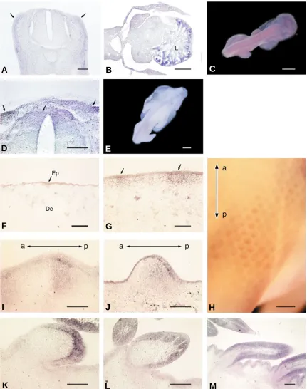
Fig. 1. Expression pattern of Hex mRNA during feather development as revealed by in situ hybridization.
Transverse sections (A,B,D) through
the dorsal ectoderm and trunk region of chick embryos and whole embryo (C,E,H) after whole mount in situ hybridization (WISH)(A-E, H).
Hex
expression
is seen in the dorsolateral region (arrows) of the mesenchyme, if any,
(A,C)
and is strong in liver (L)
(B)
at 4 days. The expression is seen from the
dorsolateral to dorsomedial region (arrows) of the ectoderm and mesenchyme
(D,E)
at 5 days. Cryostat sections
(F,G,I-M)
.
Hex
expression is seen through
the epidermis (Ep) while the expression was very little in the dermis (De) of the dorsal skin before placode formation
(F)
at 6 days and is restricted to
the epidermis and dermis of the placode region (arrows)
(G)
at 7 days. Later, at short bud stage, the stronger expression is seen in both the epidermis
and the dermis at the posterior buds
(I,J)
. At long bud stage, the signal is stronger in the epidermis than in the dermal cells in the posterior bud
(K-M)
.
In WISH
(H)
, as well as in cryostat sections,
Hex
gene expression is detectable in feather buds with stronger signal in posterior regions, but not in interbud
regions in 8-day-old chick embryo. a, anterior; p, posterior. Bars, 100
µ
m in A,D,F,G,I-M; 500
µ
m in B; 1 mm in C,E; 2 mm in H.
A
B
C
D
E
F
G
H
I
J
C
D
E
F
A
B
(arrows) (Fig. 1 A,C), but was strong in liver (B) at 4 days. The
expression was seen from the dorsolateral to dorsomedial region
(arrows) of the ectoderm and mesenchyme at 5 days (Fig. 1 D,E).
Hex expression was seen through the epidermis of the dorsal skin
before placode formation at 6 days while the expression was very
little in the dermis (Fig. 1F) and is restricted to the placode
epithelium and the dermis underneath the placode at 7 days (Fig.
1G). Later, at short bud stage, stronger
Hex expression was seen
in both the epidermis and the dermis of the posterior bud (Fig. 1
H,I,J). At long bud stage, the signal was stronger in the epidermis
than in the dermis at the distal region of the bud (Fig. 1 K,L) and
later an intense signal in the dermis at the bottom region of the bud
adjacent to the epidermis was also seen (Fig. 1 M). Surprisingly,
the
Wnt7a expression pattern in the epithelium (Fig. 2 A,B,C and
Widelitz
et al., 1999) was almost similar to that of Hex (Fig. 1
F,G,I), while less expression of
Wnt7a in the dermis relative to
that of the epidermis was observed under the placode (compare
Fig. 1G with Fig. 2C).
Immunohistochemical localization of Hex protein during
feather bud development
To know whether
Hex mRNA and Hex protein are expressed
correlatively and Hex locates in nucleus as a transcription
factor during feather bud development, we used two different
kinds of antiserum. One is antiserum raised against glutathione
S-transferase fusion protein containing the 76 COOH-terminal
amino acids of mouse Hex (Ghosh
et al., 2000), which was
Fig. 2. Expression pattern of Wnt7a mRNA during feather
develop-ment as revealed by in situ hybridization.
Sections of dorsal skin from
6- to 8-day-old chick embryos (A-C).
Wnt7a
expression is seen through the
epidermis before placode formation
(A)
at 6 days and the stronger signal
is seen in the epidermis of the placode region
(B)
at 7 days. At late placode
stage, the stronger signal is seen in the epidermis of the posterior buds
(C)
.
Bar, 50
µ
m.
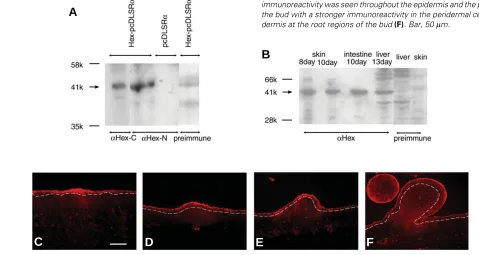
Fig. 3. Western blot and immunohistochemical staining of Hex
pro-tein during feather development.
Whole-cell lysates (50
µ
g)obtained
from HeLa cells transfected with Hex-pcDLSR
α
(A)
and chick embryonic
tissues
(B)
. See text for details. Numbers used at the left side indicate
molecular weights. Hex protein is detected by anti-N-terminal chick Hex
antiserum followed by Cy3-conjugated secondary antibody (C-F). Hex was
observed through the epidermis and the dermis with a stronger
immunore-activity at the placode
(C)
and the short bud
(D,E)
, while less
immunore-activity was observed in the dermis
(C,D,E)
. At the long bud stage, the
immunoreactivity was seen throughout the epidermis and the periderm at
the bud with a stronger immunoreactivity in the peridermal cells and the
dermis at the root regions of the bud
(F)
. Bar, 50
µ
m.
A
B
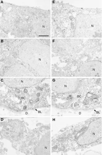
Fig. 4. Immunoelectron microscopic study of feather
bud at 10 days.
Anti-N-terminal chick Hex antiserum
(A-D)
and antibody against glutathione S-transferase
fusion protein containing the 76 COOH-terminal amino
acids of mouse Hex
(E-H)
were used. Colloidal gold
labeling represents the localization of Hex in the
epi-dermal cells of superficial layer
(A,E)
, intermediate
layer
(B,F)
, basal layer
(C,G)
and in the dermal
fibro-blasts
(D,H)
. Both antibodies indicated the same
local-ization pattern in nucleus and cytoplasm of the
epider-mis and the derepider-mis in the bud at 10 days with stronger
immunoreactivity in the cytoplasm
(A-H)
. BC, basal
cell of epidermis; BL, basal lamina; D, dermis; N,
nucleus. Bar, 1
µ
m.
A
B
C
D
E
F
G
H
kindly provided by Dr C.Bogue. The other is anti-N-terminal chick
Hex antiserum. By Western blots, they developed a specific strong
immune response against a 41k dalton band of Hex recombinant
protein (Fig. 3A), the homogenate of chick embryonic dorsal skin,
intestine, liver (Fig. 3B), or 5-day-old whole embryo (data not
shown). At placode stage and later, Hex was observed throughout
the epidermis with a stronger expression at the placode and bud
region and in the dermis under these regions (Fig. 3 C-E). At the
long bud stage, the immunoreactivity was seen at the bud with a
stronger immunoreactivity in the peridermal cells and the dermal
cells at the root regions of the bud (Fig. 3F). It is interesting to note
that, while tissue localization of Hex protein was correlated with
that of its mRNA, the extent of their expression
level was different. The periderm is sloughed at
later stages in the development. To study more
precisely about the localization of the Hex in the
cell, immunoelectron microscopic study was
per-formed with 2 different kinds of Hex antibody.
Colloidal gold labeling represents the
localiza-tion of Hex in the epidermal cells of superficial
layer (Fig. 4 A,E), intermediate layer (Fig. 4 B,F),
basal layer (Fig. 4 C,G) and in the dermal
fibro-blasts (Fig. 4 D,H) of the long bud at 10 days.
Both antibodies indicated the same localization
pattern in nucleus and cytoplasm of the
epider-mis and the derepider-mis with stronger
immunoreac-tivity in the cytoplasm (Fig. 4 A-H). Few colloidal
gold labeling was observed in the skin without
first antibody (Fig. 5).
Acknowledgements
We thank Dr G.Goodwin (Haddow Laboratories, Institute of Cancer
Research,Sutton,UK), for providing a chick Hex cDNA; Dr.C.Bogue
(Department of Pediatrics, Yale University, School of Medicine, New
Haven, Connecticut) for providing a Hex polyclonal antibody and Dr
T.Nohno (Departmennt of Molecular Biology, Kawasaki Medical School,
Kurashiki, Japan) for providing chick Wnt7a cDNA. We are grateful to Ms.
S.Matsubara and Ms. T.Shibata for their technical support. This work was
supported, in part, by Grants-in-Aid from the Ministry of Education, Science,
Sports, Culture and Technology, Japan.
References
AKIMOTO, Y., OBINATA, A., ENDO, H. and HIRANO, H. (1992). Immunohis-tochemical study of basement membrane reconstruction by an epidermis-dermis recombination experiment using cultured chick embryonic skin: Induc-tion of tenascin. J. Histochem.Cytochem. 40: 1129-1137.
ANDL, T., REDDY, S.T., GADDAPARA, T. and MILLAR, S.E. (2002). Wnt signals are required for the initiation of hair follicle development. Dev.Cell 2: 643-653.
CHUONG, C.-M., WIDELITZ, R.B., TING-BERRETH, S. and JIANG, T.-X. (1996). Early events during avian skin appendage regeneration: Dependence on epithelial-mesenchymal interaction and order of molecular reappearance. J.Invest.Dermatol. 107: 639-646.
CROWE, R., HENRIQUE, D., ISH-HOROWICZ, D. and NISWANDER, L. (1998). A new role for Notch and Delta in cell fate decisions: patterning the feather array. Development 125: 767-775.
CYGAN, J.A., JOHNSON, R.L. and MCMAHON, A.P. (1997). Novel regulatory interactions revealed by studies of murine limb pattern in Wnt-7a and En-1 mutants. Development 124: 5021-5032.
DAI, X., SCHONBAUM, C., DEGENSTEIN, L., BAI, W., MAHOWALD, A. and FUCHS, E. (1998). The ovo gene required for cuticle formation and oogenesis in flies is involved in hair formation and spermatogenesis in mice. Genes Dev. 12: 3452-3463.
DASSULE, H.R. and MCMAHON, A.P. (1998). Analysis of epithelial-mesenchymal interactions in the initial morphogenesis of the mammalian tooth. Dev.Biol. 202: 215-227.
GEHRING, W.J., AFFOLTER, M. and BUURGLIN, T. (1994). Homeodomain proteins. Annu.Rev.Biochem.63: 487-526.
GHOSH, B., GONEA, G.R., DENSON, L.A. IANNUCCI, R., JACOBS, H.C. and BOGUE, C.W. (2000). Immunocytochemical characterization of mucine Hex, a homeobox-containing protein. Pediatr.Res. 48: 634-638.
GODWIN, A.R. and CAPECCHI, M.R. (1998). Hoxc13 mutant mice lack external hair. Genes Dev. 12: 11-20.
HEADON, D. and OVERBEEK, P.A. (1999). Involvement of a novel Tnf receptor homologue in hair follicle induction. Nature Genetics 22: 370-374.
HUELSKEN, J., VOGEL, R., ERDMANN B., COTSARELIS, G. and BIRCHMEIER, W. (2001). β-catenin controls hair follicle morphogenesis and stem cell differen-tiation in the skin. Cell 105: 533-545.
HUMPHREYS, R.C., LYDON, J., O’MALLY, B.W. and ROSEN, J.M. (1997). Mammary gland development is mediated by both stromal and epithelial progesterone receptors. Mol.Endocrinol. 11: 801-811.
JAVE-SUAREZ, LF., WINTER, H., LANGBEIN, L., ROGERS, MA. and SCHWEIZER, J. (2002). HOXC13 is involved in the regulation of human hair keratin gene expression. J.Biol.Chem. 277: 3718-3726.
JIANG, T.X., LIU, Y.H., WIDELITZ, R.B. MAXON, R.E. AND CHUONG, C.M. (1999). Epidermal dysplasia and abnormal hair follicles in transgenic mice overexpressing homeobox gene Msx-2. J.Invest.Dermatol. 113: 230-237.
KAWAKAMI, Y., WADA, N., NISHIMATSU, S. and NOHNO, T. (2000). Involvement of frizzled-10 in Wnt-7a signaling during chick limb development. Dev.Growth Differ. 42: 561-569.
KENG, V.W., YAGI, H., IKAWA, M., NAGANO, T., MYNTZ, Z., YAMADA, K., TANAKA, T., SATO, A., MURAMATSU, I., OKABE, M., SOTO, M. and NOGUCHI, T. (2000). Homeobox gene Hex is essential for onset of mouse embryonic liver development and differentiation of the monocyte lineage. Biochem.Biophys.Res.Comm.276: 1155-1161.
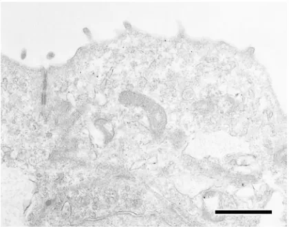
Fig. 5. Cytochemical control.
Immunoelectron microscopic study of the
superficial layer of feather bud at 10 days. To check the specificity,
anti-chick Hex antiserum was replaced with normal rabbit serum. Scarce
colloidal gold labeling was observed. Bar, 1
µ
m.
kidney(Torres & Nelson 2000) and liver (Suksaweang
et al., 2004).
Recent studies have now addressed the issue of whether
Hex is
involved in the Wnt7a signaling pathway or in the initiation of
feather bud formation (Obinata and Akimoto, 2005).
Experimental Procedures
Preparation of a digoxigenin (DIG)-labeled RNA probe
The
Hex RNA probe was prepared as described previously (Obinata et
al., 2002). For synthesis of a Wnt 7a RNA probe, a Wnt7a cDNA fragment
containing the entire coding region, which was kindly provided by Dr
T.Nohno (Kawakami
et al., 2000), was amplified and prepared using a
standard protocol.
In situ hybridization.
In situ hybridization with the DIG-labeled probe was performed as
described previously (Kosaka
et al., 2000a).
Transgene construction
A full-length
Hex cDNA containing the entire Hex coding region, which
was generously provided by Dr G.Goodwin (Haddow Labolatories, Institute
of Cancer Research, Sutton,UK), was constructed with pcDLSR
α
.
Lipofection
HeLa cells were transfected with
Hex -pcDLSR
α
using TransIT-LT1
Transfection Reagent (Invitrogen, California, USA) according to the
manufacturer’s instructions.
Western blotting
A rabbit antiserum was generated against a KLH-conjugated chick Hex
NH2 peptide (MQYQAPGAAPAAALC). Western blotting was performed
using standard protocols.
Immunostaining for light and electron microscopy
Frozen skin sections were processed for immunohistochemical staining
as described previously (Akimoto
et al.,1992; Kosaka et al., 2000b).
Microscopy
KISHIMOTO, J., BURGESON, R.E. and MORGAN, B.A. (2000). Wnt signaling maintains the hair-inducing activity of the dermal papilla. Genes Dev. 14: 1181-1185.
KNAUER, S.K., CARRA, G. and STAUBER, R.H. (2005). Nuclear export is evolutionarily conserved in CVC paired-like homeobox proteins and influences protein stability, transcriptional activation and extracellular secretion. Mol.Cell. Biol. 25: 2573-2582.
KOSAKA, Y., AKIMOTO, Y., OMOTO, Y., OBINATA, A. and HIRANO, H. (2000a). Expression of the HB9 homeobox gene concomitant with proliferation accom-panying epidermal stratification during development of chick embryonic tar-sometatarsal skin. Histochem.J. 32: 275-280.
KOSAKA, Y., AKIMOTO, Y., OBINATA, A. and HIRANO, H. (2000b). Localization of HB9 homeobox gene mRNA and protein during the the early stages of chick feather development. Biochem.Biophys.Res.Commu. 276: 1112-1117.
LI, B., MACKAY, D.R., DAI, Q., LI, T.W.H., NAIR, M., FALLAHI, M., SCHONBAUM, C.P., FANTES, J., MAHOWALD, A.P., WATERMAN, M.L., FUCHS, E. and DAI, X. (2002). The LEF1/β-catenin complex activates movo 1, a mouse homolog of Drosophila ovo required for epidermal appendage differentiation. Pros.Natl.Acad.Sci.USA 99: 6064-6069.
MAIZEL, A., TASSETTO, M., FIHOL, O., COCHET, C., PROCHIANTZ, A. and JOLIOT, A. (2002). Engrailed homeoprotein secretion is a regulated process. Development 129: 3545-3553.
MARTINEZ-BARBERA, J.P. and BEDDINGTON, R.S. (2001). Getting your head around Hex and Hesx1: forebrain formation in mouse. Int.J.Dev.Biol. 45: 327-336.
MARTINEZ-BARBERA, J.P., CLEMENTS, M., THOMAS, P., RODRIGUEZ, T., MELOY, D., KIOUSSIS, D. and BEDDINGTON, R.S. (2000). The homeobox gene Hex is required in definitive endodermal tissues for normal forebrain, liver and thyroid formation. Development 127: 2433-2445.
NORAMLY, S., FREEMAN, A. and MORGANB.A. (1999). β-catenin signaling can initiate feather bud development. Development 126: 3509-3521.
NOVEEN, A., JIANG, T-X, TING-BERRETH, S.A. and CHUONG, C-M. (1995). Homeobox genes Msx-1 and Msx-2 are associated with induction and growth of skin appendages. J.Invest.Dermatol. 104: 711-719.
OBINATA, A., AKIMOTO, Y., HIRANO, H. and ENDO, H. (1991). Stimulation by Bt2cAMP of epidermal mucous metaplasia in retinol-pretreated chick embry-onic cultured skin and its inhibition by herbimycin A, an inhibitor for protein-tyrosine kinase. Exp.Cell Res.193: 36-44.
OBINATA, A., AKIMOTO, Y., OMOTO, Y. and HIRANO, H. (2001). Increase in expression of the homeobox gene, Gbx1, in retinol-induced epidermal mucous metaplasia. Biochem.Biophys.Res.Commu. 280: 1055-1061.
OBINATA, A., AKIMOTO, Y., OMOTO, Y. and HIRANO, H. (2002). Expression of Hex homeobox gene during skin development: Increase in epidermal cell proliferation by transfecting the Hex to the dermis. Develop.Growth.Differ. 44: 281-292.
OBINATA, A. and AKIMOTO, Y. (2005). Involvement of Hex in the initiation of feather morphogenesis. Int. J. Dev. Biol. (in press).
PATEL, K., MAKARENKOVA, H. and JUNG, H.S. (1999). The role of long range, local and direct signaling molecules during chick feather bud development involving the BMPs, follistatin and the Eph receptor tyrosine kinase Eph-A4. Mech. Dev. 86: 51-62.
PAYRE, F., VINCENT, A. and CARRENO, S. (1999). ovo/svb integrates Wingless and DER pathways to control epidermis differentiation. Nature 400: 271-275.
SCAAL, M., PROLS, F., FUCHTBAUER, E-M., PATEL, K., HORNIK, C., KOHLER, T., CHRIST, B. and BRAND-SABERI, B. (2002). BMPs induce dermal markers and ectopic feather tracts. Mech.Dev. 110: 51-60.
SENGEL, P. (1976). Morphogenesis of Skin. Cambridge University Press, Cam-bridge.
SHU, W., JIANG, Y.Q., LU, M.M. and MORRESEY, E.E. (2002). Wnt7b regulates mesenchymal proliferation and vascular development in the lung. Development 129: 4831-4842.
SMOLA, H., THIEKOTTER, G. and FUSENIG, N.E. (1993). Mutual induction of growth factor gene expression by epidermal-dermal cell interaction. J.Cell Biol. 122: 417-429.
SONG, H., WANG, Y. and GOETINCK, P.F. (1996). Fibroblast growth factor 2 can replace ectodermal signaling for feather development. Proc.Natl.Acad.Sci.USA 93: 10246-10249.
SUKSAWEANG, S., LIN, C.M., JIANG, T.X., HUGHES, M.W., WIDELITZ, R.B. and CHUONG, C.M. (2004). Morphogenesis of chicken liver identification of local-ized growth zone and the role of beta-catenin/Wnt in size regulation. Dev.Biol. 266: 109-122.
TING-BERRETH, S. and CHUONG, C-M. (1996). Sonic hedgehog in feather morphogenesis: Induction of mesenchymal condensation and association with cell death. Dev.Dynamics 207: 157-170.
TORRES, M.A. and NELSON, W.J. (2000). Colocalization and redistribution of disheveled and actin during Wnt-induced mesenchymal morphogenesis. J.Cell Biol. 149: 1433-1442.
VIALLET, J.P., PRIN, F., OLIVERA-MARTINEZ, I., HIRSINGER, E., POURQUIE, O. and DHOUAILLY, D. (1998). Chick Delta-1 gene expression and the formation of the feather primordia. Mech.Dev. 72: 159-168.
WIDELITZ, R.B., JIANG, T.X., NOVEEN, A., CHEN, C.W. and CHUONG, C.M. (1996). FGF induces new feather buds from developing avian skin. J.Invest.Dermatol. 107: 797-803.
WIDELITZ, R.B., JIANG, T-X., CHEN, C.-W.J., STOTT, N.S. and CHUONG, C.-M. (1999). Wnt7a in feather morphogenesis:Involvement of anterior-posterior asymmetry and proximal-distal elongation demonstrated with an in vitro recon-stituted model. Development 126: 2577-2587.
WODARZ, A. and NUSSE, R. (1998). Mechanism of Wnt signaling in development. Annu.Rev.Cell Dev.Biol. 14: 59-88.
YATSKIEVYCH, T.A., PASCOE, S. and ANTIN, P.K. (1999). Expression of the homeobox gene Hex during early stages of chick embryo development. Mech.Dev. 80: 107-109.
ZHANG, W., YATSKIEVYCH, T.A., CAO, X. and ANTIN, P.B. (2002). Regulation of Hex gene expression by a Smads-dependent signaling pathway. J.Biol.Chem. 277: 45435-45441.