Original Article
Patterns of migration and regulation of trunk neural crest
cells in Zebrafish (Danio rerio)
JANET L. VAGLIA
1and BRIAN K. HALL
21Department of Biology. Duke University, Durham, North Carolina, USA and 2Department of Biology. Dalhousie University, Halifax, Nova Scotia, Canada
ABSTRACT Regulation is the replacement of lost, undifferentiated embryonic cells by neighboring cells in response to environmental signals. Neural crest cells, embryonic cells unique to craniates, are good candidates for studies of regulation because they are pluripotent, and thus might be able to alter their behavior in response to environmental signals. This study investigated regulation for the loss of trunk neural crest (TNC) cells, specifically pigment derivatives, in the zebrafish, Danio rerio. The first part of the study clarifies and extends what has previously been described on normal patterns of TNC migration and differentiation. These data were then used to address the hypothesis that there is regulation for loss of TNC, and that regulation would vary with the amount removed, the position or stage of removal. Zebrafish TNC cells are large and numerous. SEM and DiI labeling revealed that TNC cells undergo several successive waves of ‘sheet’ and ‘segmental’ migration, beginning as early as the 12 somite stage. DiI-labeled TNC cells often migrated several somite lengths anteriorly and posteriorly along the trunk axis to form glial cells, ganglia, pigment, ectomesenchyme and tail reticular cells. Regulation occurred on a sliding scale, ranging from complete to incomplete. Defects in development and/or pigmentation occurred if large regions of TNC cells were removed, or if cells were removed from anterior (cardiac) and posterior (tail) extremities of the trunk. Melanophores were the cell type most visibly affected by TNC extirpations. Otherwise, pigmentation was remarkably normal. We propose that the completeness of regulation largely depends upon healing of the overlying epidermis.
KEY WORDS:
trunk neural crest, migration, regulation, pigment cells, zebrafish.
0214-6282/2000/$20.00 © UBC Press
Printed in Spain www.ehu.es/ijdb
*Address correspondence to: Dr. Janet Vaglia. Department of Biology, Box 90338, Duke University, Durham, North Carolina NC 27708, USA. FAX: +1-919-660-7293. e-mail: jvaglia@duke.edu
Abbreviations used in this paper: hpf, hours post-fertilization; ss, somite stage; TNC, trunk neural crest.
Introduction
Successful proliferation and migration of cell populations are critical to shaping embryos. If errors occur during development, such as through delayed cell migration or defective proliferation, effects on the embryo can be profound. For instance, deficient neural crest cell migration to the human distal mandibular arch can result in otocephaly (absence of lower jaws, ear defects, heart anomalies) or Treacher Collins syndrome (open eyelid, ear de-fects, cleft palate; Johnston, 1975; Jones, 1990; Hall, 1999). The finding that some populations of embryonic cells can compensate for cell loss, or for actions that would otherwise lead to abnormal development is termed regulation (Bellairs, 1971; Hall and Hörstadius, 1988). More specifically, we define regulation as the replacement of lost, undifferentiated embryonic cells by other cells in response to environmental signals.
Research on regulative potential in embryos is scattered among invertebrates and vertebrates and only alludes to underlying
system and pigment cells (Le Lièvre and Le Douarin, 1975; Le Douarin, 1982; Hall and Hörstadius, 1988; Kirby, 1988a,b; Bronner-Fraser, 1995). The purpose of this study is two-fold. The first goal is to clarify and extend what has previously been described on the migration patterns of trunk neural crest (TNC) cells in the zebrafish, Danio rerio, in order to provide a framework for addressing the question of regulation. The second is to determine whether there is regulation for the loss (removal) of TNC cells.
Regulation of neural crest cells has been shown in lampreys (Newth, 1951, 1956; Langille and Hall, 1988b), teleost fish (Langille and Hall, 1988a; Raible and Eisen, 1996), amphibians (e.g. Chibon, 1970; Moury and Jacobson, 1990), birds (e.g. Kirby et al., 1983; Scherson et al., 1993; Couly et al., 1996) and mice (Snow and Tam, 1979) (also reviewed in Vaglia and Hall, 1999). The limitations and potentials associated with regulation of cranial (e.g. Yntema and Hammond, 1945, 1954 and references therein; McKee and Ferguson, 1984; Couly et al., 1996; Hunt et al., 1995; Sechrist et al., 1995) and cardiac (vagal) (e.g. Bockman and Kirby, 1984; Bockman et al., 1987; Kirby, 1987; Nishibatake et al., 1987; Kuratani et al., 1991; Waldo et al., 1996; Suzuki and Kirby, 1997) neural crest cells have been well established, especially in chicks. Regulation of TNC cells is less resolved, studies being limited to analyses of pigment cells and dorsal root ganglia in amphibians (Lehman and Youngs, 1952), and dorsal root ganglia derivatives of early- versus late-migrating populations of TNC cells in zebrafish
(Raible and Eisen, 1996). Although early-migrating TNC typically produce dorsal root ganglia, Raible and Eisen (1996) found that dorsal root ganglia still form after ablation of early-migrating cells. From this result the authors suggest that late-migrating TNC can regulate for loss of the early migrating cells by changing fate. Late-migrating TNC cells may be bi- or multipotent, but interactions with early-migrating cells prevent them from forming dorsal root ganglia under normal circumstances.
In light of the single study by Raible and Eisen (1996), the potential for TNC cells to regulate, or for other cells to regulate for loss of TNC requires a more thorough investigation. In this study, regions of TNC were extirpated from zebrafish embryos to deter-mine whether the amount of TNC removed, position along the embryonic axis, and/or timing of TNC cell extirpation affect regula-tive ability. Pigment cells (melanophores, iridophores, xanthophores) served as landmarks for analyzing the effects of extirpation in live embryos throughout embryogenesis and during the larval period. Ideally, regulation of pigment cells would be compared with regu-lation of other TNC derivatives such as dorsal root ganglia, the sensory components of the peripheral spinal nerve. Dorsal root ganglia accumulate at the mid-somite as segmentally arranged clusters of neurons and glia between the 16-19 somite stages (ss) (Laudal and Lim, 1993; Raible and Eisen, 1996). To determine whether dorsal root ganglia can be identified within the first 24-36 hours of development, embryos were labeled with one of five
antibodies: anti-acetylated-α-tubulin (6-11B-1), anti-Hu (16A1(6-11B-1), HNK-1, zn-12 or zn-5. Because the antibodies tested did not label dorsal root ganglia early in development, regulation for these TNC derivatives will not be addressed in the current study.
This study provides the first documenta-tion of a populadocumenta-tion of early-migrating TNC cells, and characterizes two major modes of migration that have received little attention in the literature. The term ‘sheet migration’ is used to describe how neural crest cells spread over the neural keel as an unsegmented, coherent sheet with cells in extensive contact with one another. ‘Segmental migration’ re-fers to more localized cell migration in streams over the lateral faces of somites, although cells still contact one another. Differences in the overall number of TNC cells that develop and temporal patterns of migration are dis-cussed with regard to existing descriptions of zebrafish TNC cells and in context with regu-lation.
As first suggested by Raible and Eisen (1996), we show that TNC cells in zebrafish can be replaced by regulation. Pigment patterns re-vealed that regulation varies spatially along the embryonic axis and temporally during development. The results strongly suggest that regulation for TNC cells is by TNC cells, rather than by an alternative cell population such as placodes or epidermal ectoderm.
in ‘yolk crawling’ by extending numerous lamellar-like processes (filopodia) amidst an extracellular matrix (Figs. 2A, 3B). By the 15 ss, sheet migration is visible in the trunk just posterior to the cardiac region (somites three-seven) and continues to spread caudally during development. Trunk neural crest cells labeled with DiI between the level of somites four to eight at the 14-15 ss gave rise to pigment cells, glia, dorsal root and enteric ganglia.
Synchronous with ongoing sheet migration in the mid- and posterior trunk, a third wave of neural crest cells emerges in the head and cardiac regions between the 16-17 ss (Figs. 2C, 3A). This third wave of neural crest cell migration is characterized by a transformation in how cells associate with one another and their environment – the lateral migratory path – and involves a transition from ‘sheet-like’ to ‘segmental’ migration. While segmental migra-tion proceeds anteriorly, a lull in TNC development is observed more posteriorly as sheet migration gradually ceases and few cells emerge from the neural keel. Little to no TNC cell migration was observed following DiI labeling at the 16 ss (level of somites five-nine), thus confirming the lull in migration demonstrated with SEM in the mid trunk at this stage (Fig. 2D).
17-23 ss
The 17 ss marks the beginning of true segmental migration in the trunk, where cells migrate in groups over the lateral face of somites directly beneath the epidermis. During the 17 ss, cells gradually separate into groups between somites three-seven and
Results
Migration of neural crest cells
10-12 ss
In fishes, neural crest cells segregate from a solid neural keel after the neuroepithelium separates from the epidermis during neurulation. Using a combination of SEM imaging and DiI injec-tions (cell labeling between somites four-eight), we found that zebrafish TNC cells begin to migrate from the neural keel at approximately the 12 somite stage (ss), and that they migrate in three or four waves. Migration of the first wave of TNC cells along a medial path in the cardiac (somites one-three/five) and anterior trunk regions (somites six-eight) (Fig. 1) supports observations made by Jesuthasan (1996).
13-16 ss
At the 13/14 ss, a second wave of neural crest cells migrates from the head (mid- and hindbrain) and cardiac regions as expan-sive sheets, concurrent with ongoing migration along the medial path in the mid-posterior trunk (Fig. 2B). During ‘sheet migration,’ compact layers of large neural crest cells migrate laterally over the somites and onto the yolk in a continuous dorso-ventral stream (Fig. 2A). As neural crest cells invade areas lateral to the neural keel, furrows between somites are concealed. At any level – cranial, cardiac or trunk – once cells reach the yolk, they engage
seven-twelve (Fig. 3 B-C). While some neural crest cells continue to bridge clefts between somites, most cells tend to aggregate over individual somites. Also by the 17 ss (Fig. 4B), somites have elevated and lie more ventral than lateral to the neural keel; in younger stages (e.g. 14 ss) somite pairs are distinct from one another and lie more lateral to the neural keel. This morphological change occurs in concert with a raising and narrowing of the neural keel. Posterior to somite 12, cells continue to migrate as a sheet, or remain over the neural keel (Figs. 3D, 4A). The first cells to leave the posterior trunk as new somites are added generally exhibit sheet migration. However, it is difficult to classify migratory pat-terns in the posterior trunk because neural crest cells from more anterior regions mix with those that have just formed. Cells migrat-ing from more anterior locations tend to overlie, and be oriented obliquely, to newly emerging or migrating cells.
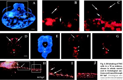
At the 18-20 ss TNC cells continue to migrate laterally in the trunk, concurrent with TNC that penetrate the medial migratory pathway between the neural keel, notochord and somites (Fig. 4B). Cells migrating along the medial path at these later stages may represent a fourth wave of TNC, or a continuation of the third wave. Classification of this phase of migration awaits further study using clonal cell culture or cell labeling. Trunk neural crest cells injected with DiI at the level of somites 7-11 in 17-21 ss embryos became pigment cells –mainly dorsal melanophores and xanthophores – and contributed to dorsal root, sympathetic and enteric ganglia, and ectomesenchyme of the aorta (Fig. 5). Labeling at these stages also revealed extensive migration, especially at somite stages 19-21.
Although lateral migration persists in the trunk beyond the 20 ss, fewer new cells emerge. Because trunk neural crest deriva-tives are ultimately present along the entire embryonic axis, the posterior trunk might be patterned by neural crest cells that emerge from the neural keel as the last few somites form, or by cells migrating from more ante-rior regions. Extensive dispersion of TNC-derived cells over the course of 36 hours post-fertilization suggests not only that large numbers of cells develop from the neural keel at specific locations and stages, but also that cells, especially pigment, divide along their migratory paths.
In summary, SEM and DiI labeling re-vealed striking patterns of TNC migration that support and extend those described by Raible et al. (1992) by demonstrating: (1) the presence of more cells than previously suggested, (2) the existence of waves of sheet-like (Fig. 2A) as well as segmental (Figs. 2C, 3 B,C) migration along much of the trunk, including the yolk (Figs. 2A, 3B) and (3) migration over larger distances.
Regulation
Results of TNC extirpations are based on somite stages (ss) (Table 1) that relate to the patterns of cell migration just described,
Fig. 3. SEM images of the head and trunk of zebrafish embryos. Head (anterior) is to the right in all images. (A) TNC cells (arrows) are layered over the neural keel prior to and during segmental migration along lateral and medial pathways at the level of somites one-six (17 ss). e, epidermis. (B) At the level of somites four-eight, TNC cells are separating into streams (between lines; arrows). nk, neural keel. The area between the two parallel vertical lines (somites four-eight) is magnified in (C) (17 ss). Arrows indicate groups of TNC cells migrating segmentally (in streams) over the somites. s, somite. (D) TNC cells overlying the neural keel in the posterior trunk are elongated antero-posteriorly (arrows) and migrate along the dorsal neural keel prior to migrating laterally over the somites (arrowheads; 18 ss).
with embryos between the 12-13 ss representing early TNC cell migration along the medial path, 13-22 ss embryos representing migration along the lateral path (simultaneous medial and lateral migration at the 18-22 ss), and embryos older than the 23 ss representing near completion of cell migration. While some data analyses include results from extirpations that overlapped with the cardiac (vagal) region (somites one-three/five), emphasis is placed on regulation of TNC cells.
Of the 107 zebrafish embryos from which cardiac and trunk neural crest cells were removed, approximately 13% died prior to one week post-hatching. Fifty-seven percent of the total mortality occurred when cells were removed from regions at the level of all or part of the first five somites in 14-15 ss embryos. The remaining 43% died following removal of large amounts of neural crest, which may or may not be associated with the fact that these extirpations included cells from the region of somites one-five (Table 2).
The presence and patterning of pigment cells (melanophores, iridophores, xanthophores) were the characteristics used to as-sess regulation of TNC cells. Because the absence of xanthophores created regions that had very pale (almost albino-like) coloration, xanthophores were classified simply as present (N=normal) or absent (A=absent) following extirpations.
Normal pigmentation
black, melanin-producing cells that form dorsal, ventral, lateral, and yolk sac stripes in the larvae (Fig. 6 A,B). Iridophores are reflecting pigment cells that appear as iridescent gold spots in association with the dorsal, ventral and yolk sac melanophore stripes. In some regions such as over the braincase, swim bladder and ventral yolk sac, iridophores merge to cover larger areas (Fig. 6C). In older juveniles, iridophores along the dorsal midline merge into a single stripe. Xanthophores produce pteridine and/or cartenoid pigments that appear as a fairly uniform greenish yellow color. These pigment cells are most prominent dorsally, extending from the head to tail and wrapping slightly over the lateral sides (Fig. 6D). Xanthophore data are presented, but these cells were rarely affected by extirpations.
Sham-operated control embryos
Two control (sham-operated) embryos per stage had only the epidermis removed from the level of somites 7-11. At four days post-hatch, pigmentation was normal in all but one of the controls in which the melanophore patterning was severely disrupted, most likely because the epidermis did not heal properly over the wound. As discussed in the following sections, regulation for the pigment derivatives of TNC cells was complete at 48 hours post-fertilization (hpf) for many of the stages analyzed. Forty-eight hpf was chosen as the first time interval to assess pigment morphology because all three types of pigment cells were visible, and patterning was underway. Regulation was complete for most stages analyzed by four days post-hatch.
Does the amount of neural crest removed affect pigmenta-tion?
In a series of preliminary experiments, it was found that zebrafish embryos regulate completely for removal of small amounts of TNC cells (three somite lengths) posterior to somite five. In order to push the limits under which regulation may occur, the amount of neural crest removed was increased to regions five somites or greater in length.
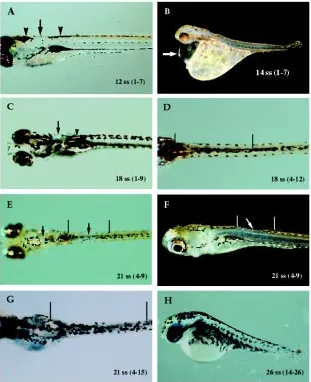
Removing larger amounts of neural crest cells (five or more somite lengths) increased mortality and generated defects ranging from missing to increased densities or disruptions of pigment cells (Table 2). These types of defects in pigment pattern represent various extremes of incomplete regulation. Embryos at the 12 ss survived extirpations up to seven somites in length, even when extirpation overlapped the cardiac region (somites one-three/five). In these embryos, patches of missing or disrupted melanophores and/or iridophores were the only evidence that cells had been removed (Table 2; Fig. 7A). Results were similar for 18-19 ss
embryos following removal of even greater amounts of TNC – up to nine somites in length (Table 2; Fig. 7 C-D). Embryos at the 21 ss or older also exhibited patches of missing or disrupted pigmen-tation, especially when TNC cells were removed over the length of five or more somites (Fig. 7 E-G). In these older embryos, regions of abnormal pigmentation consistently occurred over a length of two or more somites within, rather than peripheral to, extirpated regions. In contrast, few 13-15 ss embryos survived removal of five or more somite lengths of neural crest cells if extirpations over-lapped the cardiac region (Table 2). Survivors were underdevel-oped and exhibited different degrees of body curvature and peri-cardial deformities (Fig. 7B). If the extirpated region included the posterior tail, abnormal pigmentation was accompanied by de-layed development, a degenerating tail (Fig. 7H) and spastic muscle twitching.
No correlation was found between the amount of TNC extirpated and number of pigment cell types affected. Absence of more than one cell type (e.g., melanophores and iridophores) was just as likely to occur whether five or ten somite lengths of TNC were removed (Table 2); loss or disruption of melanophores was frequently accompanied by loss or disruption of iridophores (Fig. 7 C-D). Xanthophores were frequently present in patterns that appeared unchanged from controls.
Does the position of neural crest removal affect pigmentation? Having observed that there was decreased regulation for neural crest removed from the region of somites one-three/five, embryos of the 18-19 ss were used to more specifically test how the position
Fig. 4. Histological sections through the trunk of an 18 ss embryo. (A) In this longitudinal section taken through the trunk posterior to somite 12, TNC cells can be seen migrating along the dorsal neural keel (arrows). (B) By the 18 ss the somites (arrow-heads) have elevated and lie more ventral than lateral to the neural keel (nk). TNC cells are penetrating the medial and lat-eral pathways at the level of the mid-trunk (arrows). Bars, 50 µm
TABLE 1
RELATIONSHIP BETWEEN SOMITE STAGE AND HOURS/DAYS OF DEVELOPMENT
Stage (somites) Age (hrs. post-fertilization)
10 14
12 15
14 16
16 17
18 18
20 19
22 20
24 21
26 22
30 24
Note: Beyond the 30 somite stage (approx. 24 hrs.) embryos are staged according to
from which neural crest cells are removed affects pigmentation. Embryos at these stages are resilient to neural crest cell extirpations (there was no post-hatch mortality), and many cells are emerging from the neural keel along much of the trunk.
Regions of missing or disrupted pigment cells in 18-19 ss embryos most frequently corresponded to TNC removed from the level of somites one-five and posterior trunk. Pigmentation was normal in all embryos when TNC cells were removed just posterior to somite five (somites five-nine) (Table 3). Regardless of the axial level, melanophore disruptions most frequently occurred within and anterior to operated regions; iridophore disruptions more frequently occurred posterior to operated regions.
Does the stage of neural crest removal affect pigmentation? To determine whether the stage when neural crest cells are removed affects regulation, TNC cells were removed from the level of somites 7-11 between the 16-20 ss. A comparison of regulation across several stages is significant for determining whether regula-tion is limited or non-existent during specific times of development.
Bearing in mind that neural crest cells are not equally distributed over the neural keel through development, this comparison also indicates how TNC cells regulate at various stages of development.
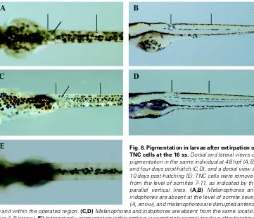
16 ss
Of the stages examined, the 16 ss exhibited relatively less regulation for pigment cells derivatives. Under these conditions, the word ‘relatively’ is used liberally because it is not practical to have sample sizes large enough for true statistical comparisons. While ‘gaps’ in melanophore pigmentation were typically ‘filled-in’ by four days post-hatch for most stages, it took longer for these cells to be replaced when extirpations occurred at the 16 ss (Fig. 8 A-E). Minor disruptions in melanophore and iridophore patterning were observed towards the anterior boundaries of extirpated regions. Xanthophores were unaffected at this and subsequent stages.
17 ss
Although development of normal pigmentation, especially later-ally positioned melanophores, was occasionlater-ally delayed in some
embryos following surgery, pigmentation was normal within four days post-hatch. Disruptions in melanophore or iridophore patterning occurred at the anterior margins of the surgery sites or anterior to where cells were removed.
18 ss
TNC extirpations at the 18 ss created disruptions in melanophore patterning such as abnormally spaced cells or increased densities of melanin in approximately 50% of the embryos within the first 48
TABLE 2
EFFECTS OF EXTIRPATING DIFFERENT LENGTHS OF TNC CELLS ON THE PRESENCE AND PATTERNING OF PIGMENT CELLS IN ZEBRAFISH EMBRYOS, AS ASSESSED ONE WEEK POST-HATCHING
Stage NC Removed Melanophores2 Iridiophores2 Xanthophores2
(ss) (somites)1 n N A D N A D N A Mortality
12 1-5 6 3 3 2 3 3 0 6 0 0
(hb-2)3 (4-6) (hb)
1-7 5 2 3 3 2 3 0 5 0 0
(hb-3) (7-8) (hb)
13-15 1-5 4 0 1 2 2 1 0 1 2 1
(5-6) (3-8) (5-10) (hb-2)
1-7 4 0 0 1 0 1 0 0 1 3
(3-10) (7-10) (hb-3)
1-9 4 0 0 1 0 1 0 0 1 3
(3-10) (7-10) (hb-3)
18-19 1-5 5 0 2 3 2 2 1 5 0 0
(1-2) (4-6) (7-11) (6-9)
1-7 6 0 2 2 1 3 0 4 0 2
(hb-2) (hb-4) (5-9)
1-9 5 0 3 2 0 3 2 5 0 0
(hb-2) (hb-11) 0 (5-9) (3-4)
1-12 4 ——- ——- ——- 4
Notes: 1Refers to the level (prs. somites) from which TNC cells were removed. 2Numbers under pigment headings indicate number of the total sample size for each stage. 3Numbers in parentheses refer to the somite levels from which cells were missing or patterning was disturbed. In some cases, the anterior extremity is the hindbrain (hb).
hours. As for earlier stages, these disruptions always occurred anteriorly, whether anterior to the surgery site or anteriorly within the surgery site. More embryos were lacking iridophores, or had slight abnormalities in patterning at four days post-hatch than at previous stages.
19/20 ss
Regulation at the 19/20 ss most resembled regulation at the 17 ss. No loss of pigmentation was observed at 48 hours; minor disruptions in melanophore and iridophore patterning were not evident by four days post-hatch.
To summarize regulation as assessed by pigment cells, the stage when TNC cells are removed from the mid-trunk does not impact regulation for pigment cells to the same extent as the amount removed and the location from which they are removed. Embryonic survival was poor when neural crest cells were re-moved from the level of somites one-five (cardiac region). Embryos that survived through hatching exhibited abnormal development, pericardial deformities and/or absence of pigmentation. On the other hand, TNC cells (posterior to somite five) exhibited extensive regulation, especially when fewer than nine somite lengths were removed. Experiments demonstrated that regulation was often
complete when TNC cells were removed from the level of somites 7-11 at various stages. At this axial level, the absence and disruption of pigmentation were indicators of incomplete regula-tion. Generally, absence of melanophores correlated with greater densities and disruptions of surrounding melanophores at all stages, regardless of the amount of neural crest cells removed. This pattern was not shared by iridophores which tended to be missing in localized patches. In most embryos, initial disturbances in pigment patterning were resolved by four days post-hatching. Xanthophores were the least affected by extirpations.
Discussion
Migration of neural crest cells
SEM and DiI were the primary techniques used to describe development and migration of TNC cells in the zebrafish, Danio rerio. These data clarify and extend what has previously been described on TNC cell migration, and provide a context for ad-dressing the question of regulation. Zebrafish neural crest cells have been described as being larger and less numerous (10-12 cells per segment) than those of other vertebrates such as the chick (Raible et al., 1992; Raible and Eisen, 1996). This observation
teral paths seen in amniotes (Lamers et al., 1981; Loring, 1987; Serbedzija et al., 1989, 1990; Erickson et al., 1992). In zebrafish, the first wave of TNC cells begins to migrate along the medial path at the level of somites one-six between the 12-13 ss (Raible and Eisen, 1996) (Fig. 1).
Between the 14-15 ss, a second wave of neural crest cells begins to migrate laterally as a uniform sheet over the somites and
yolk in the hindbrain and cardiac regions (Fig. 2A). ‘Sheet migra-tion’ proceeds in a rostral-caudal direction along the trunk during development. Sadaghiani and Vielkind (1989) describe a similar phenomenon of neural crest cells arranged as a sheet between the otic vesicle and the first somite in Xiphophorus (swordfish). How-ever, there is little mention in the literature of extensive sheet-like migration along the entire embryonic axis (Weston, 1970), or of
Fig. 8.Pigmentation in larvae after extirpation of TNC cells at the 16 ss. Dorsaland lateral views of pigmentation in the same individual at 48 hpf (A,B), and four days post-hatch (C,D), and a dorsal view at 10 days post-hatching (E). TNC cells were removed from the level of somites 7-11, as indicated by the parallel vertical lines. (A,B) Melanophores and iridophores are absent at the level of somite seven (A, arrow), and melanophores are disrupted anterior to and within the operated region. (C,D) Melanophores and iridophores are absent from the same location as in A-B (arrow). (E) Interestingly, pigmentation in this embryo is completely normal ten days after hatching. Note how melanophores have filled in the ‘gap’ observed at the younger stages.
would not seem to support the number and variety of derivatives that arise from teleost neural crest cells. While zebrafish neural crest cells are defi-nitely large, our study demonstrates the presence of more cells than previ-ously documented. SEM images and DiI labeling suggest that at least 10-15 neural crest cells emerge per segment for each wave of migration. Because there are several waves of TNC migra-tion, up to three times as many cells are produced. Development of populations of TNC containing larger numbers of cells more closely corre-sponds to the extensive migration and derivative formation observed along the length of the trunk in zebrafish.
A specific pattern and pathway of migration is associated with each ma-jor population of TNC cells that leaves the neural keel. Similar to other teleosts such as the platyfish and swordtail (Sadaghiani and Vielkind, 1990; Sadaghiani et al., 1994), populations of TNC cells in zebrafish migrate in sequential waves – firstly along a me-dial path, secondly along a lateral path, and thirdly along a medial path. The medial and lateral migratory routes correspond to the ventral and
dorsola-TABLE 3
EFFECTS OF EXTIRPATING DIFFERENT REGIONS OF TNC CELLS ON THE PRESENCE AND PATTERNING OF PIGMENT CELLS IN ZEBRAFISH EMBRYOS, AS ASSESSED ONE WEEK POST-HATCHING
Stage Neural crest Melanophores2 Iridiophores2 Xanthophores2 Other
(ss) Removed
(somites)1 n N A D N A D N A
19 1-5 5 0 3 2 3 2 0 5 0 –
(1-2)3 (1-4) (7-11)
18 5-9 7 7 0 0 7 0 0 7 0 –
18 10-14 6 1 2 3 3 2 1 6 0 3
(9-12) (2-9) (9-12) 1(7-8) short fin
18 14-18 5 1 3 1 2 2 1 2 3 _
(15-18) (13-17) (<14) (<15) (>11)
Notes: 1Refers to the level (prs. somites) from which TNC cells were removed. 2Numbers under pigment headings indicate number of the total sample size for each stage. 3Numbers in parentheses refer to the somite level from which cells were missing or patterning was disturbed (<, anterior to; >, posterior to). Abbreviations: A: absence of pigment
neural crest cell migration over the yolk (Sadaghiani and Vielkind, 1989). It remains unclear what cell types arise from sheets of migrating neural crest, but precursor pigment cells and ectomesenchymal cells of the heart and vessels are good candi-dates (Bockman et al., 1987; Erickson, 1993; Olsson, 1994; Sadaghiani et al., 1994).
At approximately the 18 ss, a third wave of neural crest begins to migrate segmentally, rather than as a sheet, along the lateral path in the head and cardiac regions (Fig. 2C). Segmental migra-tion of neural crest cells in the head is a common pattern among amniotes and teleosts (Sadaghiani and Vielkind, 1989; Lumsden et al., 1991; Bemis and Grande, 1992; Serbedzija et al., 1992), while segmental migration through somites in the trunk has been well documented for birds (Teillet et al., 1987; Serbedzija et al., 1989, 1990; Krull et al., 1995). Also beginning around the 18 ss, a new population of neural crest cells – a potential fourth wave –
begins to migrate on the medial path. Migration of a second group of neural crest cells along the medial path appears to be simulta-neous with ongoing migration on the lateral path (Fig. 3B-D). Trunk neural crest cells also migrated distances equivalent to several somite lengths in the anterior and posterior directions. Newly emerging TNC cells in the anterior trunk may migrate one-three somites anteriorly and as far posterior as somite 25.
Regulation
Much of our insight on regulation has been indirectly harnessed from neural crest cell ‘fate-mapping’ studies. The goal of such studies was to remove cells and subsequently infer missing or reduced structures as being derived from those cells. In order to prevent regulation from occurring, and to effectively deplete struc-tures, it was necessary to extirpate large regions of neural crest (e.g., Newth, 1951; Hammond and Yntema, 1964). In addition to ‘fate-mapping’ studies, more direct tests of regulation have tended to be specific to a particular population, or even derivative of neural crest cells. In this study, the development and patterning of TNC-derived pigment cells were used as measures of regulation in zebrafish. The experimental procedure involved extirpating re-gions of TNC and varying the amount removed, position and stage of removal. Regulation occurred on a sliding scale, ranging from complete to incomplete.
A combination of the amount of neural crest removed, and the position and stage of removal affects regulation
Based on earlier fate-mapping studies by Hammond and Yntema (1964) and Langille and Hall (1988a), we predicted that a sizable amount of TNC must be extirpated to produce defects in pigmen-tation. In experiments where the amount of neural crest cells removed spanned regions five somites or greater in length, defects in the amount and/or patterning of pigmentation were generated (Fig. 7). Almost no pigment defects were observed when smaller regions of neural crest cells were extirpated, unless extirpations overlapped the cardiac region (somites one-three/five). The nature and extent of the defects also correlated with the position from which cells were removed. Regulation was most affected when TNC cells were removed from anterior (cardiac) and posterior (tail) extremities of the trunk at various stages (Table 3; Fig. 7 B,H). In more extreme cases, embryos that survived extensive extirpations were underdeveloped, had minor pericardial defects and/or spinal column deformities, and were missing pigment cells.
Although heart development was not investigated following neural crest extirpations, the results suggest that neural crest cells removed from the region of somites one-three/five (cardiac) have limited regulative ability. This was no surprise; several studies have shown that precursor cardiac neural crest cells in the chick do not regulate (Bockman and Kirby, 1984; Kuratani et al., 1991; Waldo et al., 1996; Suzuki and Kirby, 1997). Cardiac neural crest cells provide both ectomesenchymal and neuronal components to the heart and great vessels, and connective tissue to thymus, thyroid and parathyroid glands (Le Lièvre and Le Douarin, 1975; Bockman et al., 1987; Kirby, 1988b; Kuratani and Kirby, 1991). The lack of regulation for cardiac neural crest cells suggests that they are specified early in development, and that timing of migration is critical for normal differentiation. Thus, any opportunity for these cells to regulate would likely fall within a narrow window of time.
Because cardiac neural crest precursors emerge and migrate as a large population of tightly layered cells, as described earlier in the paper, extirpations may target so many cells that the embryo cannot adjust for the loss. The observation that regulation differs among neural crest cell populations located only one or several somites apart is significant for determining the transition between cardiac and trunk regions, as well as for future studies on regulation of cardiac neural crest.
Regulation also was poor when TNC cells were removed from the posterior trunk/tail. In zebrafish, tail and trunk tissue contribute neural crest cells as far anteriorly as somites 11-12 (Kanki and Ho, 1997). Because cellular movements and differentiation in the tail are less well studied than in the anterior-mid trunk, interpreting how regulation functions for cells extirpated from the tail will need to be the topic of another study.
Extirpations of TNC cells at different stages affect populations with different migratory patterns, from cells that are prevalent at the 14-16 ss and destined to migrate laterally as a uniform sheet, to cells that migrate segmentally on the medial path from the 10 ss onward, or on the lateral path from the 18 ss onward. Moreover, neural crest cells are not uniformly present along the entire length of the dorsal neural keel, and therefore, may not be uniformly present within the surgery site. Bearing these factors in mind, regulation for zebrafish TNC cells did not appear to vary signifi-cantly across stages (with the exception of somite stage 16). Embryos at the 16 ss were the most likely to be missing pigment cells, namely melanophores and iridophores, at four days post-hatch (Fig. 8 A-D). Relative to the 18-21 ss where segmental migration of TNC is occurring along the lateral path, fewer cells emerge at the 16 ss. In studies of the chick, regulation for hindbrain neural crest cells changes during development, yet there is some contention as to whether optimal regulation occurs early (prior to the four ss; Saldivar et al., 1997), or later in development (approxi-mately the 10 ss; Diaz and Glover, 1996).
The nature of pigment cells and the role of extracellular matrix Trunk neural crest cells may acquire different regulative abilities over development, but our data suggest (with the exception of the cardiac region and tail) that regulation is most efficient when large numbers of TNC cells are migrating from the neural keel over a length of fewer than six consecutive somites. This would allow greater opportunity for relatively ‘plastic’ (undifferentiated) cells to compen-sate for the loss, compared to a stage where few cells are migrating, or cells have yet to migrate. In a situation where cells have not yet migrated, extirpation of TNC and neural keel cells would affect the development, migration and patterning of the next wave of cells (Weston, 1970; Nishida and Satoh, 1989; Patterson, 1990; Thibaudeau and Holder, 1998). At the other extreme, regulation is less, or at least delayed, when cells are removed from later stages (e.g. 21 ss and older; Fig. 7 E-F). Cells that have differentiated or are beginning to differentiate might be less able to regulate – early migratory neural crest are heterogeneous and may be more respon-sive to environmental signals (e.g. Frank and Sanes, 1991; Ito and Sieber-Blum, 1993; Le Douarin et al., 1994; Thibaudeau and Holder, 1998; Dorsky et al., 2000). At later stages of development, we would need to consider whether cell replacement in larvae or among differentiated cells is regulation, or a different form of wound healing. Evidence of decreased regulation in older embryos does not imply that compensation for cells removed during the embryonic
period ends with a specific stage. Following TNC cell extirpations, pigmentation was typically monitored for up to seven days post-hatching. If abnormalities in pigmentation were observed between four and seven days post-hatching, the morphology was viewed as ‘permanent’ for the individual. We later found that the absence of pigmentation in one-week old larvae was not necessarily indicative of the final juvenile/adult pigment pattern. Extirpation of TNC cells at the level of somites 7-11 in a 16 ss embryo resulted in a patch of missing melanophores and iridophores that was still evident at four days post-hatch. By 10 days post-hatching, pigmentation was perfectly normal (Fig. 8). Thus, pigment cells are able to replace themselves, or to be replaced, well into the larval period (Johnson et al., 1995; Parichy et al., 1999). Furthermore, normal pigmenta-tion in species of the salamander genus Triturus arises in part from a secondary invasion of pigment cells later in development (Niu, 1954).
In general, regions lacking pigmentation corresponded to loca-tions from which TNC cells were removed. Patches of dense and/ or disrupted pigmentation frequently occurred anterior to, and occasionally posterior to pigment-free surgery areas. In other cases, an excess of pigment cells, namely melanophores, was produced along the length of extirpated regions and persisted through at least one week post-hatching. Extirpations of both cranial and trunk neural folds in salamanders also have resulted in larvae with regions of increased pigmentation (Twitty, 1944; Lehman and Youngs, 1952) versus pigment free areas (Niu, 1947). Such uncharacteristically dense regions of pigmentation give the ap-pearance of ‘over regulation’ (i.e. excess cell division) for cell loss. Conversely, isolated patches of dense or disrupted pigmentation may represent cells that failed to migrate as a result of severely disrupted extracellular matrix (ECM).
Several studies have demonstrated that the ECM is both struc-turally and molecularly important for the migration and differentia-tion of neural crest cells (Löfberg et al., 1980; Erickson et al., 1992; Erickson, 1993; reviewed by Parichy, 1996). The importance of ECM to neural crest cell migration is exemplified in the white axolotl mutant that lacks pigmentation because the ECM is a defective substrate for migrating precursor pigment cells (Löfberg et al., 1989). If TNC cells in zebrafish lack a scaffolding/matrix upon which to migrate in either the anterior or posterior direction (Kanki and Ho, 1997), a “traffic jam” could occur at the perimeters of the surgery region where there is no matrix. Similarly, disrupted pigment cells could reflect an altered ECM that prevented cells from migrating along their normal routes.
Role of the epidermis in regulation
neural crest cells are removed, the initial absence of epidermis might prevent induction of a new population of potentially regulat-ing cells from the underlyregulat-ing neural keel. In addition, the epidermis is not immediately available as a substrate for cells to migrate into the neural crest-ablated area from surrounding regions. Because epithelium is an essential positioning signal for embryonic cells (Ho and Weisblat, 1987), some instances of incomplete regulation may result from delays in epidermal closure.
Potential sources of replacement cells – a summary
Many studies have discussed possible sources of cells that could replace extirpated neural crest. They include placodes, neural and epidermal ectoderm and neural crest itself (Kirby, 1987; 1988a,b). Placodal cells are precursors to sensory receptors and cranial ganglia. They arise in the head as ectodermal thickenings that invaginate and further delaminate following transformation from an epithelial to a mesenchymal cell type in all but the lens placode. In this study, for placode cells to be considered a source of replacement cells for cardiac and TNC cells, they would either need to be in close proximity to extirpated regions, or migrate a considerable distance. Epidermis also was not expected to be a source of replacement cells because it would first need to heal. Moreover, epidermis is necessary as an inducing factor for neural crest cells to be generated from the neural keel.
Many studies have shown that neural crest cells regulate, and that regulative potential varies with cranial versus cardiac versus trunk crest populations (reviewed in Vaglia and Hall, 1999). As alluded to in the context of fate-mapping studies, cranial neural crest cells regulate quite proficiently in organisms ranging from lampreys to chicks (e.g. McKee and Ferguson, 1984; Langille and Hall, 1988a,b; Moury and Jacobson, 1990; Scherson et al., 1993; Hunt et al., 1995; Couly et al., 1996; Diaz and Glover, 1996; Suzuki and Kirby, 1997). Cardiac neural crest cells, on the other hand, have little potential for regulation (e.g. Waldo et al., 1996; Suzuki and Kirby, 1997). Our study has shown there is regulation for TNC-derived pigment cells in zebrafish, and that TNC cells most commonly regulate for other TNC cells. The capacity for regulation of TNC-derived ganglia is unknown. Having demonstrated in zebrafish that at least a single cell lineage derived from TNC is regulated for, the next goal is to investigate possible mechanisms of replacement. Preliminary experiments using fluorescent DiI to trace cell migration and BrdU (bromodeoxyuridine) to assess levels of cell division reveal that both cell migration and increased division are important to regulation.
Materials and Methods
Zebrafish maintenance and breeding
Adult zebrafish (Danio rerio ) were purchased from either local pet stores in Halifax, Nova Scotia, Canada or from Boreal Laboratories, Ltd. (Ontario, Canada). Females and males were separated and kept in 10 gallon tanks at a temperature of 27-29ºC on a 14 hour light and 10 hour dark cycle. Breeding colonies were established with three females and three-five males. Eggs were collected in the evening and raised at 28.5ºC in Hank’s solution (Westerfield, 1995).
Scanning electron microscopy
Patterns of trunk neural crest cell migration were primarily determined using scanning electron microscopy (SEM; Nanolab 2000, Bausch and Lomb). To remove the epidermis, dechorionated embryos were incubated in an enzyme solution (0.257 g Trypsin + 0.043 g Pancreatin in 10 ml PBS)
at 4ºC for 20-30 minutes. Embryos were then transferred to room tempera-ture and covered with a thin layer of 1.0% agarose. A glass-pulled micropipette was used to tease the epidermis away from underlying cells, after which embryos were preserved immediately in Karnovsky’s fixative. Following overnight fixation, samples were rinsed in 0.2 M cacodylate buffer at pH 7.4 with 20% sucrose. They were then post-fixed in OsO4, rinsed a second time in 0.2 M cacodylate with 20% sucrose, dehydrated and critical point dried. Specimens were coated with gold using a sputter coater prior to viewing (Samsputter-2a; Tousimis Research Corp.; Sargent-Welch Vacuum Pump). Because of individual variation, and differences associated with processing specimens for SEM, several embryos were used for each stage.
DiI labeling
DiI labeling provided further detail on early patterns of TNC cell migra-tion in the anterior-mid trunk. Fluorescent DiI (SP-DiIC18(3)) (Molecular Probes, D-7777) was diluted to a concentration of 2 mg/ml in 92% Dimethylformamide (DMF) and 8% H20. Immediately prior to use, DiI aliquots were briefly centrifuged to remove any precipitate. Thick-walled glass capillaries (OD 1.2 mm, I.D. 0.69 mm with filament) were pulled into micropipettes using a vertical pipette puller (Model 700C, David Kopf Instruments). Micropipettes were backfilled with 1.5 µl DiI, followed by 1 µl 0.2 M KCL and stored in glass jars in a 37° C incubator to slow crystallization of dye during injection sessions.
For injections, embryos between somite stages 10 and 22 (14-20 hours) were embedded in 1% agarose. An empty glass-pulled micropipette was used to create an opening in the agarose and pierce the epidermis. The empty needle was replaced by a DiI-filled needle attached to a joystick micromanipulator (World Precision Instruments), and DiI was focally in-jected as a single two millisecond pulse under a Leitz Laborlux D compound microscope using a PV830 pneumatic picopump (World Precision Instru-ments) attached to a nitrogen air source. Embryos were injected at a constant tank pressure of 100 kPa and ejection pressure of approximately 30 psi. Holding pressure varied slightly according to the size of the micropipette tip opening, but generally ranged between 10 and 20 psi. For consistency, cardiac and TNC cells were labeled over the length of two somites between the level of somites 4-11. Specimens were viewed under a Leitz Aristoplan fluorescent microscope (N2.1 filter) within five-ten minutes of the initial injections, and subsequently at hourly intervals. Injections were replicated several times to account for developmental variation. Because mesodermal cells also migrate during the early stages of development, it was important to ensure that somites were not inadvert-ently labeled during DiI injections. Controls were performed by injecting DiI onto somitic cells lying dorsolateral to the neural keel (adjacent to where TNC emerge) at stages corresponding to those used for tracing TNC cell migration. DiI remained localized and no cell migration was observed in the controls (see also Smith et al., 1994).
Removal of trunk neural crest cells
Live embryos
Pigment development and patterning were traced through embryogen-esis and into the larval period in individual control and experimental embryos. To document changes in pigmentation, embryos and larvae were anesthetized using a 0.4% solution of MS-222 (Tricaine) and stabilized on slides using 3% methyl cellulose. Live embryos or larvae were photo-graphed under a Tessavar dissecting microscope at magnifications of 10x or 16x. After removing the animals from methyl cellulose, recovery was rapid and they developed as usual in Hank’s solution.
Histology
The depth of neural keel extirpation, and completeness of trunk neural crest removal were verified in sectioned embryos (Fig. 9B). For paraffin sectioning, embryos were fixed in 4% paraformaldehyde, dehydrated in a series of methanol/PBS (Dulbecco’s phosphate buffered saline, Freshney, 1987), infiltrated, embedded in low melting point wax in a vacuum oven, and cut at 5 µm. Sections were mounted on Poly-L-Lysine-coated slides and stained with HBQ (Hall and Brunt’s Quadruple Stain, Hall, 1986) or Ehrlich’s Hematoxylin (see Gurr, 1962).
Antibody staining
For whole-mount antibody staining, embryos were fixed overnight in 4% paraformaldehyde at 4°C followed by several rinses in 0.1 M PBS. Embryos were incubated for one hour in blocking solution (PBS with 0.1% Tween, 1% BSA and 20% normal goat serum for HNK-1; 20% normal sheep serum for all other antibodies), then incubated overnight in primary antibody at 4ºC (6-11B-1, anti-acetylated-∝-tubulin: 1:1000; 16A11 (anti-Hu): 1:150; HNK-1: 1:50; zn-5: 1:50; zn-12: 1:10). Following a one hour rinse with several changes in 0.1M PBS with 0.1% Tween, HNK-1-labeled embryos were incubated in fluorescent CY3-conjugated goat anti-mouse secondary an-tibody (1:500) for two hours at room temperature in the dark. Embryos labeled with other antibodies were incubated in horseradish peroxidase-conjugated sheep anti-mouse secondary antibody (1:250) for two-three hours at room temperature or overnight at 4°C. HNK-1-labeled embryos were then rinsed extensively over one-three hours with several changes in ddH20 and viewed under a Leitz Aritstoplan fluorescent microscope using an N2.1 filter. Embryos labeled with other antibodies were rinsed for one hour with several changes in PBS with Tween, washed for 10 minutes in TRIS (pH 7.6), and then incubated in 1.0 mg/2.0 ml diaminobenzidine (DAB) dissolved in 20 µl DMSO (dimethylsulfoxide) and brought to a final volume of 20 ml with TRIS buffer. One µl H2O2 was added to the final solution to develop the HRP (POD) reaction product.
Acknowledgements
This work was supported by Sigma Xi Grants-in-Aid of Research to J.L. V., the Natural Sciences and Engineering Research Council of Canada (grant #A5056) to B.K. H. and by the Izaak Walton Killam Trust of Dalhousie University. We thank Dr. Margaret Kirby for her insights and enriching discussions on this extremely complex but fascinating area of developmen-tal biology. Thanks also to Dr. Kathleen Smith for financial assistance during the final preparation of this manuscript.
References
BELLAIRS, R. (1971). Developmental Processes in Higher Vertebrates, University of Miami Press, Coral Gables.
BEMIS, W.E. and GRANDE, L. (1992). Early development of the actinopterygian head. I. External development and staging of the paddlefish Polyodon spathula. J. Morphol. 213: 47-83.
BOCKMAN, D.E. and KIRBY, M.L. (1984). Dependence of thymus development on derivatives of the neural crest. Science 223: 498-500.
BOCKMAN, D.E., REDMOND, M.E., WALDO, K., DAVIS, H. and KIRBY, M.L. (1987). Effect of neural crest ablation of development of the heart and arch arteries in the chick. Am. J. Anat. 180: 332-341.
BRONNER-FRASER, M. (1995). Origins and developmental potential of the neural
crest. Exp. Cell Res. 218: 405-417.
CHIBON, P. (1970). Capacité de régulation des excédents dans la crête neurale d’Amphibien. J. Embryol. Exp. Morphol. 24: 479-496.
COULY, G., GRAPIN-BOTTON, A., COLTEY, P. and LE DOUARIN, N. (1996). The regeneration of the cephalic neural crest, a problem revisited: the regenerating cells originate from the contralateral or from the anterior and posterior neural fold. Development 122: 3393-3407.
DÍAZ, C. and GLOVER, J.C. (1996). Appropriate pattern formation following regula-tive regeneration in the hindbrain neural tube. Development 122: 3095-3105.
DORSKY, R.I., MOON, R.T. and RAIBLE, D.W. (2000). Environmental signals and cell fate specification in premigratory neural crest. BioEssays 22: 708-716.
ERICKSON, C.A. (1993). From the crest to the periphery: control of pigment cell migration and lineage segregation. Pig. Cell Res. 6: 336-347.
ERICKSON, C.A., DUONG, T.D. and TOSNEY, K.W. (1992). Descriptive and experi-mental analysis of the dispersion of neural crest cells along the dorsolateral path and their entry into ectoderm in the chick embryos. Dev. Biol. 151: 251-272.
ETTENSOHN, C.A. and McCLAY, D.R. (1988). Cell lineage conversion in the sea urchin embryo. Dev. Biol. 125: 396-409.
FRANK, E. and SANES, J.R. (1991). Lineage of neurons and glia in chick dorsal root ganglia: analysis in vivo with a recombinant retrovirus. Development 111: 895-908.
FRESHNEY, R.I. (1987). Culture of Animal Cells, Alan R. Liss, New York.
GURR, E. (1962). Staining Animal Tissues: Practical and Theoretical. Leonard Hill Books Limited, London, UK.
HALL, B.K. (1986). The role of movement and tissue interactions in the development and growth of bone and secondary cartilage in the clavicle of the embryonic chick. J. Embryol. Exp. Morphol. 93: 133-152.
HALL, B.K. (1999). The Neural Crest in Development and Evolution, Springer-Verlag, New York.
HALL, B.K., and HÖRSTADIUS, S. (1988). The Neural Crest, Oxford University Press, New York.
HAMMOND, W.S. and YNTEMA, C.L. (1964). Depletions of pharyngeal arch carti-lages following extirpation of cranial neural crest in chick embryos. Acta Anat. 56: 21-34.
HO, R.K. and WEISBLAT, D.A. (1987). A provisional epithelium in leech embryo: cellular origins and influence on a developmental equivalence group. Dev. Biol. 120: 520-534.
HUNT, P., FERRETTI, P., KRUMLAUF, R. and THOROGOOD, P. (1995). Restora-tion of normal hox code and branchial arch morphogenesis after extensive deletion of hindbrain neural crest. Dev. Biol. 168: 584-597.
ITO, K. and SIEBER-BLUM, M. (1993). Pluripotentiality and developmentally re-stricted neural crest-derived cells in posterior visceral arches. Dev. Biol. 156: 191-200.
JESUTHASAN, J. (1996). Contact inhibition/collapse and pathfinding of neural crest cells in the zebrafish trunk. Development 122: 381-389.
JOHNSON, S.L., AFRICA, D. WALKER, C., and WESTON, J.A. (1995). Genetic control of adult pigment stripe development in zebrafish. Dev. Biol. 167: 27-33.
JOHNSTON, M.C. (1975). The neural crest in abnormalities of the face and brain. In Morphogenesis and malformation of face and brain (Ed. D. Bergsma). Vol. 11. Alan R. Liss, Inc., New York, pp. 1-18.
JONES, M.C. (1990). The neurocristopathies: reinterpretation based upon the mecha-nism of abnormal morphogenesis. Cleft Palate J 27: 136-140.
KANKI, J.P. and HO, R.K. (1997). The development of the posterior body in zebrafish. Development 124: 881-893.
KIRBY, M.L. (1987). Cardiac morphogenesis—recent research advances. Pediatr. Res. 21: 219-224.
KIRBY, M.L. (1988a). Nodose placode contributes autonomic neurons to the heart in the absence of cardiac neural crest. J. Neurosci. 8: 1089-1095.
KIRBY, M.L. (1988b). Nodose placode provides ectomesenchyme to the developing chick heart in the absence of cardiac neural crest. Cell Tissue Res. 252: 17-22.
KIRBY, M.L., GALE, T.F. and STEWART, D.E. (1983). Neural crest cells contribute to normal aorticopulmonary septation. Science 220: 1059-1061.
PNA-binding molecules. Development 121: 3733-3743.
KURATANI, S.C. and KIRBY, M.L. (1991). Initial migration and distribution of the cardiac neural crest in the avian embryo: an introduction to the concept of the circumpharyngeal crest. Am. J. Anat. 191: 215-227.
KURATANI, S.C., MIYAGAWA-TOMITA, S. and KIRBY, M.L. (1991). Development of cranial nerves in the chick embryo with special reference to the alterations of cardiac branches after ablation of the cardiac neural crest. Anat. Embryol. 183: 501-514.
LAMERS, C.H.J., ROMBOUT, J.W.H.M. and TIMMERMANS. L.P.M. (1981). An experimental study on neural crest migration in Barbus conchonius (Cyprinidae, Teleostei), with special reference to the origin of the enteroendocrine cells. J. Embryol. Exp. Morphol. 62: 309-323.
LANGILLE, R.M. and HALL. B.K. (1988a). Role of the neural crest in development of the cartilaginous cranial and visceral skeleton of the medaka, Oryzias latipes (Teleostei). Anat. Embryol. 177: 297-305.
LANGILLE, R.M. and HALL. B.K. (1988b). Role of the neural crest in development of the trabeculae and branchial arches in embryonic sea lamprey, Petromyzon marinus (L). Development 102: 301-310.
LAUDAL, T.P. and LIM, T-M. (1993). Development of the dorsal root ganglion in a teleost, Oreochromis mossambicus (Peters). J Comp Neurol 327: 141-150.
LE DOUARIN, N. (1982). The Neural Crest. Cambridge University Press, Cambridge London et al.
LE DOUARIN, N.M., DUPIN, E. and ZILLER, C. (1994). Genetic and epigenetic control in neural crest development. Curr. Opin. Genet. Dev. 4: 685-695.
LE LIÉVRE, C.S. and LE DOUARIN, N.M. (1975). Mesenchymal derivatives of the neural crest: analysis of chimaeric quail and chick embryos. J. Embryol. Exp. Morphol. 34: 125-154.
LEHMAN, H.E. and YOUNGS. L.M. (1952). An analysis of regulation in the amphibian neural crest. J. Exp. Zool. 121: 419-447.
LÖFBERG, J., AHLFORS, K. and FÄLLSTRÖM, C. (1980). Neural crest cell migration in relation to extracellular matrix organization in the embryonic axolotl trunk. Dev. Biol. 75: 148-167.
LÖFBERG, J., PERRIS, R and EPPERLEIN, H.H. (1989). Timing in the regulation of neural crest cell migration: Retarded “maturation” of regional extracellular matrix inhibits pigment cell migration in embryos of the white axolotl mutant. Dev. Biol. 131: 168-181.
LORING, J.F. and ERICKSON, C.A. (1987). Neural crest cell migratory pathways in the trunk of the chick embryo. Dev. Biol. 121: 220-236.
LUMSDEN, A., SPRAWSON, N. and GRAHAM, A. (1991). Segmental origin and migration of neural crest cells in the hindbrain region of the chick embryo. Development 113: 1280-1291.
MARTIN, P. (1996). Mechanisms of wound healing in the embryo and fetus. Curr. Topics Dev. Biol. 32: 175-203.
MCKEE, G.J. and FERGUSON, M.W.J. (1984). The effects of mesencephalic neural crest cell extirpation on the development of chicken embryos. J. Anat. 139: 491-512.
MESCHER, A. (1996). The cellular basis of limb regeneration in urodeles. Int. J. Dev. Biol. 40: 785-795.
MOURY, J.D and JACOBSON, A.G. (1990). The origins of neural crest cells in the Axolotl. Dev. Biol. 141: 243-53.
NEWTH, D.R. (1951). Experiments on the neural crest of the lamprey embryo. J. Exp. Biol. 28: 247-260.
NEWTH, D.R. (1956). On the neural crest of the lamprey embryo. J. Embryol. Exp. Morphol. 64: 105-120.
NISHIBATAKE, M., KIRBY, M.L. and VAN MIEROP. L.H.S. (1987). Pathogenesis of persistent truncus arteriosus and dextroposed aorta in the chick embryo after neural crest ablation. Circulation 75: 255.
NISHIDA, H. and SATOH, N. (1989). Determination and regulation in the pigment cell lineage of the ascidian embryo. Dev. Biol. 132: 355-367.
NIU, M.C. (1947). The axial organization of the neural crest, studied with particular reference to its pigmentary component. J. Exp. Zool. 105: 79-113.
NIU, M.C. (1954). Further studies on the origin of amphibian pigment cells. J. Exp. Zool. 125: 199-220.
OLSSON, L. (1994). Pigment pattern formation in larval Ambystomatid salamanders: Ambystoma talpoideum, Ambystoma barbouri, and Ambystoma annulatum. J.
Morphol. 220: 123-138.
PAPAN, C. and CAMPOS-ORTEGA, J.A. (1994). On the formation of the neural keel and neural tube in the zebrafish Danio (Brachydanio) rerio. Roux’s Arch. Dev. Biol. 203: 178-186.
PARICHY, D.M. (1996). Salamander pigment patterns: how can they be used to study developmental mechanisms and their evolutionary transformation? Int. J. Dev. Biol, 40: 871-884.
PARICHY, D.M., STIGSON, M. and VOSS, S.R. (1999). Genetic analysis of steel and the PG-M/versican encoding gene AxPG as candidates for the white (d) pigmen-tation mutant in the salamander Ambystoma mexicanum. Dev. Genes Evol. 209: 349-356.
PATTERSON, P.H. (1990). Control of cell fate in a vertebrate neurogenic lineage. Cell 62: 1035-38.
RAIBLE, D.W. and EISEN, J.S. (1996). Regulative interactions in zebrafish neural crest. Development 122: 501-507.
RAIBLE, D.W., WOOD, A., HODSDON, W., HENION, P.D., WESTON, J.A. and EISEN, J.S. (1992). Segregation and early dispersal of neural crest cells in the embryonic zebrafish. Dev. Dynamics. 195: 29-42.
SADAGHIANI, B. and VIELKIND, J.R. (1989). Neural crest development in Xiphophorus fishes: scanning electron and light microscopic studies. Development 105: 487-504.
SADAGHIANI, B. and VIELKIND, J.R. (1990). Distribution and migration pathways of HNK-1-immunoreactive neural crest cells in teleost fish embryos. Development 110: 197-209.
SADAGHIANI, B., CRAWFORD, B.J. and VIELKIND, J.R. (1994). Changes in the distribution of extracellular matrix components during neural crest development in Xiphophorus spp. embryos. Can. J. Zool. 72: 1340-1353.
SALDIVAR, J.R., SECHRIST, J.W., KRULL, C.E., RUFFINS, S. and BRONNER-FRASER, M. (1997). Dorsal hindbrain ablation results in rerouting of neural crest migration and changes in gene expression, but normal hyoid development. Development 124: 2729-2739.
SCHERSON, T., SERBEDZIJA, G., FRASER, S. and BRONNER-FRASER, M. (1993). Regulative capacity of the cranial neural tube to form neural crest. Development 118: 1049-1061.
SCHMITZ, B., PAPAN, C. and CAMPOS-ORTEGA, J.A. (1993). Neurulation in the anterior trunk region of the zebrafish Brachydanio rerio. Roux’s Arch. Dev. Biol. 202: 250-259.
SECHRIST, J., NIETO, M.A., ZAMANIAN, R.T. and BRONNER-FRASER, M. (1995). Regulative response of the cranial neural tube after neural fold ablation: spatiotemporal nature of neural crest regeneration and up-regulation of slug. Development 121: 4103-4115.
SERBEDZIJA, C.N., BRONNER-FRASER, M. and FRASER, S.E. (1989). A vital dye analysis of the timing and pathways of avian trunk neural crest cell migration. Development 106: 809-816.
SERBEDZIJA, G.N., BRONNER-FRASER, M. and FRASER, S.E. (1992). Vital dye analysis of cranial neural crest cell migration in the mouse embryo. Development 116: 297-307.
SERBEDZIJA, G.N., FRASER, S.E. and BRONNER-FRASER, M. (1990). Pathways of trunk neural crest cell migration in the mouse embryo as revealed by vital dye labeling. Development 108: 605-612.
SMITH, M., HICKMAN, A., AMANZE, D., LUMSDEN, A. and THOROGOOD, P. (1994). Trunk neural crest origin of caudal fin mesenchyme in the zebrafish Brachydanio rerio. Proc. R. Soc. London (Biol.) 256: 137-145.
SNOW, M.H.L. and TAM, P.P.L. (1979). Is compensatory growth a complicating factor in mouse teratology? Nature 279: 555-557.
SUZUKI, H.R. and KIRBY, M.L. (1997). Absence of neural crest cell regeneration from the postotic neural tube. Dev. Biol. 184: 222-233.
TAGHERT, P.H., DOE, C.Q. and GOODMAN, C.S. (1984). Cell determination and regulation during development of neuroblasts and neurones in grasshopper embryo. Nature 307: 163-165.
TEILLET, M.A., KALCHEIM, C. and LE DOUARIN, N.M. (1987). Formation of the dorsal root ganglia in the avian embryo: segmental origin and migratory behavior of neural crest progenitor cells. Dev. Biol. 120: 329-47.
THIBAUDEAU, G. and HOLDER, S. (1998). Cellular plasticity among axolotl neural crest-derived pigment cell lineages. Pig. Cell Res. 11: 38-44.
of the developing pigment cells. J. Exp. Zool. 100: 141-178.
VAGLIA, J.L. and HALL, B.K. (1999). Regulation of neural crest cell populations: occurrence, distribution and underlying mechanisms. Int. J. Dev. Biol. 43: 95-110.
WALDO, K.L., KUMISKI, D. and KIRBY, M.L. (1996). Cardiac neural crest is essential for the persistence rather than the formation of an arch artery. Dev. Dynamics 205: 281-292.
WESTERFIELD, M. (1995). The Zebrafish Book: A guide for the laboratory use of zebrafish (Danio rerio). University of Oregon Press, Eugene.
WESTON, J.A. (1970). The migration and differentiation of neural crest cells. Adv. Morphol. 8: 41-114.
YNTEMA, C.L. and HAMMOND, W.S. (1945). Depletions and abnormalities in the cervical sympathetic system of the chick following extirpation of neural crest. J. Exp. Zool. 100: 237-263.
YNTEMA, C.L. and HAMMOND, W.S. (1954). The origin of intrinsic ganglia of trunk viscera from vagal neural crest in the chick embryo. J. Comp. Neurol. 101: 515-541.