The role of Otx2 in organizing the anterior patterning in mouse
ANTONIO SIMEONE* and DARIO ACAMPORA
International Institute of Genetics and Biophysics, CNR, Naples, Italy and MRC Centre for Developmental Neurobiology, King’s College London, England
ABSTRACT Understanding the molecular mechanism controlling induction and maintenance of signals required for specifying anterior territory (forebrain and midbrain) of the central nervous system is a major task of molecular embryology. The current view indicates that in mouse, early specification of the anterior patterning is established at the beginning of gastrulation by the anterior visceral endoderm, while maintenance and refinement of the early specification is under the control of epiblast-derived tissues corresponding to the axial mesendoderm and rostral neuroectoderm. In vertebrates a remarkable amount of data has been collected on the role of genes contributing to brain morphogenesis. Among these genes, the orthodenticle group is defined by the Drosophila orthodenticle and the vertebrate Otx1 and Otx2 genes, which contain a bicoid-like homeodomain. Mouse models and chimera experiments have provided strong evidence that Otx2 plays an important role in the specification and maintenance of the rostral neuroectoderm destined to become forebrain and midbrain. In evolutionary terms, some of these findings lead us to hypothesize a fascinating and crucial contribution of the Otx genes to the genetic program underlying the establishment of the mammalian brain.
KEY WORDS:
Otx2, Otx1, gastrulation, AVE, head specification.
0214-6282/2000/$25.00 © UBC Press
Printed in Spain www.ijdb.ehu.es
*Address correspondence to: Dr. Antonio Simeone. International Institute of Genetics and Biophysics, CNR, Via G. Marconi, 12, 80125 Naples, Italy. Fax: +39-081-593-6123 or -725-7202. e-mail: [email protected]
Abbreviations used in this paper: AVE, anterior visceral endoderm; cer-l, cerberus-like; CNS, central nervous system; d.p.c., days post coitum; En, engrailed; gsc,
goosecoid; otd, Drosophila orthodenticle; RA, retinoic acid; VE, visceral endoderm.
Introduction
Fate and patterning of tissues depend on the activity of orga-nizer cells emanating signals to a responding tissue which under-goes morphogenetic changes resulting in a specific differentiated fate (Spemann and Mangold, 1924; Waddington, 1932; Gurdon, 1987). Indeed, in amphibians, the dorsal lip of the blastopore induces a new, ectopic secondary axis when transplanted on the ventral side of a host embryo. Because of this ability, the dorsal lip of the blastopore has been called the organizer (Spemann and Mangold, 1924). Further experiments in amphibian and chick embryos have suggested that the age of the organizer tissue influences the extension of the induced neural plate as well as its regional identity. An organizer deriving from an early gastrula induces anterior as well as posterior neural tissue, whereas a late organizer induces only posterior tissue (Gallera, 1971; Nieuwkoop et al., 1985; Storey et al., 1992).
These results suggested the possibility that head and trunk inducing ability of the organizer might be associated to different cell populations coexisting in an early organizer. In mouse, at late streak stage the node is located at the rostral end of the primitive streak and is able to induce a secondary axis. Importantly, the secondary axis lacks any anterior neural tissues (Beddington, 1994) suggesting a functional parallelism with the amphibian late organizer. Inducing properties of an early mouse node have been
tested in transplantation experiments, and also in this case it fails to induce anterior identity (Tam et al., 1997), thus indicating that, unlike the amphibian organizer, the mouse node is unable to induce rostral neuroectoderm independently of its age.
Surprisingly, a number of evidences have highlighted a crucial role for the anterior region of the visceral endoderm (AVE) in specifying anterior neural patterning in mouse (reviewed in Beddington and Robertson, 1999). The visceral endoderm (VE) is an extraembryonic tissue surrounding the epiblast cells at the pre-early streak stage. Subsequently, the AVE cells move into the extraembryonic region and are replaced by the definitive endo-derm that originates from the rostralmost portion of the primitive streak, the node. The definitive endoderm and the dorsal meso-derm constitute the so-called axial mesendomeso-derm.
presumptive antero-posterior axis of the embryo. This asymmetry is reinforced by the expression of a number of genes such as Hex, Hesx1, goosecoid (gsc), cerberus-like (cer-l), Lim1, Otx2 and nodal (Simeone et al., 1993; Thomas and Beddington, 1996; Belo et al., 1997; Varlet et al., 1997; Dattani et al., 1998; Thomas et al., 1998). Cell ablation and tissue recombination experiments have indi-cated the functional relevance of the AVE in inducing anterior patterning. Indeed, it has been shown that: i) the removal of a patch of AVE cells expressing the Hesx1 gene prevents the subsequent expression of the gene in the rostral neuroectoderm which be-comes reduced and abnormally patterned (Thomas and Beddington, 1996); ii) recombining chick epiblast with rabbit pre-streak AVE, the expression of forebrain markers is induced while chick hypoblast is unable to mediate the same response thus suggesting that chick hypoblast and murine AVE might not have shared the property of specifying anterior neural patterning (Knötgen et al., 1999), and iii) chimera and transplantation experiments as well as the analysis of mouse mutants have indicated that a number of genes, including Lim1, Otx2 and nodal are required in the AVE for proper specifica-tion of the anterior neuroectoderm (Acampora et al., 1995, 1998b; Matsuo et al., 1995; Shawlot and Behringer, 1995; Ang et al., 1996; Varlet et al., 1997; Rhinn et al., 1998; Shawlot et al., 1999). Therefore, altogether these findings support the notion that in mouse the AVE contains genetic information required to instruct the patterning of the rostral neuroectoderm.
Although the mouse node is unable to duplicate anterior struc-tures in transplantation experiments, it gives rise to tissues such as the prechordal mesoderm, the definitive endoderm and the noto-chord that are similar to those originated by the amphibian orga-nizer and expresses similar genes (Beddington, 1981; Lawson et
al., 1991). Moreover, there is clear evidence that the murine node and its derivatives are able to emanate neuralizing signals (Ruiz i Altaba, 1993, 1994; reviewed in Beddington and Robertson, 1999) and to induce the expression of the midbrain-hindbrain marker engrailed in the neuroectoderm (Rhinn et al., 1998). Indeed, early patterning of the CNS primordium is also controlled by additional mechanisms involving vertical signals emitted from the axial mesendoderm underlying the neural plate and planar signals acting through the neuroectodermal plane (Doniach, 1993; Ruiz i Altaba, 1993, 1994). Furthermore, it has been shown that in zebrafish a small group of ectodermal cells located in the anteriormost head region is required for the patterning and survival of the anterior brain (Houart et al., 1998; Ruiz i Altaba, 1998).
Therefore, these and other evidences (see below) indicate that while initiation of the anterior patterning is under the control of the AVE, epiblast-derived tissues are required to maintain and/or refine the rostral identity of the neural plate as well as to induce patterning of trunk and hindbrain. Among the genes required in the early specification of the anterior neural plate, the homeobox-containing gene Otx2 plays a remarkable role in both induction and maintenance of the rostral neural patterning (Simeone, 1998; Acampora and Simeone, 1999). These and other potential roles are now subject of intense study that takes advantage from genetically modified mouse models and embryological approaches.
Specification of anterior patterning requires Otx2
func-tion in the AVE
The orthodenticle group is defined by the Drosophila orthodenticle (otd) and the vertebrate Otx1 and Otx2 genes (Finkelstein and Boncinelli, 1994; Chen et al., 1997; Freud et al., 1997). Murine OTX1 and OTX2 gene products share extensive sequence similarities even though in OTX1, downstream of the homeodomain, these regions of homology to OTX2 are separated by stretches of addi-tional amino acids including repetitions of alanine and histidine residues (Simeone et al., 1993). In mouse Otx1 expression is first detected at the 2-5 somite stage throughout the fore- and midbrain neuroepithelium. Otx2 is already transcribed before the onset of gastrulation in the epiblast and in the visceral endoderm (VE), and at the end of gastrulation in the axial mesendoderm and rostral neural plate (Simeone et al., 1992, 1993; Ang et al., 1994). During brain regionalization, Otx1 and Otx2 show largely overlapping expression domains with a posterior border coincident with the mesencephalic side of the isthmic constriction (Simeone et al., 1992; Millet et al., 1996). Therefore, during gastrulation Otx2 is transcribed and trans-lated in the cells that are believed to emanate signals in early specification and patterning of the neural plate (AVE and axial mesendoderm) as well as in those responding to these instructing signals (epiblast and anterior neuroectoderm) (reviewed in Simeone, 1998; Acampora and Simeone, 1999) (Fig. 1). The first indication that Otx2 was responsive to inductive interactions came from explant-recombination experiments in gastrulating mouse embryos showing that a positive signal from anterior mesendoderm of headfold stage embryos is able to maintain Otx2 expression in the anterior ectoderm of early streak embryos, and that a negative signal from the posterior mesendoderm, mimicked by exogenous retinoic acid (RA), re-presses Otx2 expression in the anterior ectoderm of late streak embryos (Ang et al., 1994). Similar interactions have been also demonstrated in Xenopus (Blitz and Cho, 1995).
The possibility that RA might contribute to the early distinction between fore-midbrain and hindbrain by controlling Otx2 expres-sion is supported by the finding that administration of exogenous RA at mid-late streak stage represses Otx2 expression in both the axial mesendoderm and the posterior neural plate (Ang et al., 1994; Simeone et al., 1995; Avantaggiato et al., 1996). This repression correlates with the appearance of microcephalic em-bryos that show early anteriorization of Hoxb1 expression, hind-brain expansion (Sive and Cheng, 1991; Conlon and Rossant, 1992; Marshall et al., 1992), loss of forebrain molecular and morphological landmarks, and gain of midbrain molecular markers in the rostralmost neuroectoderm (Simeone et al., 1995; Avantaggiato et al., 1996). Moreover, Otx2 responsiveness to RA application is a common feature in different species including Xenopus and chick (Bally-Cuif et al., 1995; Pannese et al., 1995). Nevertheless, the question of whether endogenous RA plays a physiological role in rostral CNS demarcation by contributing to the establishment of the posterior border of Otx2 expression, still remains open.
The evidence that Otx2 may play a remarkable role in rostral CNS specification derives from in vivo genetic manipulation experi-ments performed in mouse and Xenopus which, to some extent, complement each other. In Xenopus, microinjection of synthetic Otx2 RNA results in an abnormal reduction of the size of tail and trunk structures, and in the appearance of a second cement gland (Blitz and Cho, 1995; Pannese et al., 1995). These phenotypes have been interpreted either with a possible Otx2-mediated inter-ference with movements of extension and convergence during gastrulation (for trunk and tail reduction) and/or with an Otx2-requirement in the specification of anteriormost head structures (for the ectopic cement gland). Moreover, by using a dexametha-sone-inducible OTX2 protein it has been shown that the Xenopus Otx2 activity is regulated by regionally restricted factor(s), and that the cement gland-specific gene XCG may represent a direct target of the Otx2 gene product (Gammill and Sive, 1997).
In mouse, Otx2 null embryos die early in embryogenesis, lack the rostral neuroectoderm fated to become forebrain, midbrain and rostral hindbrain, and show heavy abnormalities in their body plan
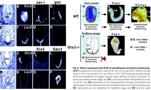
(Acampora et al., 1995; Matsuo et al., 1995; Ang et al., 1996). Heterozygous Otx2+/- embryos in an appropriate genetic back-ground show defects of the head such as serious brain abnormalities and craniofacial malformations, which are reminiscent of otocephalic phenotypes (Matsuo et al., 1995). The analysis of Otx2 null embryos revealed that at late streak stage, the rostral neuroectoderm is not specified and the primitive streak as well as the node and the axial mesendoderm are severely impaired. Therefore, one possibility is that the resulting headless phenotype might be due to the abnormal development of node-derived axial mesendoderm which lacks head organizer activity. A second possibility is that neural inducing prop-erties of the AVE are severely impaired or abolished. In embryos replacing Otx2 with the lacZ reporting gene, the first abnormality is already detected at the early streak stage (Acampora et al., 1995). Indeed, at this stage, lacZ transcription and staining are abolished in the epiblast while they remain high in the VE of Otx2-/- embryos, the goosecoid (gsc) expression is undetectable or confined to the proximal region of the mutant embryos (Acampora et al., 1995) and the presumptive AVE does not anteriorize, thus remaining confined to the distal region of the embryo (Fig. 2). Therefore, these results indicate that headless phenotype and abnormal organization of the primitive streak may be determined very early at the pre-early-streak stages by an impairment of AVE properties.
Moreover, as revealed by the primitive streak early marker Brackyury (T), epiblast cells do not migrate posteriorly at the site of the primitive streak formation but remain for an abnormal longer period in the circumpherential ring close to embryonic-extraembry-onic boundary. Only later and in a fraction of Otx2-/- embryos the presumptive AVE cells appear anteriorized. However, although abnormal, a primitive streak forms in all the Otx2-/- embryos (Acampora et al., 1995, 1998b; Ang et al., 1996). Indeed, mesodermal cells appear concentrated at the posterior third of the embryo and both the node and axial mesendoderm are heavily abnormal or absent as detected by histological and molecular analyses (Acampora et al., 1995; Ang et al., 1996).
These findings, therefore, favor a relation between primitive streak formation and anterior location of the AVE and suggest that Otx2, normally expressed in both epiblast cells and AVE, might be required in these two cell types in order to mediated proper position-ing of the AVE and normal formation of the primitive streak. However, data deduced from the analysis of chimeric embryos and further mouse models indicate that early abnormalities in both AVE and primitive streak of Otx2-/- embryos should be ascribed to Otx2 requirement in the VE (Acampora et al., 1998b; Rhinn et al., 1998; see below).
An intriguing feature of Otx2-/- embryos is that lack of anterior neuroectoderm correlates with failure of epiblast cells to express the lacZ reporter gene. Thus, since Otx2 is already transcribed from the earliest stages (unfertilized egg in Xenopus and at least morula in mouse), this observation suggests that maintenance of Otx2 tran-scription in the epiblast cells requires at least one normal allele expressed in the AVE while Otx2 transcription in the latter is indepen-dent from the presence of a normal allele (Simeone, 1998; Acampora and Simeone, 1999). Moreover, these findings support the existence of Otx2-mediated signal(s) emitted from the AVE and directed to the epiblast cells (Fig. 2). Nevertheless, it is still unclear whether the same signal operates to mediate both specification of anterior patterning and Otx2 transcription in epiblast cells or these two events are independently controlled.
The role of Otx2 in induction and maintenance of rostral CNS
In order to understand the role of Otx2 in the specification of the anterior patterning, it is of particular relevance to define its func-tional contribution to the different tissues where it is expressed during gastrulation. More direct evidence proving a role for Otx2 in the AVE has been recently provided by generating murine chimeric embryos and new mouse models (Acampora et al., 1998b; Rhinn et al., 1998; Acampora et al., unpublished results). Chimeric embryos containing Otx2-/- epiblast and wild-type VE rescue an early Otx2-/- neural plate but subsequently they fail to develop a brain, suggesting that Otx2 is required in the AVE for induction of rostral neural plate and, subsequently, in the epiblast-derived tissues for specification of forebrain and midbrain regional identi-ties. Conversely, when chimeric embryos consist of an Otx2-/- VE and an Otx2+/+ epiblast, none of the phenotypic features of Otx2-/- embryos are rescued (Rhinn et al., 1998). This latter result also argues, as previously suggested by Otx2-/- mice (Acampora et al., 1995), that impaired axial mesendoderm of Otx2-/- embryos is a consequence of Otx2 requirement at earlier stages in the VE.
Mice replacing Otx2 with Otx1 were originally generated in order to assess whether the two proteins shared functional equivalence or, alternatively, displayed unique properties specified by their limited amino acid divergence (Acampora and Simeone, 1999). Interestingly, homozygous mutant embryos replacing Otx2 with the human Otx1 (hOtx1) cDNA (hOtx12/hOtx12) recover anterior
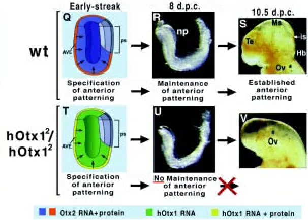
neural plate induction and normal gastrulation but show a headless phenotype from 9 days post coitum (d.p.c.) onwards. A combined analysis of both hOtx1 RNA and protein distribution during early gastrulation has revealed that while the hOtx1 mRNA is detected in the VE and epiblast, the hOTX1 protein is revealed only in VE (Fig. 3). Nevertheless, this VE-restricted translation of the hOtx1 mRNA is sufficient to recover early anterior neural plate but fails to maintain fore-midbrain identities, and consequently hOtx12/hOtx12
embryos display a headless phenotype with a normal body plan (Acampora et al., 1998b). In fact at early-streak stage, hOtx12/
hOtx12 embryos recover anteriorization of the AVE, normal
primi-tive streak and proper expression of Lim1, cer-l, Hesx1, gsc and T. At late gastrula stage, the anterior patterning of the rostral neural plate is correctly defined by the expression of fore-, mid- and hindbrain markers such as Six3, Pax2, Gbx2 and Hoxb1 and the axial-mesendoderm properly expresses Lim1, Noggin, cer-l and Hesx1 thus, leading to argue that, as deduced in chimera study, Otx2 is required in the AVE for initiating the specification of the anterior patterning and demarcation of forebrain and midbrain territories (Fig. 3 and compare to Fig. 2) (Acampora et al., 1998b). Further analysis of hOtx12/hOtx12 embryos reveals that at the
early somite stage the forebrain markers BF1 and the hOtx12 mRNA
phases both requiring Otx2: early induction of anterior neural pattern-ing that is under the control of AVE and its subsequent maintenance that is likely mediated by epiblast-derived cells (the axial mesendoderm and neuroectoderm). However, since Otx2 is normally transcribed and translated in both axial mesendoderm and rostral neuroecto-derm, while hOtx12 is transcribed but not translated neither in the
axial mesendoderm nor in the rostral neuroectoderm, it cannot be deduced whether Otx2 is required in one or both of these tissues to mediate maintenance properties of the anterior identity and whether hOtx1 is functionally equivalent to Otx2 also in these tissues. A cell-autonomous role of Otx2 in the neuroectoderm is emerged by the analysis of chimeras with a moderate contribution of Otx2-/- cells (Rhinn et al., 1999). From this analysis it has been shown that genes such as Wnt1, Hesx1 and R-cadherin are selectively not expressed in Otx2-/- cells while other genes such as En2 and Six3 are uniformly expressed in neuroectoderm cells of both genotypes. Nevertheless, this finding does not help to under-stand whether Otx2 plays a non-cell autonomous role in the axial
mesendoderm. In this context, it is noteworthy that Lim1 is correctly expressed in the anterior mesendoderm of hOtx12/hOtx12 embryos
and that Lim1-/- and hOtx12/hOtx12 embryos show impressive
phenotypic similarities. Recently, the stage and the cell type in which Lim1 is required for head specification have been approached by chimeras and tissue recombination experiments (Shawlot et al., 1999). These experiments have revealed that Lim1 is required in the AVE for inducing anterior neural pattern and, subsequently, in the axial mesendoderm to refine and maintain the anterior identity. Therefore, since maintenance of anterior pattern requires Otx2 in the axial mesendoderm or in the anterior neuroectoderm or in both tissues, it can be speculated that Lim1 might contribute to mediating the release of axial-mesendoderm signal(s) instructing maintenance of anterior character and Otx2 might confer to the neuroectoderm the competence in responding to this signal(s) emitted from the mesendoderm. This possibility is also compatible with a cell-autono-mous role within the neuroectoderm and does not exclude an additional role also in the axial mesendoderm.
A recent in vitro study supports the possibility of a direct protein-protein interaction between Otx2 C-terminal and Lim1 homeodomain (Nakano et al., 2000). Furthermore, the hOtx12 mouse model
together with findings deduced from the analysis of mice lacking the Cripto gene (Ding et al., 1998) may lead to some tentative considerations on the temporal stability of AVE signal(s). The Cripto gene encodes a membrane-associated protein containing epidermal growth factor-like motifs and is expressed shortly before the onset gastrulation in the proximal region of the epiblast where the primitive streak forms (Dono et al., 1993; Ding et al., 1998; Minchiotti et al., 2000).
Cripto-/- embryos lack a primitive streak and do not anteriorize the AVE thus failing to convert the proximo-distal into antero-posterior axis (Ding et al., 1998). Nevertheless, the AVE markers are correctly expressed but in a distal position and the epiblast does express rostral neuroectoderm markers such as Otx2 and En until 8 - 8.5 d.p.c., thus indicating that in the absence of a node and derived tissues the epiblast acquires the identity of anterior neuroectoderm. Moreover, in hOtx12/hOtx12 embryos, where the
OTX1 protein is detected only in the AVE, anterior pattern is detected until 8 d.p.c. Similar results have been deduced from the analysis of chimeras composed of Otx2-/- epiblast and wild-type VE. Together these findings indicate that i) the AVE, even in an ectopic position may confer the anterior character to the sur-rounding epiblast cells; ii) in the absence of a node and axial mesendoderm, signal(s) emitted from the AVE may be efficient in initiating and maintaining the anterior specification until late streak-early somite stage and iii) Otx2 codes for an important genetic component of the pathway leading the AVE to instruct the anterior patterning.
Otx genes and the evolution of brain complexity
In our opinion, one of the most interesting observations is the existence of a differential post-transcriptional control of the hOtx1 mRNA between the VE and the epiblast cells. In heterozygous hOtx12/Otx2embryos the Otx2 mRNA and protein colocalize while
the hOtx1 mRNA is translated only in the VE, thus suggesting that the hOtx1 mRNA detected in epiblast cells is post-transcriptionally regulated by an Otx2-independent cis-acting control (Acampora et al., 1998b).
In Otx2+/- embryos, the same Otx2 region that is replaced by the hOtx1 cDNA is substituted with the lacZ gene fused to the SV40 polyA site (Acampora et al., 1995). In these embryos the lacZ mRNA is correctly detected in VE and epiblast while the staining is heavily reduced in the epiblast at early-mid streak stage (Acampora et al., 1995). Furthermore, it is noteworthy that recently a new mouse model replacing the same Otx2 region with the Drosophila otd cDNA generates a phenotype displaying impressive similari-ties with that of the hOtx12/hOtx12 embryos. Also in this case, the
otd mRNA is transcribed in epiblast and VE but the protein is detected only in the VE and the VE-restricted OTD protein is sufficient to rescue specification of anterior patterning and normal gastrulation but fails in maintenance of anterior patterning (Acampora et al., unpublished results). Therefore, three different mouse models replacing the same Otx2 genomic region with three different genes (lacZ, hOtx1 and otd) converge towards the possi-bility that VE and epiblast cells are characterized by different post-transcriptional control properties.
Moreover, these mouse models also suggest that the Otx2 replaced region, possibly the 3' UTR, might contain regulatory element(s) required for the Otx2 translation in the epiblast cells. However, such molecular element(s) is actually unknown and in vivo experiments aimed at generating new mouse models specifi-cally addressing this issue will certainly contribute towards un-masking eventual Otx2 post-transcriptional control element(s) that at the moment can be only hypothesized. Nevertheless, since the Otx2 locus is heavily engineered and does not contain introns, 3' UTR and part of the 5' UTR, it could be possible that the loss of mRNA translation in the epiblast might be also influenced by abnormal molecular event(s) affecting RNA stability, processing and transport of the chimeric knock-in transcripts. However, what-ever the impairments are, since in the VE the mRNA is correctly translated, the absence of protein should be considered a peculiar event occurring in epiblast cells and their derivatives.
The finding that Otx2 escapes this post-transcriptional control suggests that this is necessary for the maintenance of fore-midbrain territory specified by the VE (Acampora et al., 1998b) and might have evolutionary implications. Indeed, the architectural components of the vertebrate brain (telencephalon, diencephalon and mesencephalon) are less clear in protochordates and a vertebrate-type brain, which is composed of a midbrain and six prosomers, firstly appears in Petromyzontoidea. It may be hypoth-esized that the specification of this prosomeric brain might have required the presence of a sufficient level of OTX2 protein in the early neuroectoderm cells and this event might have been acquired by modifying post-transcriptional control of Otx2 transcripts rather that its coding sequence which would have retained common functional properties among the otd-related genes throughout evolution.
Comparative studies have demonstrated the existence of otd-related genes in all chordates (Simeone et al., 1992; Li et al., 1994; Bally-Cuif et al., 1995; Mercier et al., 1995; Pannese et al., 1995) including urochordates (Wada et al., 1996), cephalochordates (Williams and Holland, 1996), and agnates, where they are ex-pressed in the rostralmost CNS independently of the complexity acquired by this area during evolution. In urochordates and cephalochordates, only one Otx gene has been identified so far that may be related to Otx2 (Wada et al., 1996; Williams and Holland, 1998). Indeed, in addition to similarities in amino acid sequence and expression, they are both expressed during gastru-lation in endoderm cells which suggests that their oldest and primary role might be to mediate signals required to specify anterior neuroectoderm. Therefore, although this conservation in expres-sion pattern and coding sequence might favor a remarkable role and functional equivalence of otd/Otx genes, it is unclear why the brain territory of protochordates has been so deeply and suddenly modified in a prosomeric territory that has been maintained in its basic topography until mammals (Rubenstein et al., 1998).
(Acampora et al., 1998a); ii) the human Otx1 and Otx2 rescue most of the otd defects in flies (Leuzinger et al., 1998; Nagao et al., 1998; Sharman and Brand, 1998); iii) Otx1 rescues Otx2 requirements in VE (Acampora et al., 1998b); iv) Otx2 rescues most of the Otx1-/ - defects (Acampora et al., 1999); v) the Drosophila otd, similarly to Otx1, rescues Otx2 requirements in the VE (Acampora et al., unpublished results), indicate that Otx1, Otx2 and otd genes show a high degree of functional equivalence in the tissues and body regions where they are properly expressed. Therefore, these data support the notion that otd/Otx functions have been established in a common ancestor of fly and mouse and retained throughout evolution, while copy number and regulatory control of their ex-pression have been modified and re-adapted by evolutionary events that have led to the specification of the vertebrate brain (Sharman and Brand, 1998; Simeone, 1998; Acampora and Simeone, 1999; Hirth and Reichert, 1999; Reichert and Simeone, 1999). Gene duplication and modification of the regulatory control of gene expression may greatly contribute to increase the complex-ity of the body plan and these events appear particularly relevant in the vertebrate evolution from protochordates (Garcia-Fernandez and Holland, 1994; Holland et al., 1994; Williams and Holland, 1998). In fact, drastic evolutionary modifications in copy number and/or expression pattern might likely represent the most rapid and efficient mechanisms to confer morphological changes during embryonic development. Hence, gene duplication may allow the duplicated gene to acquire new specific function(s) either retaining or losing ancestral properties, and similarly, modification of the regulatory control of gene expression may establish new expres-sion patterns which might alter preexisting cell-fates by generating new specialized cellular functions.
A likely consequence of both increased genomic complexity and modification of regulatory control of gene expression may result in an increase in the number of molecular interactions. Such increase in molecular interactions may contribute towards modifying rel-evant morphogenetic processes that in turn may confer a change in shape and size of the body plan as well as in the generation of cell-types with new developmental potentialities. On this basis, Otx gene duplication and subsequent or contemporary modification of regulatory control might have contributed to the evolution of the mammalian brain (e.g. by increasing the extent of neuroectoder-mal territory recruited to form the brain). This event might involve an improvement of proliferative activity of early neuronal progeni-tors (Acampora et al., 1998a, 1999) and/or the positioning of the mes-met boundary (Acampora et al., 1997, 1998b; Suda et al., 1997). Additional property(ies) may be conferred to the duplicated gene by altering its coding sequence. Thus, the limited amino acid divergence between OTX1 and OTX2 might underlie modifications of their functional properties.
In this context, it is noteworthy that mice replacing Otx1 with Otx2 (Acampora et al., 1999) rescued epilepsy, cortical impairments, eye and lachrymal/Harderian gland complex abnormalities, while they never recovered the lateral semicircular canal of the inner ear (Acampora et al., 1996; Morsli et al., 1999), strongly suggesting that, even though Otx1 shares an extended functional equivalence to Otx2 and otd, the ability to specify the lateral semicircular canal of the inner ear might represent a unique property of the Otx1 coding sequence. Further mouse models will assess whether AVE-re-stricted equivalence of Otx1 and otd to Otx2 may be also extended to Otx2 function in epiblast and derived tissues.
Conclusions
A remarkable amount of data indicates that, in mouse, early specification of the anterior patterning is under the control of AVE and that maintenance of this early specification may be contributed by both non-cell autonomous function(s) from the axial mesendoderm and cell autonomous function(s) of the anterior neuroectoderm. This review dealt with the role of Otx2 during early events controlling specification and maintenance of anterior iden-tity. Mouse models (Otx2-/-; hOtx12/hOtx12; otd2/otd2) and chimera
experiments have largely contributed to defining differential roles of Otx2 in the AVE and in epiblast-derived cells. These studies now indicate that Otx2 is a major genetic requirement for early specifi-cation of the anterior identity. Future studies aimed at dissecting the genetic cascade, functional domains and regulatory control will certainly contribute to the understanding of the molecular basis governing development and evolution of the mammalian brain.
Acknowledgments
We thank J. P. Martinez-Barbera for comments and A. Secondulfo for skilful secretarial assistance. This work was supported by the “MURST-CNR Biotechnology Programme Legge 95/95”, the ““MURST-CNR Target Project on Biotechnology” and the EC BIOTECH programme.
References
ACAMPORA, D. and SIMEONE, A. (1999). Understanding the roles of Otx1 and Otx2 in controlling brain morphogenesis. TINS 22: 116-122.
ACAMPORA, D., AVANTAGGIATO, V., TUORTO, F. and SIMEONE, A. (1997). Genetic control of brain morphogenesis through Otx gene dosage requirement. Development 124: 3639-3650.
ACAMPORA, D., AVANTAGGIATO, V., TUORTO, F., BARONE, P., PERERA, M., CORTE, G. and SIMEONE, A. (1999). Differential transcriptional control as the major molecular event in generating Otx1-/- and Otx2-/- divergent phenotypes. Development 126: 1417-1426.
ACAMPORA, D., AVANTAGGIATO, V., TUORTO, F., BARONE, P., REICHERT, H., FINKELSTEIN, R. and SIMEONE, A. (1998a). Murine Otx1 and Drosophila otd genes share conserved genetic functions required in invertebrate and vertebrate brain development. Development 125: 1691-1702.
ACAMPORA, D., AVANTAGGIATO, V., TUORTO, F., BRIATA, P., CORTE, G. and SIMEONE, A. (1998b). Visceral endoderm-restricted translation of Otx1 mediates recovering of Otx2 requirements for specification of anterior neural plate and proper gastrulation. Development 125: 5091-5104.
ACAMPORA, D., MAZAN, S., AVANTAGGIATO, V., BARONE, P., TUORTO, F., LALLEMAND, Y., BRÛLET, P. and SIMEONE, A. (1996). Epilepsy and brain abnormalities in mice lacking Otx1 gene. Nat. Genet. 14: 218-222.
ACAMPORA, D., MAZAN, S., LALLEMAND, Y., AVANTAGGIATO, V., MAURY, M., SIMEONE, S. and BRÛLET, P. (1995). Forebrain and midbrain regions are deleted in Otx2-/- mutants due to a defective anterior neuroectoderm specification during gastrulation. Development 121: 3279-3290.
ANG, S.-L., CONLON, R.A., JIN, O. and ROSSANT, J., (1994). Positive and negative signals from mesoderm regulate the expression of mouse Otx2 in ectoderm explants. Development 120: 2979-2989.
ANG, S.-L., JIN, O., RHINN, M., DAIGLE, N., STEVENSON, L. and ROSSANT, J. (1996). Targeted mouse Otx2 mutation leads to severe defects in gastrulation and formation of axial mesoderm and to deletion of rostral brain. Development 122: 243-252.
AVANTAGGIATO, V., ACAMPORA, D., TUORTO, F. and SIMEONE, A. (1996). Retinoic acid induces stage-specific repatterning of the rostral central nervous system. Dev. Biol. 175: 347-357.
BEDDINGTON, R.S.P. (1981). An autoradiographic analysis of the potency of embry-onic ectoderm in the 8th day postimplantation mouse embryos. J. Embryol. Exp. Morphol. 64: 87-104.
BEDDINGTON, R.S.P. (1994). Induction of a second neural axis by the mouse node. Development 120: 613-620.
BEDDINGTON, R.S.P. and ROBERTSON, E.J. (1999). Axis development and early asymmetry in mammals. Cell 96: 195-209.
BELO, J.A., BOUWMEESTER, T., LEYNS, L., KERTESZ, N., GALLO, M., GOLLETTIE, M. and DE ROBERTIS, E.M. (1997) Cerberus-like is a secreted factor with neuralizing activity expressed in the anterior primitive endoderm of the mouse gastrula. Mech. Dev. 68: 45-57.
BLITZ, I.L. and CHO, K.W.Y. (1995). Anterior neuroectoderm is progressively induced during gastrulation: the role of the Xenopus homeobox gene orthodenticle. Devel-opment 121: 993-1004.
CHEN, S., WANG, Q.-L., NIE, Z., SUN, H., LENNON, G., COPELAND, N.G., GILBERT, D.J., JENKINS, N.A. and ZACK, D.J. (1997). Crx, a novel Otx-like paired-homeodomain protein, binds to and transactivates photoreceptor cell-specific genes. Neuron 19: 1017-1030.
CONLON, R.A. and ROSSANT, J. (1992). Exogenous retinoic acid rapidly induces anterior ectopic expression of murine Hox-2 genes in vivo. Development 116: 357-368.
DATTANI, M.T., MARTINEZ-BARBERA, J.-P., THOMAS, P.Q., BRICKMAN, J.M., GUPTA, R., MÅRTENSSON, I.-L., TORESSON, H., FOX, M., WALES, J.K.H., HINDMARSH, P.C., KRAUSS, S. BEDDINGTON, R.S.P. and ROBINSON, I.C.A.F. (1998). Mutations in the homeobox gene HESX1/Hesx1 associated with septo-optic dysplasia in human and mouse. Nat. Genet. 19: 125-133.
DING, J., YANG, L., YAM, Y.-T., CHEN, A., DESAI, N., WYNSHAW-BORIS, A. and SHEN, M.M. (1998) Cripto is required for correct orientation of the anterior-posterior axis in the mouse embryo. Nature 395: 702-707.
DONIACH, T. (1993). Planar and vertical induction of antero-posterior pattern during the development of the amphibian central nervous system. J. Neurobiol. 24: 1256-1276.
DONO, R., SCALERA, L., PACIFICO, F., ACAMPORA, D., PERSICO, M.G. and SIMEONE, A. (1993). The murine cripto gene: expression during mesoderm induction and early heart morphogenesis. Development 118: 1157-1168.
FINKELSTEIN, R. and BONCINELLI, E. (1994). From fly head to mammalian forebrain: the story of otd and Otx. Trends Genet. 10: 310-315.
FREUD, C.L., GREGORY-EVANS, C.Y., KURUKAWA, T., PAPAIOANNOU, M., LOOSER, J., PLODER, L., BELLINGHAM, J., NG, D., HERBRICK, J.-A.S., DUNCAN, A., SCHERER, S.W., TSUI, L.-C., LOUTRADIS-ANAGNOSTOU, A., JACOBSON, S.G., CEPKO, C.L., BHATTACHARYA, S.S. and McINNES, R.R. (1997). Cone-rod dystrophy due to mutations in a novel photoreceptor-specific homeobox gene (CRX) essential for maintenance of the photoreceptor. Cell 91: 543-553.
GALLERA, J. (1971). Primary induction in birds. Adv. Morph. 9: 149-180.
GAMMILL, L.S. and SIVE, H. (1997). Identification of otx2 target genes and restric-tions in ectodermal competence during Xenopus cement gland formation. Devel-opment 124: 471-481.
GARCIA-FERNANDEZ, J. and HOLLAND, P.W.H. (1994). Archetypal organization of the amphioux Hox gene cluster. Nature 370: 536-566.
GURDON, J.B. (1987). Embryonic induction - molecular prospects. Development 99: 285-306.
HIRTH, F. and REICHERT, H. (1999). Conserved genetic programs in insect and mammalian brain development. BioEssays 21: 677-684.
HOLLAND, P.W.H., GARCIA-FERNANDEZ, J., WILLIAMS, N.A. and SIDOW, A. (1994). Gene duplications and the origins of vertebrate development. Dev. Suppl. 125-133.
HOUART, C., WESTERFIELD, M. and WILSON, S.W. (1998). A small population of anterior cells patterns the forebrain during zebrafish gastrulation. Nature 391: 788-792.
KNÖTGEN, H., VIEBAHN, C. and KESSEL, M. (1999). Head induction in the chick by primitive endoderm of mammalian, but not avian origin. Development 126: 815-825.
LAWSON, K.A., MENESES, J.J. and PEDERSEN, R.A. (1991). Clonal analysis of the epiblast during germ layer formation in the mouse embryo. Development 113: 763-769.
LEUZINGER, S., HIRTH, F., GERLICH, D., ACAMPORA, D., SIMEONE, A., GEHRING, W., FINKELSTEIN, R., FURUKUBO-TOKUNAGA, K. and REICHERT. H. (1998). Equivalence of the fly orthodenticle gene and the human OTX genes in embryonic brain development of Drosophila. Development 125: 1703-1710.
LI, Y., ALLENDE, M.L., FINKELSTEIN, R. and WEINBERG, E.S. (1994). Expression of two zebrafish orthodenticle-related genes in the embryonic forebrain. Mech. Dev. 48: 229-244.
MARSHALL, H., NONCHEV, S., SHAM, M.H., MUCHAMORE, I., LUMSDEN, A. and KRUMLAUF, R. (1992). Retinoic acid alters hindbrain Hox code and induces transformation of rhombomeres 2/3 into 4/5 identity. Nature 360: 737-741.
MATSUO, I., KURATANI, S., KIMURA, C., TAKEDA, N. and AIZAWA, S. (1995). Mouse Otx2 functions in the formation and patterning of rostral head. Genes Dev. 9: 2646-2658.
MERCIER, P., SIMEONE, A., COTELLI, F. and BONCINELLI, E. (1995). Expression patterns of two Otx genes suggest a role in specifying anterior body structures in zebrafish. Int. J. Dev. Biol. 39: 559-573.
MILLET, S., BLOCH-GALLEGO, E., SIMEONE, A. AND ALVARADO-MALLART, R.-M. (1996). Is the caudal limit of Otx2 gene expression a marker of the midbrain/ hindbrain boundary? A study using a chick-Otx2 riboprobe and chick/quail homo-topic grafts. Development 122: 3785-3797.
MINCHIOTTI, G., PARISI, S., LIGUORI, G., SIGNORE, M., LANIA, G., ADAMSON, E.D., LAGO, C.T. and PERSICO, M.G. (2000). Membrane-anchorage of Cripto protein by glycosylphosphatidylinositol and its distribution during early mouse development. Mech. Dev. 90: 133-142.
MORSLI, H., TUORTO, F., CHOO, D., POSTIGLIONE, M.P., SIMEONE, A. and WU, D.K. (1999). Otx1 and Otx2 activities are required for the normal development of the mouse inner ear. Development 126: 2335-2343.
NAGAO, T., LEUZINGER, S., ACAMPORA, D., SIMEONE, A., FINKELSTEIN, R., REICHERT, H. and FURUKUBO-TOKUNAGA, K. (1998). Developmental rescue of Drosophila cephalic defects by the human Otx genes. Proc. Natl. Acad. Sci USA 95: 3737-3742.
NAKANO, T., MURATA, T., MATSUO, I. and AIZAWA, S. (2000). OTX2 directly interacts with Lim1 and HNF-3β. Biochem. Bioph. Res. Comm. 267: 64-70. NIEUWKOOP, P.D., JOHNEN, A.G. and ALBERS, B. (1985). The Epigenetic Nature
of Early Chordate Development. Inductive Interactions and Competence. Oxford University Press, Oxford.
PANNESE, M., POLO, C., ANDREAZZOLI, M., VIGNALI, R., KABLAR, B., BARSACCHI, G. and BONCINELLI, E. (1995). The Xenopus homologue of Otx2 is a maternal homeobox gene that demarcates and specifies anterior body regions. Development 121: 707-720.
REICHERT, H. and SIMEONE, A. (1999). Conserved usage of gap and homeotic genes in patterning the CNS. Curr. Opin. Neurobiol. 9: 589-595.
RHINN, M., DIERICH, A., LE MEUR, M. and ANG, S.-L. (1999). Cell autonomous and non-cell autonomous functions of Otx2 in patterning the rostral brain. Development 126: 4295-4304.
RHINN, M., DIERICH, A., SHAWLOT, W., BEHRINGER, R.R., LE MEUR, M. and ANG, S.-L. (1998). Sequential roles for Otx2 in visceral endoderm and neuroectoderm for forebrain and midbrain induction and specification. Development 125: 845-856.
RUBENSTEIN, J.L.R., SHIMAMURA, K., MARTINEZ, S. and PUELLES, L. (1998). Regionalization of the prosencephalic neural plate. Annu. Rev. Neurosci. 21: 445-477.
RUIZ I ALTABA, A. (1993). Induction and axial patterning of the neural plate: planar and vertical signals. J. Neurobiology 24: 1276-1304.
RUIZ I ALTABA, A. (1994). Pattern formation in the vertebrate neural plate. Trends Neurosci. 17: 233-243.
RUIZ I ALTABA, A. (1998). Deconstructing the organizers. Nature 391: 748-749.
SHARMAN, A.C. and BRAND, M. (1998). Evolution and homology of the neuron system: cross-phylum rescues of otd/Otx genes. TIG 14: 211-214.
SHAWLOT, W. and BEHRINGER, R.R. (1995). Requirement for Lim-1 in head organizer function. Nature 374: 425-430.
SHAWLOT, W., WAKAMIYA, M., KWAN, M.K., KANIA, A., JESSELL, T.M. and BEHRINGER, R.R. (1999). Lim1 is required in both primitive streak-derived tissues and visceral endoderm for head formation in the mouse. Development 126: 4925-4932.
SIMEONE, A., ACAMPORA, D., GULISANO, M., STORNAIUOLO, A. and BONCINELLI, E. (1992). Nested expression domains of four homeobox genes in developing rostral brain. Nature 358: 687-690.
SIMEONE, A., ACAMPORA, D., MALLAMACI, A., STORNAIUOLO, A., D’APICE, M.R., NIGRO, V. and BONCINELLI, E. (1993). A vertebrate gene related to orthodenticle contains a homeodomain of the bicoid class and demarcates anterior neuroectoderm in the gastrulating mouse embryo. EMBO J. 12: 2735-2747.
SIMEONE, A., AVANTAGGIATO, V., MORONI, M.C., MAVILIO, F., ARRA, C., COTELLI, F., NIGRO, V. AND ACAMPORA, D. (1995). Retinoic acid induces stage-specific antero-posterior transformation of rostral central nervous system. Mech. Dev. 51: 83-98.
SIVE, H. and CHENG, P. (1991). Retinoic acid perturbs the expression of Xhox.lab genes and alters mesodermal determination in Xenopus laevis. Genes Dev. 5: 1321-1332.
SPEMANN, H. and MANGOLD, H. (1924). Über induktion von Embryonanlagen durch Implantation artfremder Organisatoren. Wilhelm Roux Arch. Entw. Mech. Organ. 100: 599-638.
STOREY, K.G., CROSSLEY, J.M., DE ROBERTIS, E.M., NORRIS, W.E. and STERN, C.D. (1992). Neural induction and regionalisation in the chick embryo. Development 114: 729-741.
SUDA, Y., MATSUO, I. and AIZAWA, S. (1997). Cooperation between Otx1 and Otx2 genes in developmental patterning of rostral brain. Mech. Dev. 69: 125-141.
TAM, P.P.L., STEINER, K.A. and QUINLAN, G.A. (1997). Lineage and functional analyses of the mouse organizer. In Cold Spring Harbor Symposia on Quantitative Biology LXII, pp. 135-144, Cold Spring Harbor: Cold Spring Harbor Laboratory Press.
THOMAS, P. and BEDDINGTON, R. (1996). Anterior primitive endoderm may be responsible for patterning the anterior neural plate in the mouse embryo. Curr. Biol. 6: 1487-1496.
THOMAS, P., BROWN, A. and BEDDINGTON, R.S.P. (1998). Hex: a homeobox gene revealing peri-implantation asymmetry in the mouse embryo and an early transient marker of endothelial cell precursors. Development 125: 85-94.
VARLET, I., COLLIGNON, J. and ROBERTSON, E.J. (1997). nodal expression in the primitive endoderm is required for specification of the anterior axis during mouse gastrulation. Development 124: 1033-1044.
WADA, S., KATSUYAMA, Y., SATO, Y., ITOH, C. and SAIGA, H. (1996). Hroth, an orthodenticle-related homeobox gene of the ascidian, Halocynthia roretzi: its expression and putative roles in axis formation during embryogenesis. Mech. Dev. 60: 59-71.
WADDINGTON, C.H. (1932). Experiments on the development of chick and duck embryos, cultivated in vitro. Phil. Trans. Roy. Soc. Lond. B 221: 179-230.
WILLIAMS, N.A. and HOLLAND, P.W.H. (1996). Old head on young shoulders. Nature 383: 490.