Regeneration and pattern formation
-an interview with Sus-an Bry-ant
MICHAEL K. RICHARDSON*
,1and CHENG-MING CHUONG
21Institute of Biology, Leiden University, Leiden, The Netherlands and
2Department of Pathology, Keck School of Medicine, University of Southern California, Los Angeles, California, USA
ABSTRACT Susan Bryant is one of the leading researchers in regeneration and pattern formation. Born in England in 1943, she studied biology at King’s College, London (UK). After a Ph.D. with Angus Bellairs on caudal autotomy and regeneration in lizards, she researched urodele regenera-tion in Marcus Singer's lab at Case Western Reserve University. Then, at the University of California, Irvine, she adopted the axolotl as a research model for limb regeneration and pattern formation. Her work supported models involving the intercalation of positional values in a polar coordinate system. Fibroblasts, often regarded as "junk" cells, are seen by Susan Bryant as central to patterning. She argues that fibroblasts express positional values needed for regeneration. She also argues that vertebrate species capable of regeneration have evolved steps to plug back into developmental programmes. Susan Bryant thinks that regeneration is essential for a full under-standing of development, and believes that developmental biology has suffered though not embracing regeneration. She also believes that deeper knowledge of pattern formation will bring advances in emerging field of tissue engineering. Since 2000, she has served as Dean of Biological Sciences and more recently, as Vice Chancellor for Research, at UC Irvine (USA). She is an advocate of equal opportunities for women and other under-represented groups in academia. She lives in California with husband David Gardiner, her scientific partner for over 20 years. They have two children. We interviewed Susan Bryant in her office in Irvine on October 5th, 2007.
KEY WORDS:
pattern formation, urodele, regeneration, interview, positional information
Sue, we’d love to know a little about where you came from and where you grew up.
I was born, and grew up, in Northern England in Rotherham and Sheffield, in a working class family. Along the way, I was fortunate to have encountered a couple of inspirational teachers. The first was in elementary school. This teacher took us on trips to collect plants that we subsequently pressed and drew. That was the beginning of my lifelong interest in both biology and art. Later I had to choose between them. In high school I had a teacher who taught about the big ideas in biology and that really inspired me to want to do research, so in high school I decided that I wanted to pursue biology.
I was the first person in my family to go to college. I went to King’s College, University of London (UK), to study zoology (with a minor in psychology), and one of my professors there was Lewis Wolpert. His style of teaching was both Socratic and inspirational. Instead of feeding us facts, he gave us data to make us think. Lewis was also one of my tutors and I chose regeneration as one
BIOLOGY
www.intjdevbiol.com*Address correspondence to: Michael K. Richardson. Institute of Biology, Leiden University, Kaiserstraat 63, Leiden, 2311GP, The Netherlands. Tel: +31-71-527-5215. Fax: +31-71-527-4900. e-mail: [email protected]
Published online: 22 June 2009.
ISSN: Online 1696-3547, Print 0214-6282
© 2009 UBC Press Printed in Spain
of my essays for him. I dug out all the literature in the library— these were the days before photocopiers — and I came back with an essay that discussed everything we knew at that time about regeneration. Lewis threw the essay on the table and said ‘if you think this is what regeneration is all about, how is it that we still don’t know how regeneration works?’ It was a combination of that
essay and Lewis’ ground-breaking work on pattern formation that got me interested in regeneration. He was very influential in my thinking.
Were Lewis, you and others at that time making an explicit link between limb development and limb regeneration?
people working in regeneration, and that has allowed develop-ment to go off in a different direction, which doesn’t make any sense because regeneration is development also. I was once
at a conference in the late eighties, at a time when the universality of developmental mechanisms was really becom-ing graphically clear, and everyone in developmental biology was reading the Drosophila literature. Some graduate students
at that conference, from chick limb labs, told me that they were
"so glad regeneration had nothing to do with development"
because that meant that they didn’t have to also read all the regeneration literature! If a thought-leader like Lewis had championed and incorporated regeneration, I think both fields would be in different places than they are now.
That took you to thinking about a Ph.D.?
Right. I wanted to do a Ph.D. in regeneration and I went to the lab of Angus Bellairs1, a first class comparative anatomist,
who was working on caudal autotomy [lit. self-severing, Ed.] in lizards. I got a grounding in the basic histology, anatomy and evolution of vertebrates. Meanwhile, Angus was interested in regeneration in the adult and embryonic lizard tail, so I was able to work on that system (Bellairs and Bryant 1968; Bryant and Bellairs 1970). I was married to Pete Bryant at that time — yes, he is also a Brit. We had met as undergraduates at King’s College, and he completed a D.Phil. at Sussex. He and I were looking for postdoc positions in the same geographical loca-tion. The one place where we both found terrific mentors was at Case Western Reserve University in Cleveland, Ohio (USA). I joined Marcus Singer2, a regeneration pioneer whose work
demonstrated the role of the nerve in regeneration in amphib-ians. Singer was working on newts (Notophthalmus viridescens)
— I didn’t switch to axolotls (Ambystoma mexicanum) until
later. The Singer lab was my introduction to regeneration in amphibians (Bryant et al. 1971; Singer and Bryant 1969). Pete
joined Howard Schneiderman3, a pioneer in promoting
Droso-phila as a genetic model for developmental biology, and in his
lab did some of the first developmentally motivated experi-ments4 on Drosophila — working on imaginal discs. A couple
of years after we arrived in California, Howard was recruited as Dean of Biological Sciences at UC, Irvine — both Pete and I moved with Howard, joined the faculty, and made our careers at Irvine. We both remarried over 20 years ago.
What was it like at Irvine back in the early days?
The Developmental Biology Center at Irvine was the place
to be for developmental biology at that time. It was physically located slightly off-campus; it was special because it was isolated, and it was all developmental biology, all the time. The front door opened into the seminar room so you couldn’t get in or out gracefully without participating in group activities; and that was where we all ate lunch together and talked about ideas and science. At Irvine, I started to work on axolotls. The reasons were the same then as now: they are easy to breed in captivity; there is a federally-funded resource5; and the
ani-mals do not need to be collected from the wild, thereby avoiding contributing to the decline of wild populations. Newts are difficult to breed and there are no genetic resources for them. We work on axolotls because it is the ‘lab rat’ of regeneration.
You emphasize in your research that regeneration is linked to pattern formation, and you particularly championed intercalation — in some respects an alternative to morpho-gen-based positional information models.
I have never found a reason to give up on intercalation6.
[intercalation is a type of regeneration in which missing positional values are restored where the amputation surfaces are in contact – Ed.] Local cell-cell interactions continue to provide the best fit to the data. And as time has gone on, the idea that there is long-range chemical signalling has just evaporated. It has not found the evidence that was needed, but because of its simplicity and elegance, it survived as a dominant view for a very long time. Work on amphibian regeneration has led us to the idea that the pattern of the body is encoded in a 2-dimensional folded sheet of fibroblasts into which other cell types are recruited — includ-ing specialized cell types such as muscle, nerves and blood vessels. The 2D fibroblast grid controls the patterning and
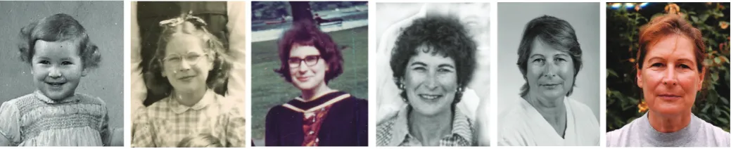
Fig. 1 Susan Bryant. From left to right: Second birthday (1945), taken in a studio in a department store in Sheffield, UK. School photo at Oakwood Primary School, Sheffield, UK (1952). Celebrating after graduating from King’s College University of London with a B.Sc. in Zoology (1964); the ceremony was held in the Royal Albert Hall. From a group photo of the Developmental Biology Center at UC Irvine, when Howard Schneiderman was the Director (c. 1978). Taken while Susan served as Chair of the Department of Developmental and Cell Biology (early 1990’s). Susan in her garden in Newport Beach (early 2000’s) . Photos courtesy Susan Bryant.
Notes: 1Angus d’Albini Bellairs (1918–1990). See obituary by Bryant (1991b). 2 Marcus Singer (1914-1994). See (Nordlander et al. 1995). 3 Howard A.
Schneiderman (1927-1990); for obituary, see Bryant (1991a). 4 See Bryant et al. (1978); Bryant and Schneiderman (1969); Kirby et al. (1982); Kirby et
al. (1983); Poodry et al. (1971); Schneiderman and Bryant (1971). 5 Ambystoma Genetic Stock Center, University of Kentucky (Smith et al. 2005). 6See
behaviour of non-fibroblasts. In amphibian regeneration, fibro-blasts alone, plus epidermis are sufficient to form a normally patterned but muscle-less limb.
You published a paper with Dave Gardiner and Ken Muneoka where you highlighted a major contribution of the dermal fibroblasts in the axolotl regenerating limb (Gardiner et al. 1986). This is different from the chick limb bud where the progress zone is doing the work.
Right, but where does the progress zone come from? The chick limb progress zone comes from the superficial layer of the flank, so I think we are talking about the same thing in both cases: a layer of superficially located mesenchyme cells. In the embryo, this layer is called the lateral plate mesoderm, and in the regenerate, it is called the superficial dermis. As the limb bud grows out, I would argue that it does so as we have described in regeneration — from a zone beneath the apical epidermis at the centre of the wound or centre of the limb field, where cells with different positional identities in the circumference and along the proximal-distal axis are in contact and are interacting and making the pattern. The recent evidence that fibroblasts can be converted into pluripotent cells is perfectly in line with the idea that fibro-blasts, which have always been regarded as the ‘junk’ cells of the body, are actually the master pattern formation cells. That’s what we have been saying from the earliest days of regeneration because that is their role in regeneration, and hence why would they not also have the same role in development?
What we think is happening in regeneration is that the dermal fibroblasts are forming the early part of the blastema, and then the de-differentiated precursors of specialised cell types move in. But
the first cells to migrate are the dermal fibroblasts and they form the niches needed by all other cells. If you regenerate an entire limb from fibroblasts, it doesn’t develop muscles, it just has muscle sheaths. So the view we developed is that the fibroblasts have the instructions for the pattern. They create this 2D sheet and, as in the polar coordinate model, they provide positional cues.
I don’t see why this should be different in development. In chick limbs what is different is that you can’t tell what the cells are
because they are undifferentiated. But they are all mesenchymal, Fig. 2. Susan Bryant, with axolotl. June, 2002, in Susan’s lab in McGaugh Hall, University of California, Irvine. Photo: copyright ©2008, Paul R. Kennedy.
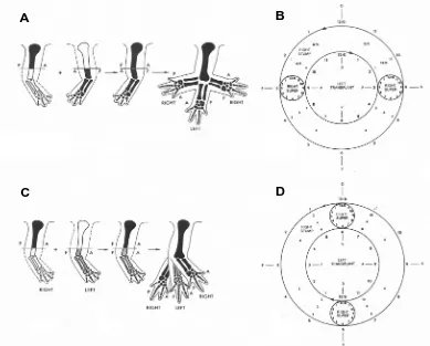
B
C
D
A
Fig. 3. Polar coordinate model appliedand so what we are talking about in the embryo limb bud is that the mesenchymal precursors of fibroblasts are controlling the pat-tern. Expression profiling (Rinn et al. 2006) is showing that
fibroblasts from different parts of the body have different patterns of gene expression, making it feasible that they could have sufficient diversity to provide the patterning template of the body.
When people juxtapose different chick limb bud regions, they sometimes get a duplication. Could that be evidence of intercalation?
Yes, because you get growth in between. The thing about grafting experiments is that interactions are very local. All pattern formation models that exclude growth are missing one of the major events of development.
For the last 25 years or so, I have had a collaborative research program with my husband David Gardiner. The experiments going on now are funded by a team grant from the Defense Advanced Research Projects Agency (DARPA); David is a prin-cipal investigator and Ken Muneoka, at Tulane University, is head of the team.
One of the things that has held back regeneration research is that the animals that do it best, do it whenever they are injured. What Dave and his group have developed is an axolotl system in which regeneration can be broken down into a series of steps, making it possible to look for factors that promote to the next step
(Endo et al. 2004). For example, there is now an assay to help
identify factors that can substitute for the nerve in the formation of a blastema.
What about the zebrafish; do you think that zebrafish caudal
fin regeneration is a good model?
I don’t think it is as useful as regenerating appendages in axolotls, based on the biology. Zebrafish fin regeneration is a more limited kind of regeneration — equivalent to regenerating the tips of digits, a feat that it shares with mice and humans — rather than regenerating an entire appendage. Ken Muneoka has been using mouse digit tips as a mammalian model because they do regenerate digit tips, and have the necessary genetics (Han et al. 2003; Reginelli et al. 1995). Zebrafish may well prove important
in contributing to figuring out common mechanisms. But person-ally, I have always been more interested in understanding how to regenerate a limb.
What is the Holy Grail in your field? To trigger the formation of a human blastema perhaps?
Yes, and I now believe it is possible. I didn’t believe that in the early days. But current advances in genomics and technology are so profound that it must be possible. Its clear that there are a few steps right at the beginning of regeneration that make the decision between regeneration and wound healing. I don’t believe there necessarily will be special ‘regeneration genes’, and I don’t think regeneration is ‘primitive’. I think the difference between urodeles and mammals is in the way that genes are deployed. I think there is something special about urodeles; they have evolved steps that have allowed them to plug back in to developmental pathways. There are a few steps at the beginning of wound healing and regeneration that are not like development, however, and so it is the linkage back to development that they have evolved. I think
regeneration is adaptive in the simple sense that having a missing part grow back has to be useful. In mammals, fibroblasts don’t participate in regeneration; they form scar tissue, so other factors need to be present if they are to make the choice between regeneration and repair.
In mammals we have found that hair can regenerate in the centre of mouse wounds (Chuong, 2007), but we don’t know where the cells come from.
This is an exciting result that suggests that the underlying fibroblasts can interact with the overlying keratinocytes to induce re-development (regeneration) of hair follicles. It indicates that mammalian fibroblasts may have hidden potentials.
Xenopus regeneration ability drops off at metamorphosis. What is the situation concerning metamorphosis and regen-eration in urodeles?
Well, axolotls do not usually go through metamorphosis, Fig. 4. Susan Bryant, photographed November, 2006. Photo:
copy-right ©2008, Paul R. Kennedy.
1943 Born in Halfway (England)
1961-64 Undergraduate in Zoology at King’s College, London (England).
1964-1967 Ph.D. research in the Angus Bellairs’ lab, St. Mary’s Hospital Medical School, London (England)
1967-1969 Postdoctoral research at Case Western Reserve in the Marcus Singer lab.
1969-present University of California, Irvine, Department of Developmental and Cell Biology, and Developmental Biology Center:
Assistant Professor, 1969-1976 Associate Professor, 1976-1980 Professor, 1980-present
Dean of Biological Sciences, 2000-2006 Vice Chancellor for Research, 2006-present
BOX 1
although they can be induced to do so. But other urodeles —
Notophthalmus, for example — do go through metamorphosis,
and can regenerate perfectly. Therefore, regeneration has nothing to do with the presence or absence of metamorphosis. Again, lizard tail regeneration is another completely different type of regeneration, and it occurs in the adult. I actually think that anurans are not too dissimilar from birds and mammals: they can regulate and recover from damage before differentia-tion. What they don’t know how to do very well is to dedifferen-tiate, so they don’t generate the stem cells needed for regen-eration.
developmental pathway.
What about tissue engineering?
By studying urodeles, I hope we can identify the necessary triggers for regeneration, and apply them to humans. But if we can’t activate mammalian fibroblasts in the same way that we can activate amphibian ones, then maybe we will have to piece bits together: some regeneration, some tissue engineering. Success-ful regeneration will require both the ingenuity of engineers, as well as the knowledge of the developmental mechanisms that control pattern formation.
Fig. 5. Dr. Susan Bryant (middle) with Michael Richardson (left) and Cheng Ming Chuong (right). Vice Chancellor’s office at UC Irvine during this interview. October, 5th, 2007.
1967, 70 First publications on regeneration (Bryant, 1970 5466 /id) (Bellairs and Bryant 1968; Bryant and Bellairs 1970)
1969 Nature paper with Marcus Singer, on Schwann cell behaviour (Singer and Bryant 1969)
1971 Ultrastructural study of the effects of denervation in newt limb regeneration (Bryant et al. 1971)
1975-77 Studies with Laurie Iten on regeneration in Notophthalmus, with evidence for intercalary regeneration (Bryant and Iten 1976; Bryant and Iten 1977; Iten and Bryant 1975; Iten and Bryant 1976a; Iten and Bryant 1976b)
1976,81 Science papers (with Vernon French and Peter Bryant) putting forward the polar coordinate model and its relation to the phenomenon of intercalation
(French et al. 1976)
1982 Nature paper with Ken Muneoka arguing for common mechanisms in regeneration and development (Muneoka and Bryant 1982)
1976-80 Nature paper and follow-on studies of regeneration in symmetrical limbs with Nigel Holder, Pat Tank and Gary Krasner (Bryant 1976; Holder et al. 1980; Krasner and Bryant 1980)
1984-89 Research with Ken Muneoka into cellular contribution to supernumerary limb formation in axolotl regeneration (Gardiner and Bryant 1989; Muneoka et al. 1984; Muneoka et al. 1985; Muneoka et al. 1986a; Muneoka et al. 1986b; Muneoka and Bryant 1984a; Muneoka and Bryant 1984b)
1986 Evidence of a role for dermal fibroblasts in axolotl limb regeneration (Gardiner et al. 1986; Muneoka et al. 1986a)
1986-92 Programme of research into development and regulation in mouse embryo limbs, with development of new experimental tools to study mouse limb development in utero
(Gardiner et al. 1992; Muneoka et al. 1986c; Muneoka et al. 1989; Wanek et al. 1989)
1997 Evidence for a link between cell cycle length and positional specification in the chick limb (Ohsugi et al. 1997)
1988 Heterochronic transplants suggest that the decline in Xenopus regeneration capacity is cell autonomous and not related to metamorphosis, and that Xenopus and axolotl have compatible limb patterning mechanisms
(Sessions and Bryant 1988)
1991-2001 Nature paper on role of retinoic acid in limb development, and a series of studies on the roles of retinoic acid and Hox genes in limb regeneration
(Carlson et al. 2001; Gardiner et al. 1995; Hayamizu et al. 1994; Hayamizu and Bryant 1994; Torok et al. 1998)
1999 Sonic hedgehog expression is found to be expressed in the posterior region of the axolotl limb blastema, and to be activated ectopically during intercalary regeneration
(Torok et al. 1999)
2004-present Step-wise model of regeneration, and induction of blastemas by nerves (Endo et al. 2004; Satoh et al. 2007)
BOX 2
SUSAN BRYANT: SELECTED RESEARCH LANDMARKS
Based on what we know of regeneration in different taxa, it seems that regeneration is an adaptation to re-use the existing developmen-tal pathways. What is evolved is the linkage back to these pathways, and that is why regen-eration has different characteristics in different species groups that display it — they may all be independently evolved. Although both urodeles and lizards regenerate their tails, the processes differ greatly. Maybe it is the evolution of the links to re-access developmental programs that are unique, and lead to different outcomes.
Do you have any advice to young scientists?
Focus on the big ideas! Developmental biology has become so detail-orientated that it is hard to see that there is a big picture. It is also helps to have a great teacher. Even though Lewis Wolpert and I have had different views about the way pattern formation works, he still was my main inspiration. I admire his life-long struggle to understand pattern formation (see interview with Lewis Wolpert by M. Richardson in Richardson, 2009) . This was the gift he gave to me, and in that I will always feel a kinship with him. We came to different conclusions about mechanisms, but that is mainly because I work on the regeneration of pattern in adult vertebrates, and his focus has been on the development of pattern in embryos.
What do you see as the future of regeneration research? I am really excited about stem cell biology (Bryant et al. 2002).
Although people are growing stem cells and treating them to form different cell types, they are not necessarily focused on their fibroblast niches. If we are lucky, replacement cells, derived from stem cells and transplanted into a damaged tissue, may find enough of a niche in the surviving tissue remnant to undergo regeneration. Stem cell research today reminds me of the the early days of tissue culture in general. Cultured cells could be caused to differentiate in particular directions, and this became the focus of developmental biology. But of course patterning is about organising the differentiation, and that is why fibroblasts are
so interesting to me — but maybe not to everyone!
References
BELLAIRS, A. A. and BRYANT, S. V. (1968). Effects of amputation of limbs and digits of lacertid lizards. Anat. Rec. 161: 489-495.
BRYANT, P. J. (1991a). In memoriam Howard A. Schneiderman (1927-1990). Dev.
Biol. 146: 1-3.
BRYANT, P. J., ADLER, P. N., DURANCEAU, C., FAIN, M. J., GLENN, S., HSEI, B., JAMES, A. A., LITTLEFIELD, C. L., REINHARDT, C. A., STRUB, S. and SCHNEIDERMAN, H. A. (1978). Regulative interactions between cells from
different imaginal disks of Drosophila melanogaster. Science 201: 928-930.
BRYANT, P. J. and SCHNEIDERMAN, H. A. (1969). Cell lineage, growth, and
determination in the imaginal leg discs of Drosophila melanogaster. Dev. Biol.
20: 263-290.
BRYANT, S. V. (1976). Regenerative failure of double half limbs in Notophthalmus
viridescens. Nature 263: 676-679.
BRYANT, S. V. (1991b). In Memoriam: Angus D’Albini Bellairs 1918-1990.
Ameri-can Zoologist 31: 279-280.
BRYANT, S. V. and BELLAIRS, A. D. (1970). Development of regenerative ability in the lizard, Lacerta vivipara. Am. Zool. 10: 167-173.
BRYANT, S. V., ENDO, T. and GARDINER, D. M. (2002). Vertebrate limb regen-eration and the origin of limb stem cells. Int. J. Dev. Biol. 46: 887-896.
BRYANT, S. V., FYFE, D. and SINGER, M. (1971). The effects of denervation on the ultrastructure of young limb regenerates in the newt, Triturus. Dev. Biol. 24:
577-595.
BRYANT, S. V. and ITEN, L. E. (1976). Supernumerary limgs in amphibians: experimental production in Notophthalmus viridescens and a new interpretation of their formation. Dev. Biol. 50: 212-234.
BRYANT, S. V. and ITEN, L. E. (1977). Intercalary and supernumerary regeneration
in regenerating the mature limbs of Notophthalmus viridescens. J. Exp. Zool.
202: 1-16.
CARLSON, M. R. J., KOMINE, Y., BRYANT, S. V. and GARDINER, D. M. (2001). Expression of Hoxb13 and Hoxc10 in developing and regenerating axolotl limbs and tails. Dev. Biol. 229: 396-406.
CHUONG, C. M. (2007). Regenerative biology: new hair from healing wounds.
Nature 447: 265-266.
ENDO, T., BRYANT, S. V. and GARDINER, D. M. (2004). A stepwise model system for limb regeneration. Dev. Biol. 270: 135-145.
FRENCH, V., BRYANT, P. J. and BRYANT, S. V. (1976). Pattern regulation in
epimorphic fields. Science 193: 969-981.
GARDINER, D. M., BLUMBERG, B., KOMINE, Y. and BRYANT, S. V. (1995. Regulation of HoxA expression in developing and regenerating axolotl limbs.
Development 121: 1731-1741.
GARDINER, D. M. and BRYANT, S. V. (1989). Organization of positional informa-tion in the axolotl limb. J. Exp. Zool. 251: 47-55.
GARDINER, D. M., GAUDIER, C. and BRYANT, S. V. (1992). Mouse limb bud cells respond to retinoic acid in vitro with reduced growth. J. Exp. Zool. 263: 406-413.
GARDINER, D. M., MUNEOKA, K. and BRYANT, S. V. (1986). The migration of
dermal cells during blastema formation in axolotls. Dev. Biol. 118: 488-493.
HAN, M., YANG, X., FARRINGTON, J. E. and MUNEOKA, K. (2003). Digit
regeneration is regulated by Msx1 and BMP4 in fetal mice. Development 130:
5123-5132.
HAYAMIZU, T. F. and BRYANT, S. V. (1994). Reciprocal changes in Hox D13 and RAR-beta 2 expression in response to retinoic acid in chick limb buds. Dev. Biol.
166: 123-132.
HAYAMIZU, T. F., WANEK, N., TAYLOR, G., TREVINO, C., SHI, C., ANDERSON, R., GARDINER, D. M., MUNEOKA, K. and BRYANT, S. V. (1994). Regeneration
of HoxD expression domains during pattern regulation in chick wing buds. Dev.
Biol. 161: 504-512.
HOLDER, N., TANK, P. W. and BRYANT, S. V. (1980). Regeneration of
symmetri-cal forelimbs in the axolotl, Ambystoma mexicanum. Dev. Biol. 74: 302-314.
ITEN, L. E. and BRYANT, S. V. (1975). The interaction between the blastema and stump in the establishment of the anterior—posterior and proximal—distal
organization of the limb regenerate. Dev. Biol. 44: 119-147.
ITEN, L. E. and BRYANT, S. V. (1976a). Regeneration from different levels along
the tail of the newt, Notophthalmus viridescens. J. Exp. Zool. 196: 293-306.
ITEN, L. E. and BRYANT, S. V. (1976b). Stages of tail regeneration in the adult
newt, Notophthalmus viridescens. J. Exp. Zool. 196: 283-292.
KIRBY, B. S., BRYANT, P. J. and SCHNEIDERMAN, H. A. (1982). Regeneration following duplication of imaginal wing disc fragments of Drosophila melanogaster. Dev. Biol. 90: 259-271.
KIRBY, B. S., BRYANT, P. J. and SCHNEIDERMAN, H. A. (1983).
Transdetermination of imaginal wing disc fragments of Drosophila melanogaster.
Dev. Biol. 97: 19-26.
KRASNER, G. N. and BRYANT, S. V. (1980). Distal transformation from double-half
forearms in the axolotl, Ambystoma mexicanum. Dev. Biol. 74: 315-325.
MUNEOKA, K. and BRYANT, S. V. (1982). Evidence that patterning mechanisms
in developing and regenerating limbs are the same. Nature 298: 369-371.
MUNEOKA, K. and BRYANT, S. V. (1984a). Cellular contribution to supernumerary
limbs in the axolotl, Ambystoma mexicanum. Dev. Biol. 105: 166-178.
MUNEOKA, K. and BRYANT, S. V. (1984b). Cellular contribution to supernumerary limbs resulting from the interaction between developing and regenerating tissues in the axolotl. Dev. Biol. 105: 179-187.
MUNEOKA, K., FOX, W. F. and BRYANT, S. V. (1986a). Cellular contribution from
dermis and cartilage to the regenerating limb blastema in axolotls. Dev. Biol.
116: 256-260.
MUNEOKA, K., HOLLER-DINSMORE, G. V. and BRYANT, S. V. (1985). A quantitative analysis of regeneration from chimaeric limb stumps in the axolotl.
J. Embryol. Exp. Morphol. 90: 1-12.
MUNEOKA, K., HOLLER-DINSMORE, G. V. and BRYANT, S. V. (1986b). Pattern discontinuity, polarity and directional intercalation in axolotl limbs. J. Embryol. Exp. Morphol. 93: 51-72.
MUNEOKA, K., WANEK, N. and BRYANT, S. V. (1986c). Mouse embryos develop
normally exo utero. J. Exp. Zool. 239: 289-293.
MUNEOKA, K., WANEK, N. and BRYANT, S. V. (1989). Mammalian limb bud development: in situ fate maps of early hindlimb buds. J. Exp. Zool. 249: 50-54.
mexicanum. Dev. Biol. 105: 240-245.
NORDLANDER, R. H., EGAR, M. W. and BRYANT, S. V. (1995). In memoriam: a
remembrance of Marcus Singer. (1914-1994). Dev. Biol. 169: iv-vi.
OHSUGI, K., GARDINER, D. M. and BRYANT, S. V. (1997). Cell cycle length
affects gene expression and pattern formation in limbs. Dev. Biol. 189: 13-21.
POODRY, C. A., BRYANT, P. J. and SCHNEIDERMAN, H. A. (1971). The
mechanism of pattern reconstruction by dissociated imaginal discs of
Droso-phila melanogaster. Dev. Biol. 26: 464-477.
REGINELLI, A. D., WANG, Y. Q., SASSOON, D. and MUNEOKA, K. (1995). Digit tip regeneration correlates with regions of Msx1 (Hox 7) expression in fetal and
newborn mice. Development 121: 1065-1076.
RICHARDSON M. (2009). Diffusible gradients are out - an interview with Lewis Wolpert. Int. J. Dev. Biol. 53: 659-662. (doi: 10.1387/ijdb.072559mr).
RINN, J. L., BONDRE, C., GLADSTONE, H. B., BROWN, P. O. and CHANG, H. Y. (2006). Anatomic demarcation by positional variation in fibroblast gene
expres-sion programs. PLoS. Genet. 2: e119.
SATOH, A., GARDINER, D. M., BRYANT, S. V. and ENDO, T. (2007). Nerve-induced ectopic limb blastemas in the axolotl are equivalent to
amputation-induced blastemas. Dev. Biol. 312: 231-244.
SCHNEIDERMAN, H. A. and BRYANT, P. J. (1971). Genetic analysis of
develop-mental mechanisms in Drosophila. Nature 234: 187-194.
SESSIONS, S. K. and BRYANT, S. V. (1988). Evidence that regenerative ability is an intrinsic property of limb cells in Xenopus. J. Exp. Zool. 247: 39-44.
SINGER, M. and BRYANT, S. V. (1969). Movements in the myelin Schwann sheath
of the vertebrate axon. Nature 221: 1148-1150.
SMITH, J. J., PUTTA, S., WALKER, J. A., KUMP, D. K., SAMUELS, A. K., MONAGHAN, J. R., WEISROCK, D. W., STABEN, C. and VOSS, S. R. (2005). Sal-Site: integrating new and existing ambystomatid salamander research and
informational resources. BMC. Genomics 6: 181
TOROK, M. A., GARDINER, D. M., IZPISUA-BELMONTE, J. C. and BRYANT, S. V. (1999). Sonic hedgehog (shh) expression in developing and regenerating axolotl limbs. J. Exp. Zool. 284: 197-206.
TOROK, M. A., GARDINER, D. M., SHUBIN, N. H. and BRYANT, S. V. (1998).
Expression of HoxD genes in developing and regenerating axolotl limbs. Dev.
Biol. 200: 225-233.
WANEK, N., MUNEOKA, K., HOLLER-DINSMORE, G., BURTON, R. and BRYANT,
S. V. (1989). A staging system for mouse limb development. J. Exp. Zool. 249:
41-49.
For all the latest on
Pattern Formation research,
see our latest Special Issue
edited by C.-M. Chuong and M.K. Richardson.
Further Related Reading, published previously in the Int. J. Dev. Biol.
See our Special Issue Developmental Morphodynamics edited by Lev Beloussov and Richard Gordon at: http://www.ijdb.ehu.es/web/contents.php?vol=50&issue=2-3
See our Special Issue Limb Development edited by Juan Carlos Izpisúa-Belmonte and Juan Hurlé at: http://www.ijdb.ehu.es/web/contents.php?vol=46&issue=7
Acquisition of plastid movement responsiveness to light during mesophyll cell differentiation
Joanna Augustynowicz, Weronika Krzeszowiec and Halina Gabrys Int. J. Dev. Biol. (2009) 53: 121-127
Diverse miRNA spatial expression patterns suggest important roles in homeostasis and regeneration in planarians
Cristina González-Estévez, Varvara Arseni, Roshana S. Thambyrajah, Daniel A. Felix and A. Aziz Aboobaker Int. J. Dev. Biol. 53: 493-505 (doi: 10.1387/ijdb.082825cg)
Analyses of regenerative wave patterns in adult hair follicle populations reveal macro-environmental regulation of stem cell activity
Maksim V. Plikus, Randall B. Widelitz, Rob Maxson and Cheng-Ming Chuong Int. J. Dev. Biol. 53: 851-862 (doi: 10.1387/ijdb.072564mp)
Planarian regeneration: achievements and future directions after 20 years of research
Emili Saló, Josep F. Abril, Teresa Adell, Francesc Cebriá, Kay Eckelt, Enrique Fernández-Taboada, Mette Handberg-Thorsager, Marta Iglesias, M Dolores Molina and Gustavo Rodríguez-Esteban
Int. J. Dev. Biol. 53: doi: 10.1387/ijdb.072414es (in press)
Origin and proliferation of blastema cells during regeneration of Drosophila wing imaginal discs
Manel Bosch, Jaume Baguñà and Florenci Serras Int. J. Dev. Biol. (2008) 52: 1043-1050
Two msh/msx-related genes, Djmsh1 and Djmsh2, contribute to the early blastema growth during planarian head regeneration
Linda Mannini, Paolo Deri, Vittorio Gremigni, Leonardo Rossi, Alessandra Salvetti and Renata Batistoni
Int. J. Dev. Biol. (2008) 52: 943-952
Distinct patterns of MMP-9 and MMP-2 activity in slow and fast twitch skeletal muscle regeneration in vivo
Malgorzata Zimowska, Edyta Brzoska, Marta Swierczynska, Wladyslawa Streminska and Jerzy Moraczewski
Int. J. Dev. Biol. (2008) 52: 307-314
Origin and proliferation of blastema cells during regeneration of Drosophila wing imaginal discs
Manel Bosch, Jaume Baguñà and Florenci Serras Int. J. Dev. Biol. (2008) 52: 1043-1050
Wnt signaling in hydroid development: ectopic heads and giant buds induced by GSK-3beta inhibitors
Werner Müller, Uri Frank, Regina Teo, Ofer Mokady, Christina Guette and Günter Plickert Int. J. Dev. Biol. (2007) 51: 211-220
Vertebrate limb regeneration and the origin of limb stem cells.
Susan V Bryant, Tetsuya Endo and David M Gardiner Int. J. Dev. Biol. (2002) 46: 887-896
Molecular mechanisms in the control of limb regeneration: the role of homeobox genes.
D M Gardiner and S V Bryant Int. J. Dev. Biol. (1996) 40: 797-805