A REVIEW ON ANTIOXIDANT METHODS
SUNITHA DONTHA*
Department of Pharmaceutical Chemistry, Malla Reddy College of Pharmacy, Maisammaguda, Secunderabad, Telangana, India. Email: [email protected]
Received: 26 May 2016, Revised and Accepted: 28 June 2016 ABSTRACT
To provide an outlook of the various available methods of antioxidant activity.Various available in vitro and in vivo methods are listed and the procedure to perform the method, its mechanism is also explained in brief. 1,1-diphenyl-2-picrylhydrazyl method was found to be used mostly for the
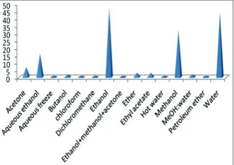
in vitro antioxidant activity evaluation purpose while lipid peroxidation was found as mostly used in vivo antioxidant assay. An ethanol was with the highest frequency as a solvent for extraction purpose. Summarized information on the various methods available provides with reliable information to confirm the benefits of antioxidant effects.
Keywords: Antioxidant activity, Reactive oxygen species, Free radical, 1,1-diphenyl-2-picrylhydrazyl, Flavonoid.
INTRODUCTION
Antioxidants became a vital part of our lives today since antioxidants neutralizes or destroys “reactive oxygen species” (ROS) or free radicals before they damage cells. The oxidation induced by ROS results in cell membrane disintegration, membrane protein damage, and DNA mutations, which results in aging and further initiates or propagates the development of many diseases such as arteriosclerosis, cancer, diabetes mellitus, liver injury, inflammation, skin damages, coronary heart diseases, and arthritis.
The chemical compounds, which decrease the rate of lipid oxidation reaction in food systems, are called antioxidants. By definition, a substance that opposes oxidation or inhibits reactions promoted by oxygen or peroxides; many of these substances being used as preservatives in various products are antioxidants. Biologically antioxidants are defined as synthetic or natural substances added to products to prevent or delay their deterioration by the action of oxygen in air. For example, enzymes or other organic substances such as vitamin E or β-carotene.
Antioxidants are chemical compounds which bind to free oxygen radicals and prevents these radicals from damaging healthy cells.
This review focuses mainly on the types of damaging free radicals generated in metabolic processes and also gives an insight of mechanistic aspect of various in vitro and in vivo methods for the evaluation of antioxidant capacity (Fig. 1).
By the normal use of oxygen [1], free radicals are produced continuously by the body. Oxygen is an element indispensable for life. When cells use oxygen to generate energy, free radicals are produced by the mitochondria. These by-products are generally ROS as well as reactive nitrogen species (RNS) that result from the cellular redox process. The free radicals have a special affinity for lipids, proteins, carbohydrates, and nucleic acids [2].
A free radical is a chemical species, capable of independent existence possessing one or more unpaired electron. The free radicals are less stable than non-radicals and are capable of reacting indiscriminately with molecules. Once radicals are formed, they can either react with another radical or with another non-radical molecule by various interactions. When two radicals collide with their unpaired electron,
radicals. A radical donates its unpaired electron to the other molecules, or takes one electron from it, thus transforming its radical character. At the same time, a new radical is formed [3,4]. ROS/RNS are present in the atmosphere as pollutants and can be generated (i) during ultra-violet (UV) light irradiation, by X-rays and gamma rays; (ii) during metal catalyzed reactions; (iii) by neutrophils, esinophils and macrophages during inflammatory cell activation [5,6]; (iv) as by-products of mitochondrial catalyzed electron transport reactions; (v) by cytochrome P450 metabolism and the enzyme xanthine oxidase, which catalyzes the reaction of hypoxanthine to xanthine and xanthine to uric acid [7].
Depending on the environment and concentration of ROS, it is both harmful and beneficial in biological systems [8,9]. For example, the physiological roles in cellular responses to noxia such as defense against infectious agents, and in the function of a number of cellular signaling systems and gene expression. In contrast, at high concentrations, ROS mediates damage to cell structures including lipids and membranes, proteins, and nucleic acids; which is known as “oxidative stress” [10].
Oxidative stress is defined as an imbalance between the production of free radicals and reactive metabolites, so-called oxidants or ROS, and their elimination by protective mechanisms referred to as antioxidants. This imbalance leads to damage of important biomolecules and cells, with potential impact on the whole organism [11]. The harmful effects of ROS are balanced by the action of antioxidants, example like enzymes present in the body [12]. Despite the presence of the cell’s antioxidant defense system to counteract oxidative damage from ROS, oxidative damage accumulates during the life cycle and has been implicated in diseases, aging and age-dependent diseases such as cardiovascular disease, cancer, neurodegenerative disorders, and other chronic conditions [13].
ROS is classified into oxygen-centered radicals and oxygen-centered non-radicals.
i. Oxygen-centered radicals are superoxide anion (∙O2–), hydroxyl radical (∙OH), alkoxyl radical (RO∙), and peroxyl radical (ROO∙). Other reactive species are nitrogen species such as nitric oxide (NO·), nitric dioxide (NO2∙), and peroxynitrite (OONO–).
ii. Oxygen-centered non-radicals are hydrogen peroxide (H2O2) and
singlet oxygen (1O
2), hypochlorous acid and ozone [14,15].
ROS, which consist of free radicals such as superoxide anion (O2−) and hydroxyl (HO∙) radicals and non-free radical species, such as
Review Article
ROS are produced by all aerobic organisms and can easily react with most biological molecules including proteins, lipids, lipoproteins, and DNA. Thus, the generation of ROS proceeds to a variety of pathophysiological disorders such as arthritis, diabetes, inflammation, cancer, and genotoxicity. Therefore, living organisms possess a number of protective mechanisms against the oxidative stress and toxic effects of ROS. Antioxidants regulate various oxidative reactions naturally occurring in tissues. Furthermore, terminates or retards the oxidation process by scavenging free radicals, chelating free catalytic metals and also by acting as electron donors.
A diet high in foods of animal origin and saturated fats increases the risk of cardiovascular diseases and some cancers [16], which has generated interest in promoting the consumption of plant-derived proteins [17,18]. Legumes such as cereals, fruits, and vegetables have health-promoting compounds and nutritional value [19]. The nutritional quality and nutraceutical content associated with the antioxidant activity of legumes such as common bean are important sources of nutritional components (proteins, carbohydrates, fiber, vitamins, and some minerals) [20,21].
Hence legumes are considered as nutraceutical food, due to the presence of wide variety of phytochemicals such as phenolic compounds, flavonoids, tannins, and unsaturated fatty acids. Nutraceutical foods are preferred because they prevent degenerative diseases and maintain good health [22]. From the epidemiological and pharmacological evidence, it was found that nutraceutical properties of active compounds in edible plants have increased, contribution for the prevention and reduction of heart disease, diabetes, hypertension, Alzheimer’s disease and arteriosclerosis, etc. [23-26].
ROLE OF ANTIOXIDANTS
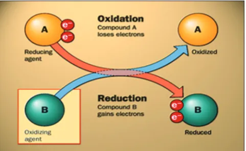
An antioxidant is a molecule capable of inhibiting the oxidation of another molecule. It breaks the free radical chain of reactions by sacrificing their own electrons to feed free radicals, without becoming free radicals themselves (Fig. 2).
ANTIOXIDANTS PREVENTS AGAINST FREE RADICAL DAMAGE Antioxidants are nature’s way of defending cells against attack by ROS. Our body naturally circulates a variety of nutrients for their antioxidant properties and manufactures antioxidant enzymes to control these destructive chain reactions. Forexample, vitamin C, vitamin E, carotenes, and lipoic acid.
Oxidative stress is defined as the state in which the free radicals in the body outnumber our antioxidant defenses. They also decrease the telomere length of the chromosome (Fig. 3).
Oxidation is a chemical reaction that transfers electrons from a substance to an oxidizing agent. Free radicals produced by these oxidation reactions, start chain reactions that damage cells. Antioxidants terminate these chain reactions by removing free radical intermediates and inhibit other oxidation reactions by being oxidized themselves. Hence, antioxidants are often reducing agents like thiols or polyphenols.
Oxidation reactions are important for life, but they are also damaging cells. Hence, plants and animals maintain complex systems of multiple types of antioxidants such as glutathione (GSH), vitamin C, and vitamin E as well as enzymes such as catalase (CAT), superoxide dismutase (SOD), and various peroxidases. Low levels of antioxidants, or inhibition of antioxidant enzymes, causes oxidative stress and damages or kill cells.
These oxidants damage cells by chain reactions such as lipid peroxidation (LPO), or by oxidizing DNA or proteins. Damage to DNA causes mutations and possibly cancer, if not reversed by DNA repair mechanisms, while damage to proteins causes enzyme inhibition, denaturation and protein degradation. The brain is vulnerable to oxidative injury, by LPO due to its high metabolic rate and elevated levels of polyunsaturated lipids. Antioxidants prevent oxidative stress in neurons and prevent apoptosis and neurological damage [27-29].
CLASSIFICATION OF ANTIOXIDANTS Antioxidants can be categorized into two types.
Non-enzymatic antioxidants
Non-enzymatic antioxidants interrupt free radical chain reactions. For example, vitamin E interrupts a chain of free radical activity after only five reactions. Other examples include vitamin C, plant polyphenols, carotenoids, Se, and GSH.
GSH (cysteine containing natural antioxidant) is called as the “master antioxidant” and is found in every single cell of your body, maximizing the activity of all the other antioxidants. GSH is a tripeptide with a gamma peptide linkage between the amine group of cysteine (which is attached by a normal peptide linkage to a glycine) and the carboxyl group of the glutamate side-chain [30].
GSH exists in both reduced (GSH) and oxidized (GSSG) states. In the reduced state, the thiol group of cysteine is able to donate a reducing equivalent (H++ e−) to other unstable molecules such as ROS. In donating an electron, GSH itself becomes reactive but readily reacts with another reactive GSH to form GSH disulfide (GSSG). Such a reaction is probable due to the relatively high concentration of GSH in cells (up to 5 mM in the liver).
GSH is regenerated from GSSG by the enzyme GSH reductase (GSR) [31]. In healthy cells and tissue, more than 90% of the total GSH pool is in the reduced form (GSH) and <10% exists in the disulfide form (GSSG). An increased GSSG-to-GSH ratio is considered indicative of oxidative stress.
Enzymatic antioxidants
Enzymatic antioxidantswork by breaking down and removing free radicals. In general, these antioxidant enzymes flush out dangerous oxidative products by converting them into hydrogen peroxide, then Fig. 1: Oxidation and reduction process
into water, in a multi-step process that requires a number of trace metal cofactors (copper, zinc, manganese, and iron). These enzymatic antioxidants cannot be supplemented orally but must be produced in our body.
The principle enzymatic antioxidants are the following.
SOD
Assisted by copper, zinc, manganese and iron, SOD breaks down superoxide (which plays a major role in lipid peroxidation) into oxygen and hydrogen peroxide. SOD is present in nearly all aerobic cells and extracellular fluids.
CAT
Converts hydrogen peroxide into water and oxygen (using iron and manganese cofactors), hence finishing up the detoxification process that SOD started.
Selenoproteins
These selenium-containing enzymes help break down hydrogen peroxide and organic peroxides into alcohols and are particularly abundant in liver. Selenium is an essential trace element having fundamental importance to human health as it is a constituent of the small group of selenocysteine-containing selenoproteins (over 25 different proteins) which are important for structural and enzymatic functions. Selenoproteins include several forms of the enzymes GSH peroxidase (GSHpx), thioredoxin reductase and iodothyronine deiodinase.
GSHpx
Catalyzes the elimination of hydrogen peroxide as well as organic peroxides (R-O-OH) by the oxidation of GSH [25].
GSR
Catalyzes the reduction of GSH disulfide (GSSG) to the sulfhydryl form GSH, which is a critical molecule in resisting oxidative stress and maintaining the reducing environment of the cell.
WATER-SOLUBLE (HYDROPHILIC) AND LIPID-SOLUBLE (LIPOPHILIC) ANTIOXIDANTS
Another categorization of antioxidants is based on whether they are soluble in water (hydrophilic) or in lipids (hydrophobic). The interior of our cells and the fluid between them are composed mainly of water, but cell membranes are made largely made of lipids [32].
The lipid-soluble antioxidants (such as vitamins E and A, carotenoids, and lipoic acid) are primarily located in the cell membranes, whereas the water-soluble antioxidants (such as vitamin C, polyphenols, and GSH) are present in aqueous body fluids such as blood and the fluids within
and around the cells (the cytosol, or cytoplasmic matrix). Free radicals can strike the watery cell contents or the fatty cellular membrane, so the cell needs defenses for both. The lipid-soluble antioxidants are the ones that protect the cell membranes from LPO.
Natural and artificial antioxidants
Antioxidants are divided into two groups according to their origin as “natural antioxidants” and “synthetic antioxidants.” Most of the synthetic antioxidants are of the phenolic type. The differences in their antioxidant activities are related to their chemical structures, which also influence their physical properties such as volatility, solubility, and thermal stability.
Natural phenolic compounds are widely distributed in plants and are the main contributors to the antioxidant activities of food [33]. Many disorders, i.e., cancer, Parkinson’s and Alzheimer’s diseases, atherosclerosis or heart failure are connected with oxidative stress. Therefore, the increasing interest in elucidating the antioxidant activity of different natural compounds [34,35].
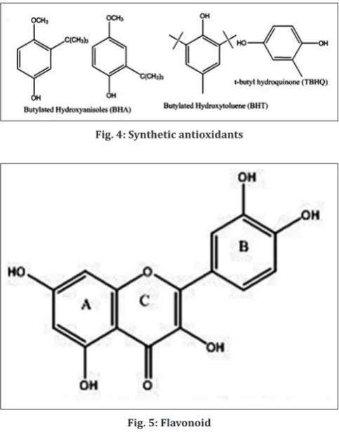
The commercially available and currently used synthetic antioxidants are butylated hydroxyanisole (BHA), butylated hydroxytoluene (BHT), and tertbutyl hydroquinone (TBHQ) (Fig. 4).
In recent years, there is an increasing interest in natural antioxidants and subsequently looking through the literature; it is recognized that the replacement of synthetic antioxidants by natural ones may have several benefits and much of the research on natural antioxidants has focused on phenolic compounds, in particular, flavonoids as potential sources of natural antioxidants [36-38].
Numbers of naturally existing antioxidant compounds present in fruits, vegetables, and dietary supplements are ascorbic acid, α-tocopherol, phenolic acids (benzoic acid, trans-cinnamic acid, and hydroxycinnamic acid), coumarins, lignans, stilbenes (in glycosylated form), flavonoids, isoflavonoids, and phenolic polymers (tannins) [39].
Flavonoids as antioxidants
Flavonoids are secondary plant products recognized as the characteristic red, blue and purple anthocyanin pigments of plant tissues. Apart from their physiological roles in the plants, flavonoids as important components in human diet but never considered as nutrient [40]. The basic structure of flavonoid is a phenylated benzopyrone consists of 3 rings A, B and C (Fig. 5).
Aluminum chloride colorimetrilac estimation is commonly used to quantify flavonoid content of plant extracts [42]. Total flavonoid contents can be determined by reaction with sodium nitrite, followed by the development of colored flavonoid-aluminum complex formation using aluminum chloride in alkaline condition which can be monitored spectrophotometrically at a maximum wavelength of 510 nm [43].
CLASSIFICATION OF ANTIOXIDANT METHODS [44] In vitro antioxidant methods
Antioxidant activity is not concluded based on a single antioxidant test model. There are several in vitro test procedures for evaluating antioxidant activities with the samples of interest. Another aspect is that antioxidant test models vary in different respects. Therefore, it is difficult to compare fully one method to another one. In general, in vitro
antioxidant tests using free radical traps are relatively straightforward to perform. Among free radical scavenging methods, 1,1-diphenyl-2-picrylhydrazyl (DPPH) method is furthermore rapid, simple (i.e. not involved with many steps and reagents) and inexpensive in comparison to other test models. On the other hand, 2, 2-azinobis (3-ethyl benzothiazoline-6-sulfonic acid) diamonium salt (ABTS) decolorization assay is applicable for both hydrophilic and lipophilic antioxidants. In this article, all in vitro methods are described and it is important to note that no one method is absolute in nature rather than an example. All in vitro antioxidant methods are listed in Table 1.
Based on the chemical reaction involved between the antioxidant compounds and the free radicals, antioxidant capacity assays are broadly classified into two types.
1. Hydrogen atom transfer (HAT) reaction based assays 2. Electron transfer (ET) reaction based assays.
ET-based assays
These assays measure the reducing capacity of the antioxidant compounds. It is based on the simple redox reaction, where antioxidant compounds reduce the free radicals and get themselves oxidized. Reduction by antioxidant compounds results in the color change of
the reagent, which correlates with the antioxidant capacity, which is measured by the change in absorbance.
X•+AH�X−+AH•+
AH•++H
2O�A•+H3O+
X−+H
3O+�XH+H2O
HAT-based assays
These assays measures/quantify the hydrogen atom donating ability of the antioxidant compounds by a proton-coupled ET reaction, where it measures the chain breaking antioxidant capacity. These assays based on the reaction between synthetic free radical generator, oxidisable molecular probe, and an oxidant where reaction kinetics is derived from the kinetic curve.
X•+AH�X−+AH•+
ET-based assays
1. DPPH free radical scavenging assay 2. Superoxide anion radical scavenging assay 3. Ferric ion reducing antioxidant power (FRAP)
4. Trolox equivalence antioxidant capacity (TEAC), using ABTS 5. Cupric ion reducing antioxidant capacity (CUPRAC) assay Fig. 4: Synthetic antioxidants
Fig. 5: Flavonoid
Table 1: List of in vitro anti-oxidant methods
Serial number Name of the antioxidant method
1. In vitro antioxidant methods
1.1. ET based assays
1.1.1. DPPH free radical scavenging assay 1.1.2. Superoxide anion radical scavenging assay
1.1.3. FRAP
1.1.4. TEAC, using ABTS
1.1.5. CUPRAC assay
1.1.6. FCR, the total phenols assay
1.1.7. Reducing power assay
1.1.8. DMPD assay
1.1.9. Nitric oxide radical inhibition activity
1.1.10. TBARS assay
1.2. HAT based assays
1.2.1. ORAC
1.2.2. ABTS radical scavenging method 1.2.3. Crocin Bleaching Assays
1.2.4. TRAP
1.2.5. Hydroxyl radical scavenging activity
1.2.6. HORAC
1.2.7. LPIC assay
1.2.8. Scavenging of H2O2 radicals
1.2.9. IOC
1.2.10. PCL Assay
1.2.11. β-carotene–linoleic acid (linoleate) assay 1.3. Other in vitro antioxidant methods 1.3.1. Ascorbic acid content assay
1.3.2. CAA
1.3.3. EPR spectroscopy investigations 1.3.4. Phosphomolybdenum assay 1.3.5. Xanthine oxidase method 1.3.6. Metal chelating activity ET: Electron transfer, HAT: Hydrogen atom transfer, DPPH
6. Folin-Ciocalteu reagent (FCR), the total phenols assay 7. Reducing power assay
8. N,N-dimethyl-p-phenylenediamine (DMPD) assay 9. NO radical inhibition activity
10. Thiobarbituric acid reactive substances (TBARS) assay.
HAT-based assays
1. Oxygen radical absorbance capacity (ORAC), 2. ABTS radical scavenging method
3. Crocin bleaching assays (CBA),
4. Total radical-trapping antioxidant parameter (TRAP), 5. Hydroxyl radical scavenging activity
6. Hydroxyl radical averting capacity (HORAC) 7. LPO inhibition capacity (LPIC) assay 8. Scavenging of H2O2 radicals
9. Inhibited oxygen uptake (IOC) 10. Photochemiluminescence (PCL) assay 11. β-carotene–linoleic acid (linoleate) assay.
Antioxidant testing of natural products has increasing interest in recent years, mainly due to the fact that antioxidants can neutralize the harmful free radicals in vitro, thus suggesting that an antioxidant-rich diet, provides health benefits.
ET-based assays
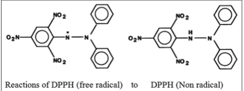
DPPH radical scavenging activity (Fig. 6)
The DPPH is one of the most stable free radicals and is frequently used in the evaluation of radical scavengers in natural foods [45]. DPPH assay method is very simple and is also quick for manual analysis of antioxidant contents. This method can be used for solid or liquid samples and is not only specific to any particular antioxidant but also applies to the overall antioxidant capacity of the sample.
The DPPH test is based on the ability of the stable 2, 2-diphenyl-1-picrylhydrazyl free radical to react with hydrogen donors [46,47].
DPPH assay method is based on the reduction of DPPH, a stable free radical. The free radical DPPH with an odd electron gives a maximum absorption at 517 nm (purple). When antioxidants react with DPPH, the stable free radical becomes paired off in the presence of a hydrogen donor (e.g., a free radical-scavenging antioxidant) and is reduced to the DPPHH and as consequence the absorbances decreased from the DPPH. Radical to the DPPH-H form, results in decolorization (yellow) with respect to the number of electrons captured. More the decolorization, more is the reducing ability. This test has been the most accepted model for evaluating the free radical scavenging activity of any new drug. The DPPH radical displays an intense UV-visible (UV-Vis) absorption spectrum. When a solution of DPPH is mixed with that of a substance that can donate a hydrogen atom, then this gives rise to the reduced form (diphenyl picryl hydrazine; nonradical) with the loss of violet (pale yellow of the picryl group present) [48].
In this test, a solution of radical is decolorized after reduction with an antioxidant (AH) or a radical (R) according to the following equation [49]:
(DPPH) + H-A → DPPH-H + A or DPPH + R→ DPPH-R
(Purple) (Yellow)
About 4.3 mg of DPPH was dissolved in 3.3 ml methanol and protected from light by covering the test tubes with aluminum foil. 150 ml DPPH solution was added to 3 ml methanol, and the absorbance was taken immediately at 517 nm for control reading. 50 ml of various concentrations of compounds as well as standard compound (e.g., ascorbic acid) were taken, and the volume was made uniformly to 150 ml using methanol. Each of the samples was then further diluted with methanol up to 3 ml and to each 150 ml DPPH was added. The
absorbance was taken after 15 minutes at 517 nm using methanol as blank on UV-vis spectrometer Shimadzu, UV-1601. The IC50 values for
each drug compounds as well as standard preparation were calculated. The free radical scavenging activity was calculated using the following formula:
% scavenging=[Absorbance of control−Absorbance of test sample/ Absorbance of control]×100
The effective concentration of sample required to scavenge DPPH radical by 50% (IC50 value) was obtained by linear regression analysis of
dose-response curve plotting between % inhibition and concentrations.
The better way of comparison of antioxidant activity between the samples is using IC50 values. Inhibition concentration (IC50) values
defined as the concentration of sample required for 50% inhibition of free radicals. IC50 is determined from the plot between the remaining
absorbance of free radical and concentration with each analysis in triplicates. In this test, quercetin, 6-hydroxy-2,5,7,8-tetramethyl chroman-2-carboxylic acid (trolox), tocopherol, and ascorbic acid are used as positive controls [50,51].
The DPPH analysis is a fast and an uncomplicated test ensuring reliable result. Furthermore, it requires only a UV-Vis spectrophotometer to perform, which explains its widespread use in screening antioxidant properties. However, the method is sometimes complicated when test compounds have spectra that overlap with DPPH at 515 nm.
Modifications
DPPH is stable nitrogen radical that bears no similarity to the highly reactive and transient peroxyl radicals involved in LPO.
a. Conventional cuvette assay of radical scavenging activity is replaced by 96-well plate titer assay from past couple of years. Cuvette assay method uses UV-Vis spectrophotometer to see the absorbance, whereas 96-well plate method uses ELISA plate reader for absorbance. The first method is very tedious, time-consuming method, allows only 1 sample to read a time and requires a high quantity of reagent, whereas the second method is time saving and it reads about 96 samples at a time, with a small amount of reagent. b. Thin layer chromatography (TLC) autography technique
The antiradical screening by TLC autography technique provides an easy, effective and rapid way to study plant extract profiles. No sample purification is needed as this technique provided a simultaneous separation and radical scavenging activity measurement of antioxidative compounds in plant extract. Qualitative as well as semi-quantitative analysis of antioxidants can be done by this technique.
Qualitative analysis
To detect the antioxidant activity, a method based on the reduction of 2,2-diphenyl-1- Picrylhydrazyl (DPPH) can be carried out. DPPH is a free radical stable at room temperature, which produces a violet solution in methanol. When the free radical reacts to an antioxidant, its free radical property is lost due to chain breakage and its color changes to light yellow. In this assay by TLC, the extracts that produced yellow or white spots in the purple background were considered as antioxidants.
Procedure
The extracts dissolved in the solvent were spotted on the silica-gel 60F 254 plates and developed the chromatogram in adequate solvent systems. Then, all the plates are sprayed with a methanolic solution of DPPH (2 mg/ml). Thus, antioxidants appear as yellow bands on a light purple background. After spotting the extracts on the TLC plates, even uneluted plates also can be used to determine the qualitative antioxidant analysis. The uneluted plates also can immerse in 0.2% DPPH methanol solution and sample spots were evaluated for radical scavenging activity.
The same method is also implemented to detect total phenolic and total flavonoid content just by changing the mobile phase solvent system and visualizing agent. Vanillin/H2SO4 reagent is sprayed on the plate
and heating it at 110°C, for 5 minutes and observed to detect different groups of compounds. Orange-yellow spots indicate polyphenolic compounds. The silicagel plate was sprayed with natural products-PEG reagent and observed at UV-365 nm, to detect the flavonoids as they appear as yellow-orange fluorescent spots.
A disadvantage of DPPH method is the fact that many antioxidants that react quickly with the radical peroxide are almost or entirely inert to DPPH. Despite having the above limitations, DPPH is stable, is commercially-available and does not have to be generated before carrying out assays like ABTS. For these reasons, it is considered as an easy and useful spectrophotometric method with regard to screening or measuring the antioxidant activity.
Super oxide free radical scavenging activity
Superoxide anion (O2·−)isan oxygen molecule with an extra electron
that can damage mitochondria, DNA, and other molecules. Superoxide generated both in vivo and in foods can undergo several reactions including dismutation to give H2O2.
O2·−+O2·−+2H+�H2O2+O2
Superoxide is biologically important as it can form singlet oxygen and hydroxyl radical. Overproduction of superoxide anion radical contributes to redox imbalance and associated with harmful physiological consequences.
Superoxide anions are generated in phenazine methosulfate-nicotinamide adenine dinucleotide (NADH) system by the oxidation of NADH and assayed by the reduction of nitro-blue tetrazolium (NBT) resulting in the formation of blue formazan.
Procedure
A value of 100 ml of riboflavin solution (20 mg), 200 ml ethylenediaminetetraacetic acid (EDTA) solution (12 mM), 200 ml methanol, and 100 ml NBT solution (0.1 mg) were mixed in test tube and reaction mixture was diluted up to 3 ml with phosphate buffer (50 mM). The absorbance of the solution was measured at 590 nm using phosphate buffer as blank after illumination for 5 minutes. This is taken as control. 50 ml of different concentrations of compounds as well as standard preparation were taken and diluted up to 100 ml with methanol. To each of these, 100 ml Riboflavin, 200 ml EDTA, 200 ml methanol, and 100 ml NBT were mixed in test tubes and further diluted up to 3 ml with phosphate buffer. The absorbance was measured after illumination for 5 minutes at 590 nm on UV-Vis spectrometer. The IC50
values for each compound as well as for the standard preparation were calculated using the following formula [52-60].
% Inhibition=[Absorbance of control−Absorbance of test sample/ Absorbance of control]×100
FRAP assay
FRAP is another method employed for the determination of total antioxidant activities. Primarily used for determining antioxidant
activity of plasma, later successfully applied to measure the antioxidant activity of a number of biological samples and pure substances [61-63]. Since antioxidant and antiradical properties are mainly attributed to the presence of phenolic compounds [64,65], it is expected that the effectiveness of a fraction is proportional to its phenolic concentrations. FRAP assay is widely-used to directly test the total antioxidant potential of several foods and plant extracts based on the reduction of complexes of 2,4,6-tripyridyl-s-triazine (TPTZ) with ferric chloride hexahydrate (FeCl3·6H2O), which are almost colorless. The solution will eventually
turn slightly brownish forming blue ferrous complexes after complete reduction.
FRAP assay uses antioxidants as reductants in a redox-linked colorimetric method, employing an easily reduced oxidant system present in stoichiometric excess. The test solutions are mixed with a FRAP reagent (10 mM of TPTZ solution in 40 mM HCl, 20 mM FeCl3, and 0.3 M acetate buffer at pH 3.6) followed by spectrophotometric measurement of the absorbance of the reaction mixture after incubation at 37°C for 10 minutes at 593 nm against the blank. The final results can be expressed as the concentration of antioxidants having a ferric reducing ability equivalent to that of 1 mM FeSO4 used as the standard
solution.
FRAP method has limitations, especially for measurements below non-physiological pH values, i.e., at pH 3.6. In addition, this method is unable to detect slowly-reacting polyphenolic compounds and thiols [66]. Furthermore, any compounds (even without antioxidant properties) with redox potential lower than that of the redox pair Fe(3+)/Fe(2+) can
theoretically reduce Fe(3+) to Fe(2+) contributing to an increase in the
FRAP value and thus inducing false positive results [67]. On the other hand, not all antioxidants reduce Fe(3+) at a rate fast enough to allow
its measurement within the observation time (typically 4 minutes). Indeed, many polyphenols react more slowly and require longer reaction times (30 minutes) for total quantification. Moreover, some polyphenolic compounds such as quercetin, caffeic, ferulic, and tannic acids have slower reactions, requiring a longer time (approximately 30 min) until the complex reduction process was completed. When used to determine the antioxidant potential of polyphenols in water and methanol, the change in absorbance continued after 4 minutes [68]. Therefore, the FRAP values for these compounds cannot be accurately determined in 4 minutes. Hence for this reason, the ideal reaction time should be at least 10 minutes.
Procedure
An aliquot of 0.20 ml of each sample at appropriate concentration was mixed with 0.5 ml of phosphate buffered saline (0.2 M; pH 6.6) and 0.5 ml of 1% potassium ferricyanide (K3Fe(CN)6). The mixture was
incubated at 50°C for 30 minutes and 0.5 ml of 10% trichloroacetic acid (TCA) was added. After centrifugation for 10 minutes at 3000 rpm, the supernatant (0.5 ml) was mixed with distilled water (0.5 ml) and 0.1% ferric chloride (0.1 ml). The absorbance was measured at 700 nm; ascorbic acid was used as positive control. Absorbance increasing relatively to that of concentration represented the reducing capacity of tested sample.
Statistical analyses
Data were expressed as a mean±standard deviation. Analyses of variance (ANOVA) were performed for the comparison of results using Fischer’s test. A statistical significance was set at p˂0.05.
TEAC assay
TEAC measures the antioxidant activity of a given substance, as compared to the standard, trolox [69]. The three TEAC tests developed at different periods - namely, TEAC assay I (ABTS) enzymatically-generated with met-myoglobin and hydrogen peroxide, TEAC II radical generation with filtration over the manganese dioxide (MnO2) oxidant, and TEAC III (with potassium
one another - are interchangeably-used, when using different solvent media. This leads to significant variability in the measurements of the antioxidants [70,71].
The “pre-addition technique” (employed by adding antioxidants before radical generation) for TEAC I could result in an overestimation of antioxidant capacity because many substances interfere with the formation of the free radical; TEAC I measured the ability of delaying radical formation as well as scavenging of the radical [71]. Since the reagent is soluble in both aqueous and organic solvent media, the advantages of ABTS/TEAC are reported to be operational simplicity, reproducibility, diversity and the most important of all, flexible usage in multiple media to determine both hydrophilic and lipophilic antioxidant capacity of food extracts and physiological fluids [72].
CUPRAC Assay
CUPRAC is an ET-based assay which is widely and popularly used method to determine the complete scavenging of free radicals, i.e. total antioxidant capacity of a compound. This method is based on the simple redox reaction between antioxidant and the free radicals, where the antioxidant activity can be measured by reduction of cupric ions to cuprous ions by antioxidants [73,74].
The novel in vitro antioxidant method based on an electron-transfer mechanism was named as CUPric ion reducing antioxidant capacity, abbreviated as the CUPRAC method. These CUPRAC method is widely used to measure the antioxidant capacity assays in food, plants, human serum, biological samples, dietary polyphenols, vitamins C and E, etc. This assay is a simple, reliable, versatile and has low cost. Antioxidant capacity was compared and found advantageous over other methods. CUPRAC assays are popular, due to their high speed and sensitivity and also extensively validated and provides useful information about the reducing capacity of the sample.
Chemistry behind CUPRAC assay
Neocuproine is an aromatic heterocyclic compounds used for the spectroscopic determination of copper. Neocuproine, a methylated phenanthroline derivative chelates with the copper from cuprous chloride and forms a chromogenic redox reagent bis(neocuproine) copper II) chloride, which is a novel reagent for the CUPRAC antioxidant capacity assays. This assay is based on reduction of Cu (II)-neocuproine complex to highly colored Cu(I)-neocuproine complex, which is measured at 450 nm absorbance.
The generation of cuprous ions (Cu1+) from cupric ions (Cu2+) is due to
reduction of cupric chloride from cuprous chloride in the chromogenic redox reagent bis(neocuproine) copper(II) chloride by antioxidants compounds. This copper(I) is highly selective toward neocuproine, the resulting Cu(I)-neocuproine complex consists of two molecules of neocuproine with one cuprous ion, with a maximum absorption at 454 nm [75] (Fig. 7).
CUPRAC methodology
This method involves mixing the antioxidant solution with aqueous copper(II) chloride, alcoholic neocuproine, and ammonium acetate
aqueous buffer at pH 7, and subsequently measuring the developed absorbance at 450 nm after 30 minutes.
Preparation of Solutions
1. CuCl2 solution 1.0×10⁻² M (10 mM is prepared by dissolving 0.4262 g
CuCl2. 2H2O in water, and diluting to 250 mL
2. Ammonium acetate buffer at pH=7.0, 1.0 M is prepared by dissolving 19.27 g NH4Ac in water and diluting to 250 mL
3. Neocuproine (Nc) solution, 7.5×10⁻³ M (7.5 mM) is prepared by dissolving 0.039 g Nc in 96% ethanol, and diluting to 25 mL with ethanol. This solution should be prepared freshly
4. Trolox, 1.0×10⁻³ M, (1 mM) is prepared by dissolving 0.00626 g of the compound in 96% ethanol, and diluting to 50 mL.
The standard CUPRAC procedure involves the addition of cuprous chloride along with neocuproine reagent, bathocuproine, a chromogenic Reducing agent, which reacts with the antioxidant molecules, and converts cupric to cuprous, to form a Cu(I)-Nc complex.
The method is summarized
Add 1 mL of 10 mM CuCl2 solution, 1 mL 7.5 mM neocuproine, 1 mL 1 M NH4Ac, and x mL of antioxidant neutral solution then makeup the final
volume to 4.1 mL using distilled water. Incubate the reaction mixture under normal condition (room temperature) for 30 minutes, after incubation the absorbance was read at 450 nm.
Expressing units for CUPRAC assay
The antioxidant activity evaluated by different antioxidant methods can be expressed as millimole/micromole equivalent of reference standard used. The antioxidant compounds used as reference standard includes vitamin E, ascorbic acid, gallic acid, BHA, BHT, trolox, etc. TE (trolox equivalent [TE]) is a parameter widely used to measure the antioxidant activity where trolox is used as the standard reference to measure antioxidant capacity of the samples.
EC50 and IC50 are also used to evaluate the antioxidant activity of the
sample, which were determined using the liner regression graph of concentration against the mean radical scavenging percentage of the antioxidant sample.
Advantages of CUPRAC method
The novel reagent for the CUPRAC antioxidant capacity assay is bis(neocuproine) copper(II) chloride. This CUPRAC reagent was easily accessible, stable, selective low-cost, and responding to all antioxidants which induce redox reaction with the antioxidant compounds and determines the antioxidant capacity of various compounds.
CUPRAC method is advantageous over other electron-transfer based methods as follows:
1. The CUPRAC assay proved to be efficient for GSH and thiol-type antioxidants due to electronic structure of Cu(II) facilitate faster kinetics, whereas FRAP method which is carried out at low pH found to be insensitive to thiol group of antioxidants due to chemical inertness by the half filled d-orbitals of high spin Fe(III).
2. CUPRAC is superior with respect to pH which is close to the physiological pH condition (pH 7) unlike FRAP which is carried out
at acidic condition, folins methods which are carried out at alkaline conditions. The reaction at acidic and alkaline conditions will suppress or enhances the reduction capability of the sample thereby altering the reducing capability of the antioxidant compounds. Thus, CUPRAC gives definite values under physiological conditions. 3. CUPRAC method found to be superior method to analyze biological
fluids over widely used Folin–Ciocalteu, whereas this method is applicable to both lipophilic serum antioxidants as well as hydrophilic serum antioxidants. Hence, this method is useful in determining the total antioxidant capacity of the biological fluids. 4. Colored reagent used in the antioxidant assay is usually sensitive
toward light, humidity, pH, solvents like DPPH reagent, whereas cuprac reagent-colored chelate of Cu(I)-Nc found to be stable and insensitive to the all external parameters.
5. Proxidants are species that causes oxidation of biological macromolecule, and results in oxidative stress. In the ferric ion based assays such as FRAP produces Fe2+, which can act as prooxidant by producing OH radicals by reacting with H2O2.
Application of CUPRAC method in food, biological fluids, and plant extracts
Beneficial influence of many foodstuffs, fruits, vegetables, and beverages including, tea, coffee is attributed due to their plant-derived antioxidants. To determine the complete profiling of antioxidant capacity in foodstuffs, beverages nutraceutical, dietary supplements, etc., there is a need to develop standardized antioxidant capacity methods. Cuprac assay is the most commonly used in vitro determination of antioxidant activity of food constituents.
CUPRAC method is widely applicable for measuring the antioxidant capacity in various food compounds namely vitamins (vitamins C and E), dietary polyphenols, flavonoids, ascorbic acid in food extract. Measuring the antioxidant capacity of plant-derived antioxidant by cuprac method in the wide range of matrices such as food, beverages, biological fluids, and plant extracts give the more appropriate biological activity information as it measures total antioxidant capacity of the matrices.
Antioxidant activity of biological fluids like serum, plasma antioxidants can be carried out using CUPRAC assay. These tests measures the combined effect of nonenzymatic antioxidants present in the biological fluids, hence it is of the most important by providing the complete picture on the antioxidant status of the organism and their ability to counter act with free radicals/ROS. There is no single widely acceptable specific method for biological samples but cuprac method found to be useful in determining the biologically important molecules such as Ascorbic acid, α-tocopherol, reduced GSH, uric acid, bilirubin, and albumin-carotene [76,77].
Cuprac method results when correlated with other spectrophotometric assays such as FRAP, TEAC, and DPPH are found to be advantages compared to other methodologies for antioxidant capacity assessment of plasma and urine samples. CUPRAC method is prominent to measure the total antioxidant capacity of biological fluids along with other commonly used methods.
Polyphenols are a group of chemicals found in many fruits, vegetables, and other plants such as tea leaves and grapes. They possess antioxidant properties due to their phenolic –OH group. Polyphenol profiling is done using CUPRAC methods were various polyphenol groups which differ on number and position of –OH groups can be reduced/oxidized by CUPRAC reagent. Polyphenols, flavonoid containing plant extracts can be measured easily and antioxidant profiling of different plant extracts can be done [78-80].
FCR, the total phenols assay
FCR is a mixture of tungsten and molybdenum oxides. This method was previously used for the analysis of proteins like tyrosine [81]
containing a phenolic group but later applied for analyzing the total phenolic content in wine. It is a sensitive, quantitative, and relatively independent method for proteins, nucleic acids, and ascorbic acid.
The product of metal oxide reduction produces a blue with a broad light absorption at 765 nm (750-770 nm). Most of the phenolic compounds are in dissociated form (as conjugate bases or phenolate anions) at the working pH~10, they can be easily oxidized with the FCR.
The molybdenum center in the complex reagent is reduced from Mo (VI) to Mo (V) with an e-donated by an antioxidant to produce a blue. The intensity of light absorption at that wavelength is proportional to the concentration of phenols and results are expressed in gallic acid equivalents (GAE). Phenols stoichiometrically reduce phosphomolybdic/phosphotungstic acid [82]. The FC chromophore which is a multivalent charged phospho-tungsto-molybdate (V) having a great affinity for water was found to be incapable of measuring lipophilic antioxidants, but the reagent was modified and standardized to enable simultaneous measurements of lipophilic and hydrophilic antioxidants in NaOH added isobutanol-water medium by Apak et al. [83].
Modification
The modified procedure was successfully applied to the total antioxidant capacity assay of trolox, quercetin, ascorbic acid, gallic acid, catechin, caffeic acid, ferulic acid, rosmarinic acid, GSH, and cysteine aswell as of lipophilic antioxidants such as α-tocopherol (Vitamin E), BHA, BHT, TBHQ, lauryl gallate, and β-carotene.
Heteropolyphosphotungstate molybdate Phenol
Reduced forms
− = →
(Tungstate series P2W18O62−7→ H4P2W18O62−8)
(Molybdate series H2P2Mo18O62−6 → H6P2Mo18O 62−6)
Reducing power assay
Potassium ferricyanide reducing power assay
This method is based on the reduction of ferric (Fe3+) to ferrous (Fe2+),
in the presence of antioxidants. Substances having a reduction potential react with potassium ferricyanide forming potassium ferrocyanide which further reacts with FeCl3 to form an intense prussian blue
complex having maximum absorbance at 700 nm. The amount of complex formed is directly proportional to the reducing power of test sample [84].
The reducing power was determined according to the method of Oyaizu [85]. Various concentrations of extracts (2.5 ml) were mixed with 2.5 ml of 200 mmol/l sodium phosphate buffer (pH 6.6) and 2.5 ml of 1% potassium ferricyanide. The mixture was incubated at 50°C for 20 minutes. After 2.5 ml of 10% TCA (w/v) were added, the mixture was centrifuged at 650 rpm for 10 minutes. The upper layer (5 ml) was mixed with 5 ml deionized water and 1 ml of 0.1% of ferric chloride, and the absorbance was measured at 700 nm: Higher absorbance indicates higher reducing power. The assays were carried out in triplicate and the results are expressed as mean values±standard deviations. The extract concentration providing 0.5 of absorbance (EC50) was calculated from the graph of absorbance at 700 nm against extract concentration. BHA and tocopherol were used as standards. In this assay, the yellow of the test solution changes to various shades of green and blue, depending on the reducing power of each compound. The presence of reducers (i.e., antioxidants) causes the reduction of the Fe3+/ferricyanide
complex to the ferrous form. Therefore, measuring the formation of Perl’s Prussian blue at 700 nm can monitor the Fe2+ concentration.
Procedure
hexacyanoferrate (K3Fe(CN)6) (1%, w/v), followed by incubating
at 50°C in a water bath for 20 minutes. The reaction was stopped by adding 0.75 ml of TCA solution (10%) and then centrifuged at 800 g for 10 minutes. 1.5 ml of the supernatant was mixed with 1.5 ml of distilled water and 0.1 ml of ferric chloride solution (0.1%, w/v) for 10 minutes. The absorbance was measured at 700 nm as the reducing power. Higher the absorbance of the reaction mixture, greater the reducing power.
DMPD assay
DMPD is an improved decolorization method developed by Verde [86] for measuring the antioxidant activity of samples.
The purple colored DMPD radical cation (DMPD·+) generated through a reaction between DMPD and potassium persulfate is reduced in the presence of H donating antioxidants [87]. The determination of anti-oxidant potential is done at pH 5.25 using 0.1 M acetate buffer. One μL of DMPD·+ (stable up to 12 hrs) solution and 50 μL antioxidant solution
were mixed continuously for 10 minutes at 25°C, then the absorbance of the solution was taken at 517.4 nm.
The advantage of this method, compared to earlier methods, is Fe(II) ions involved in generation of radical cation through Fenton’s reaction causes negative deviation in the antioxidant activity of food extracts. This assay is equally applied to both lipophilic and hydrophilic antioxidants. This method is rapid and inexpensive and reproducible, therefore used in screening a large number of fruit samples [88].
NO free radical scavenging activity
NO is involved in a variety of biological functions including neurotransmission, vascular homeostasis, antimicrobial, and antitumor activities. It also leads to oxidative damage. NO reacts with superoxide and forms the peroxynitrite anion, which is a potential oxidant that decompose and produces OH and NO.
Procedure
Sodium nitroprusside in aqueous solution at physiological pH generates NO which interacts with oxygen to produce nitrite ions, estimated using Griess reagent. Scavengers of NO compete with oxygen, leading to reduced production of nitrite ions. Large amounts of NO leads to tissue damage.
About 50 ml of each of the concentrations of compounds dissolved in DMSO and ascorbic acid (standard compound) were taken in separate tubes, and the volume was uniformly made up to 150 ml with methanol. To each tube, 2.0 ml of sodium nitroprusside (10 mM) in phosphate buffer saline was added. The solutions were incubated at room temperature for 150 minutes. The similar procedure was repeated with methanol as blank which served as control. After incubation, 5 ml of Griess reagent was added to each tube including control. The absorbance of chromophore formed was measured at 546 nm on UV-Vis spectrometer Shimadzu, UV-1601. Ascorbic acid was used as a positive control. The IC50 values for each test compound as well as standard
preparation were calculated [89-93].
% scavenging=[Absorbance of control−Absorbance of test sample/ Absorbance of control]×100
TBARS assay
The measurement of TBARS is a well-established method for screening and monitoring LPO [94]. The assay measures the inhibition of production of TBARS from sodium benzoate under the influence of the free oxygen radicals derived from Fenton’s reaction. A solution of 1 mmol/L uric acid was used as standard. A standardized solution of Fe-EDTA complex reacts with hydrogen peroxide by a Fenton type reaction, leading to formation of hydroxyl radicals.
The ROS degrade benzoate, resulting in the release of TBARS. Antioxidants from the added sample cause suppression of the
production of TBARS. At low pH and elevated temperature (90-100°C), MDA readily participates in nucleophilic addition reaction with 2-thiobarbituric acid (TBA), generating a red, fluorescent 1:2 MDA:TBA adduct [95]. The reaction is measured colorimetrically at 530-540 nm or fluorometrically at an excitation wavelength of 530 nm and emission wavelength of 550 nm [96,97].
HAT-based assays ORAC assay
ORAC assay is a method for quantifying the antioxidant strength of substances, which involves combining the sample to be tested (i.e., the antioxidant) with a fluorescent compound as well as a compound that generates free radicals at a known rate. As free radicals are being generated, the fluorescent compound (e.g., fluorescein) is damaged and subsequently loses its fluorescence.
When antioxidants are present, it mops up the free radicals being produced, and therefore, inhibits the loss of fluorescence. The stronger the antioxidant property of a substance and the higher is the degree of inhibition on the loss of fluorescence. The measurement is standardized trolox which has a known ORAC value and is reported in terms of TE (µM TE). This method serves as an excellent way to quantify the ability of various compounds to quench free radicals. ORAC assay is carried out using a modified [98-100] procedure by comparing ORAC in vitro antioxidant activity of polyphenols with the total phenolics concentrations.
The free radicals in ORAC method are produced by 2,2′-azobis (2-amidino-propane) dihydrochloride (AAPH) followed by the oxidation of the fluorescent indicator protein phycoerthrin (β-PE). The loss of fluorescence can be inhibited by antioxidants and was monitored using a microplate fluorescence reader. All reagents are prepared in phosphate buffer (75 mM), pH 7.0 with trolox (0-4 µM), which is used as a standard. Before use, the samples are suitably diluted in the phosphate buffer. Quercetin dehydrate (1 µM) (positive control) is dissolved in methanol followed by a dilution with buffer (1:249, v/v). Methanol is used in the control sample, blank and standard without having an effect on the 1:1 relationship between trolox and ORAC value. The reaction mixtures consisted of 1 mL of β-PE (0.92 nM) which has been pre-incubated for 15 minutes at 37°C, 60 µL of test compound, 40 µL of 75 mM phosphate buffer (pH 7.0), and 100 µL of AAPH (500 mM). After adding AAPH, the plate is automatically shaken for 3 seconds and the fluorescence was measured every 2 minutes for 70 minutes with emission and excitation wavelengths at 565 and 540 nm, respectively, using a micro plate fluorescence reader FL600 (BioTek, Inc., VT), maintained at 37°C. The ORAC values are calculated according to [98] and expressed as µM TE/g. ANOVA with post-hoc comparisons using Tukey’s test is performed to compare the ORAC readings shown by the different samples using SAS Software [101].
Utilization of a β-PE method provides an additional advantage as the substrate “self-prevents” free radical generations due to its oxidation. Therefore, it is good to determine the capacity of hydrophilic and hydrophobic samples simply by changing the generating source of radicals and the solvent. One limitation of this method is that the protein photo bleached under plate-reader conditions has large inter-batch differences and interacts with polyphenols due to non-specific protein binding, and therefore, may loss fluorescence even without the addition of a free radical generator.
ABTS assay
or absence of antioxidants. This concludes that the faster-reacting antioxidants might also contribute to the reduction of the ferryl myoglobin radical [106]. A more appropriate assay method is using a decolorization technique because the directly generated radical is stable prior to reacting with the putative antioxidants. This improved technique for the generation of ABTS involves the direct production of the blue/green ABTS chromophore via the reaction between ABTS and potassium persulfate which has absorption maxima at wavelengths 645 nm, 734 nm, and 815 nm [106-108] with the more commonly used maximum absorbance reported to be at 415 nm [109] (Fig. 8).
The addition of antioxidants to the pre-formed radical cation reduces ABTS on a time-scale to a certain extent, depending on the antioxidant activity of the samples analyzed, the concentration of the antioxidant and the duration of each reaction. Thus, the extent of decolorization as percentage inhibition of the ABTS radical cation is determined as a function of both concentration and time and is calculated relative to the reactivity of trolox standard under similar conditions. A modification of the method utilized to determine the antioxidant capacity of was also developed [110]. For the evaluation of antioxidant activity, ABTS solution is diluted with ethanol (96%) to obtain an absorbance of 0.700 (±0.020) at 734 nm. 2 mL of ABTS solution are mixed with 100 µL of the sample solution in a cuvette and the decrease in the absorbance is measured after 6 minutes. The reagent blank is prepared by adding 100 mL of ethanol instead of the sample. Ascorbic acid was used as the standard at different concentrations (0-100 mg/L) prepared in 96% ethanol and assayed under a similar procedure as that conducted on the samples with the means of the three values expressed as mg ascorbic acid equivalents/100 g.
ABTS assay is beneficial as it reduces labor time, material cost, and sample volume. A some of the assays are adapted for a more convenient mass screening using quantitative spectrophotometer as well as applied in agriculture and food industries. Although this method has been reported and commercialized by CAYMAN [111], it does not incorporate any blank samples which could result in further inaccuracies in the measurements.
CBA
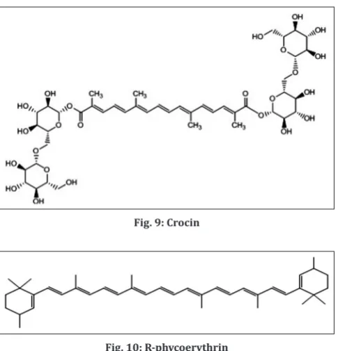
Crocin (C44H64O24) is a naturally occurring carotenoid obtained from
dried stigma of culinary spice Crocus sativus L. (Saffron).
The CBA [112] is suitable for screening radical scavenging activity. Originally, an inhibition of crocin bleaching by a range of substances was monitored by competition kinetics in the presence of photolytically produced alkoxyl radicals. In CBA, abstraction of hydrogen atoms and/or addition of the radical to the polyene structure of crocin results in a disruption of the conjugated system accounting for crocin bleaching. The latter is recorded as a reduction of absorbance at 440 nm in the presence or absence of radical scavengers. The pro-oxidant activity was taken as a ratio of decrease in crocin absorbance at 5 minutes and the relevant oxidant concentration [113,114]. Later, Bors et al. [115] found that the absolute rate of crocin bleaching depends on the sort of radical attacking the polyene structure (Fig. 9).
In the latter, peroxyl radical formation was achieved using azo-initiators (hydrophilic or lipophilic) [116]. In this way, Tubaro et al. [117] made an effort to average antioxidant and pro-oxidant effects of the constituents of complex natural mixtures. Results were expressed with reference to α-tocopherol (for lipophilic molecules) or trolox (for hydrophilic ones).
TRAP Assay
This method uses R-phycoerythrin (R-PE) as the fluorescent probe. The reaction progress of R-PE with AAPH was monitored fluorometrically (λex=495 nm and λem=575 nm). R-PR is the brightest fluorescent dye identified and is originally isolated from red algae Gracilaria (Fig. 10).
Fig. 8: 2,2-azinobis (3-ethyl benzothiazoline-6-sulfonic acid) diamonium salt reaction
Fig. 9: Crocin
Fig. 10: R-phycoerythrin
TRAP values are calculated from the length of the lag phase caused by the antioxidant compared to that of trolox. Luminol is also used as the chemiluminescent substance [118].
Dye-substrate oxidation method
Advantages
It provides concurrent multisample analysis with automated data storage, regression analyses, and calculation of oxidation inhibition rates. For screening, crude extracts and typical assay results are presented.
Hydroxyl radical scavenging activity
Hydroxyl radical is one of the potent ROS in the biological system that reacts with polyunsaturated fatty acid moieties of cell membrane phospholipids and causes damage to cell. The scavenging ability of hydroxyl radicals is measured by the method of Kunchandy and Rao in 1990 [109]. The reaction mixture (1.0 mL) consist of 100 μL of 2-deoxy- D-ribose (28 mM in 20 mM KH2PO4-KOH buffer, pH 7.4), 500 μL of the extract, 200 μL EDTA (1.04 mM) and 200 μM FeCl3 (1:1 v/v), 100 μL of H2O2 (1.0 mM), and 100 μL ascorbic acid (1.0 mM) which is
incubated at 37°C for 1 hr. 1 mL of thiobarbituric acid (1%) and 1.0 mL of TCA (2.8%) are added and incubated at 100°C for 20 minutes. After cooling, absorbance is measured at 532 nm, against a blank sample.
The scavenging activity for hydroxyl radicals was measured using the reaction mixture contained 60 μl of 1.0 mM FeCl3, 90 μl of 1mM 1,10-phenanthroline, 2.4 ml of 0.2 M phosphate buffer (pH 7.8), 150 μl of 0.17 M H2O2, and 1.5 ml of extract at various concentrations. Adding H2O2 started the reaction. After incubation at room temperature for
5 minutes, the absorbance of the mixture at 560 nm was measured with a spectrophotometer. The hydroxyl radicals scavenging activity was calculated according to the following equation:
% inhibition = [(A0−A1)/A0]×100
Where A0 was the absorbance of the control (blank, without extract)
and A1 was the absorbance in the presence of the extract.
HORAC
The hydroxyl radical is generated by a Co2+ mediated Fenton-like reaction,
and the hydroxyl radical formation under the experimental condition is indirectly confirmed by the hydroxylation of p-hydroxybenzoic acid. The fluorescence decay curve is monitored in the absence and presence of antioxidant which is the index of the hydroxyl radical prevention capacity. Gallic acid is chosen as a reference standard and activity is measured in terms of GAE. The hydroxyl radical prevention capacity is due to the metal-chelating capability of the compounds.
Procedure
The scavenging capacity for hydroxyl radical was determined according to the modified method [119]. The assay was performed by adding 0.1 ml of EDTA, 0.01 ml of ferric chloride, 0.1 ml of hydrogen peroxide, 0.36 ml of deoxyribose, 1.0 ml of test solutions (5-100 μg/ml) in distilled water, 0.33 ml of phosphate buffer (50 mM, pH 7.4), and 0.1 ml of ascorbic acid were dissolved in sequence. Then, the mixture was incubated for 1 hr at 37°C and 1.0 ml portion of the incubated mixture was mixed with 10% TCA and 1.0 ml of 0.5% TBA to develop the pink chromogen and measured at 532 nm.
LPIC assay
LPO, a well-established mechanism of cellular injury in plants and animals, is used as an indicator of oxidative stress in cells and tissues. Lipid peroxides are unstable and decompose to form a complex series of compounds including reactive carbonyl compounds. Polyunsaturated fatty acid peroxides generate malondialdehyde (MDA) and 4-hydroxyalkenals (HAE) on decomposition and the measurement of MDA and HAE are used as an indicator of LPO.
The assay is based on the reaction of a chromogenic reagent, N-methyl-2-phenylindole, with MDA and HAE at 45°C. 1 molecule of either MDA or 4-hydroxyalkenal reacts with 2 molecules of N-methyl-2-phenylindole to yield a stable chromophore (carbocyanine dye) with maximal absorbance at 586 nm [120].
Procedure
Mice liver was freshly excised and processed to get 10% homogenate in cold phosphate buffered saline (pH 7.4) and clear homogenate is obtained by filtration. LPO was analyzed by estimating the TBARS using standard method with some modifications [121]. The analogues at different concentrations (25-200 μmol/l in DMSO) were added to liver homogenate. LPO was initiated by adding 100 μl of 15 mmol/l ferrous sulfate solution to 3 ml tissue homogenate. After 30 minutes, 100 μl of the reaction mixture was taken to a tube containing 1.5 ml 10% TCA. Tubes were centrifuged after 10 minutes and the supernatant was separated and mixed with 1.5 ml of 0.67% TBA in 50% acetic acid. The mixture was heated in boiling water bath for 30 minutes. The pink obtained was measured at 535 nm. The results are expressed as percentage inhibition and compared with ascorbic acid.
Scavenging of H2O2 radicals
Hydrogen peroxide scavenging activity of the extract was estimated by replacement titration [122]. Aliquot of 1.0 ml of 0.1 mM H2O2 and 1.0 ml
of various concentrations of extracts were mixed, followed by 2 drops of 3% ammonium molybdate, 10 ml of 2 M H2SO4 and 7.0 ml of 1.8 M KI. The mixed solution was titrated with 5.09 mM NaS2O3 until yellow
disappeared. The percentage of scavenging of hydrogen peroxide was calculated as follows:
% inhibition=[(V0−V1)/V0]×100
Where V0 is the Volume of NaS2O3 solution used to titrate the control
sample in the presence of hydrogen peroxide (without extract), V1 is the
volume of NaS2O3 solution used in the presence of extract.
Hydrogen peroxide is a weak oxidizing agent and inactivates a few enzymes directly, usually by oxidation of essential thiol (-SH) groups. H2O2 crosses cell membranes rapidly, once inside the cell, H2O2 can
probably react with Fe2+, and possibly Cu2+ ions to form hydroxyl radical
and this leads to many of its toxic effects. It is therefore biologically advantageous for cells to control the amount of H2O2 that is allowed to accumulate. The extract inhibits H2O2 in dose-dependent manner. The
IC50 of the extracts were determined.
IOC/total oxidant scavenging capacity (TOSC)
This method [123] permits quantification of the absorbance capacity of antioxidants specifically toward three potent oxidants, that is, hydroxyl radicals, peroxyl radicals, and peroxynitrite [124]. It evaluates different antioxidants with different biologically relevant radical sources. The substrate that is oxidized in this assay is R-keto-γ-methiolbutyric acid, forms ethylene. The time course of ethylene formation is followed by headspace analysis of the reaction cell by gas chromatography, and the antioxidant capacity is quantified by the ability of the antioxidant to inhibit ethylene formation relative to a control reaction. The method uses an area under the curve that best defines the experimental points during the reaction time, which can be up to 300 minutes. Linear dose-response curves for antioxidants are generated from kinetics of the reaction.
Advantages of the TOSC assay
It permits the quantification of the antioxidant capacity toward three oxidants, that is, hydroxyl radicals, peroxyl radicals, and peroxynitrite.
Disadvantages
The method is not adaptable for high-throughput analyses required for quality control in that it requires multiple injections from a single sample into a gas chromatograph to measure the production of ethylene. The kinetics of the TOSC assay concludes that there is no linear relationship between the percentage inhibition of TOSC by the antioxidant source and antioxidant concentration or dilution [125]. Thus, calculated dilution factors for 20%, 50%, and 80% TOSC are determined, and a DT50 is calculated, which is the first derivative of
PCL assay
In the PCL assay, the photochemical generation of free radicals is combined with the sensitive detection using chemiluminescence [126,127]. The reaction is induced by optical excitation of a photosensitiser S, which results in the generation of the superoxide radical O2·−.
S + hυ + O2 �[S*O2] � S·+ + O2·−
The free radicals are visualized with a chemiluminescent detection reagent. Luminol (5-amino-2, 3-dihydro-1,4-phthalazinedione) acts as photosensitiser and oxygen radical detection reagent. Luminol on excitation gives L* an intermediate and triplet oxygen 3O
2. Once
the O2·− and luminol radicals are generated, they proceed through a
series of reactions resulting in the production of blue luminescence. In the presence of any exogenous antioxidant species the O2·− radical out compete the luminal radical via a HAT reaction leading to halt in luminescence until the concentration of antioxidant is exhausted. The resultant lag/log relationships of antioxidant compounds are compared with an effectiveness of standards.
Procedure
A value of 1.5 ml of buffer solution of pH 10.5, 1 ml of distilled water, 25 μl of photo sensitizer, and 10 μl of standard solution was mixed and measured; the antioxidant potential was assayed using the lag phase at different concentrations.
β-carotene–linoleic acid (linoleate) assay/conjugated diene assay
In the β-carotene-linoleic acid coupled oxidation model system, the linoleic acid free radical (LOO·) formed attacks the highly unsaturated β-carotene molecules. In the absence of an antioxidant rapidly bleaches the typically orange of β-carotene which is monitored spectrophotometrically at 450 nm. The extracts reduced the extent of β-carotene bleaching by neutralising the linoleate-free radical and other free radicals formed in the system [128].
A solution of β-carotene was prepared by dissolving 2 mg of β-carotene in 10 ml of chloroform. 2 mL of the solution were pipetted into a 100 ml round-bottom flask. After chloroform was removed under vacuum, using a rotary evaporator at 40°C, 40 mg of purified linoleic acid, 400 mg of Tween 40 emulsfier, and 100 ml of aerated distilled water were added to the flask with vigorous shaking. Aliquots (4.8 ml) of this emulsion were transferred into a series of tubes con-taining 100 or 200 ml of the extract (in methanol) so that the final concentrations of the extract in the assay media were 100 and 200 ppm. The total volume of the systems was adjusted to 5 ml with methanol. BHA and trans-sinapic acid were used for comparative purposes. As soon as the emulsion was added to each tube, the zero time absorbance was measured at 470 nm using a Hew-Let Packard diode array spectrophotometer (Model 8452A, Hewlett-Packard Co., Mississauga, ON). Sub-sequent absorbance readings were recorded over a 2-hr period at 15 minutes intervals by keeping the samples in a water bath at 50°C. Blank samples, devoid of β-carotene, were prepared for background subtraction [129]. Antioxidant index (AI) was calculated using the following equation:
AI=(β-carotene content after 2 hr of assay/initial β-carotene content)×100
This is a rapid method to screen antioxidants, which is mainly based on the principle that linoleic acid (an unsaturated fatty acid), gets oxidized by “ROS” produced by oxygenated water. The products formed will initiate the β-carotene oxidation, which will lead to discoloration. Antioxidants decrease the extent of discoloration, which is measured at 434 nm and the activity is measured.
Procedure
β-carotene (0.5 mg) in 1 mL of chloroform is added to 25 lL of linoleic acid and 200 mg of tween-80 emulsified mixture. Chloroform is
evaporated at 40°C, 100 mL of distilled water saturated with oxygen is slowly added to the residue and the solution is vigorously agitated to form a stable emulsion. 4 mL of this mixture is added into the test tubes containing 200 lL of sample prepared in methanol at final concentrations (25, 50, 100, 200 and 400 μg/mL). Immediately, as soon as the emulsified solution is added to the tubes, zero time absorbance is measured at 470 nm. The tubes are incubated for 2 hr at 50°C. Vitamin C is used as standard.
Other in vitro antioxidant methods Ascorbic acid content assay
Determination of ascorbate by high performance liquid chromatography (HPLC) is based on the methods developed by Lee and Coates [130]. Triplicate extracts are prepared by diluting 5 g of sample to 10 mL with dithiothreitol solution (4.2 mM in 0.1 M K2HPO4, pH 7.0) followed
by a through mixing. In this test, 1 mL of extract and 1 ml of 4.5% m-phosphoric acid are mixed followed by a 20 µL injection into the HPLC system [100]. The stationary phase of the HPLC is a 150 mm; 3.9 mm i.d., 5 µM XTerra RP18 (Waters, MA) column. A linear gradient is generated using 50 mM KH2PO4 (pH 4.5) (solvent A) and methanol
(solvent B) starting at 100% A and decreasing to 70% A in 8 minutes. The selected flow rate was 0.8 mL/minutes with detection done at 263 nm.
Cellular antioxidant activity (CAA)
In vitro CAA can be assessed using a Light-Scattering Properties (turbidity) of Human Erythrocytes. It relies on differences in scattering properties between lysed and intact human erythrocytes. AAPH, a peroxyl radical generator is used to enhance LPO. The consequent hemolysis triggered a loss of the light-scattering ability in the lysed erythrocytes. When an antioxidant is added, the area under the absorbance decay curve was linearly proportional to the concentration of the antioxidant compound.
Modification
The erythrocyte CAA (ERYCA) method is found to be relatively fast, sensitive, accurate, and repeatable, when using erythrocytes from different donors and for different storage times [131]. The ERYCA assay has the advantage of assessing different mechanisms of antioxidant protection including direct scavenging of free radicals in the surrounding medium and cell-mediated antioxidant protection (Cell-MAP), in one step.
Cell-MAP addresses the following: The physiochemical properties of antioxidants such as their lipo-solubility, the ability of both lipid and water-soluble compounds to diffuse effectively into lipoproteins and cell membranes and eventually enhance from there, the erythrocytes defenses through mediation of both, plasma membrane redox system, and the antioxidative defense enzyme system [132].
Electron paramagnetic resonance (EPR) spectroscopy investigations
To test the antioxidant efficacy of the prepared plant extracts, to generate the hydroxyl radical (HO˙), the Fenton (Haber–Weiss) reaction was used. Ferrous sulfate reacts with hydrogen peroxide in the following manner:
Fe2++H
2O2�Fe3++HO˙+HO−
The generated HO˙ radical reacts rapidly with either the added antioxidant or the nitrogenic spin trap, 5,5-dimethyl-1-pyrroline N-oxide (DMPO); the resultant DMPO-HO˙ radical adduct is a stable spin trap that is detectable by EPR spectroscopy [133]. DMPO and H2O2 were prepared in a 0.1 M phosphate buffer, pH 7.2,