TOPICAL SOLID LIPID NANOPARTICLES BASED GEL OF LAVENDER ESSENTIAL OIL FOR
ANTI‑INFLAMMATORY ACTIVITY
PALLAVI M CHAUDHARI*, VAISHNAVI M BIND
Department of Pharmaceutics, Dr. D. Y. Patil College of Pharmacy, Pune, Maharashtra, India. Email: [email protected] Received: 24 August 2019, Revised and Accepted: 15 October 2019
ABSTRACT
Objective: The main objective of the study was to formulate and evaluate and perform an optimization study of lavender essential oil loaded solid lipid nanoparticles (SLNs) based gel.
Materials and Methods: SLNs were prepared by the hot homogenization technique. A total of eight formulations were formulated as per 23 factorial design by design expert 11 software. The formulated SLNs were further evaluated for particle size, entrapment efficiency, drug release profile. After evaluation, the optimized batch was further used for formulating gel. The formulated gel was further subjected to ex vivo studies.
Results: After the evaluation of all the parameters, batch 7 was found to be optimized. Batch 7 was found to have the lowest particle size of 30.91±0.30, higher entrapment efficiency of 89.99±0.87, and higher drug release of 90.41±0.55. It was further used for formulating gel which was found to be consistent, homogenous, smooth, and spreadable. The % inhibition of the formulated SLN based gel was found to be 28±0.1%.
Conclusion: The SLNs were prepared and were formulated into the gel. The gel showed anti-inflammatory activity.
Keywords: Anti-inflammatory activity, Lavender essential oil, Solid lipid nanoparticles.
INTRODUCTION
Inflammation is one of the pathophysiological conditions of living tissues that leads to the accumulation of plasmatic fluid near the injury site. It is a so-called host defense mechanism of the body. It is the immune response that allows the body to survive during infection or injury and upholds tissue homeostasis in injurious situations. Inflammation is the major and the multifaceted response of the body in contradiction of infection on the tissue damage [1].
Mediators are the substances that are released as plasma proteins or the cells which are released from mast cells, platelets, neutrophils, and monocytes or macrophages. They are activated due to allergic or chemical irritation, injury/damage or infection. Depending on the extent of the injury, these factors define the severity of the injury, and they are labeled as pro-inflammatory fundamental factors. These substances binds to specific target receptors present on the cells and may increase vascular permeability, promote neutrophil chemotaxis, stimulate smooth muscle contraction, increase direct enzymatic activity, and induce pain. Examples of chemical mediators include nitric oxide, prostaglandins, leukotrienes, histamine, serotonin, and cytokines such as interleukin-3 (IL-3), IL-4, IL-5, IL-6, IL-10, and IL-13 [1,2].
Today, scientists are facing various challenges in the formulation of drugs with low solubility, low bioavailability, and stability issues [3,4]. To overcome these challenges, it is necessary to formulate a drug into a novel drug delivery system. One such novel approach is nanotechnology. Solid lipid nanoparticles (SLN), which were invented in 1991, are one of the conventional approaches of nanotechnology for developing new therapeutics with various applications. They are mostly involved in controlled and targeted drug delivery systems. The skill to integrate drugs into nanocarriers sets a unique example in drug delivery system that could be used for drug targeting. Hence, SLNs hold great potential for attainment of the goal of controlled and site-specific drug delivery systems and hence have attracted extensive attention from researchers [4,5].
There are various essential oils such as lavender oil, neem oil, garlic oil, eucalyptus oil, rosemary oil, and sesame oil, which possess different medicinal properties and can have lesser side effects as compared to synthetic drugs [6-9].
MATERIALS AND METHODS Materials
SLNs were prepared using cocoa butter (New Neeta Chemicals) as solid lipid, Tween 80 (Laba Chemie Pvt., Ltd.) as surfactants, and LEO (New Neeta Chemicals) as an active ingredient. For formulating SLN into gel Carbopol 940 (Laba Chemie Pvt., Ltd.) as a gel base and triethanolamine (Laba Chemie Pvt., Ltd.) as a viscosity modifier, propylene glycol, and glycerol (Laba Chemie Pvt., Ltd.) as a stabilizer.
Methods
Preparation of SLN
LEO loaded SLNs were prepared by the hot homogenization technique. Optimization studies were performed using Design Expert 11 software. 23 factorial design was selected for the optimization studies. 23 factorial design includes 8 runs and 2 levels and 3 factors. Optimization was performed to find out the level of independent variables that would yield a minimum value of particle size (Response 1), maximum entrapment efficiency (Response 2), and high drug release (Response 3).
Initially, cocoa butter which was used as solid lipid was melted. LEO was added to it so as to form lipid phase. The surfactant was added to hot distilled water, which forms aqueous phase. Both the phases were mixed together and were subjected to high shear homogenizer by maintaining temperature conditions for 3 h. After 3 h they were allowed to cool down [9,10].
Independent variables and their corresponding levels of LEO loaded SLN preparation for 23 factorial design are stated in Table 1 and the formula table of preparing LEO based SLNs is stated in Table 2. © 2019 The Authors. Published by Innovare Academic Sciences Pvt Ltd. This is an open access article under the CC BY license (http://creativecommons. org/licenses/by/4. 0/) DOI: http://dx.doi.org/10.22159/ajpcr.2019.v12i11.35459
Preparation of SLN based gel
About 1% lavender oil loaded SLN was prepared. About 0.2% of Carbopol was dispersed in water for 24 h to avoid the formation of lumps. After 24 h add 1% SLN dispersion and stir it using mechanical stirrer. Add a sufficient quantity of propylene glycol and glycerol to it. Add 2–3 drops of triethanolamine. As soon as, the triethanolamine is added the solution turns viscous and gel is formed [11].
Characterization of SLN
Particle size
Particle size analysis was determined using a digital microscope (Labomed Lx 300 Binocular). The software used was PixelPro. The microscope was cleaned properly. Calibration of the microscope was done using a stage micrometer. A drop of SLN dispersion was placed on a clean glass slide. This slide was placed on the stage. The microscope was adjusted accordingly to visualize the SLNs. The image was captured. The size of nanoparticles was counted using PixelPro software. The particle size was checked in a triplicate manner [12,13].
Entrapment efficiency
Entrapment efficiency can be stated as the ratio of actual drug content to the theoretical drug content. It was determined using a cooling centrifuge method. Initially, the LEO was dissolved in ethanol and filled in vials. It was centrifuged. The supernatant was taken and diluted up to 10 ml with ethanol. The absorbance was measured on ultraviolet (UV)-spectrophotometer “Shimadzu UV 1700, Japan” and accordingly, concentration was calculated. This is the initial weight of the LEO. The SLN dispersion was filled into vials of the cooling centrifuge. Then, the dispersion was centrifuged at 9000 rpm for 1 h. The supernatant was collected and was diluted to 10 ml with ethanol. The absorbance was measured, and accordingly concentration was calculated. This is the incorporated concentration of the LEO. The study was done in a triplicate manner [9,14].
The formula for calculating entrapment efficiency is as follows:
Entrapment efficiency (%) = Actual drug loading/Theoretical drug loading * 100
In vitro drug release studies
Drug release studies were done using the artificial cellophane membrane of molecular weight 12,000. Apparatus used was vertical
Franz diffusion cell. Phosphate buffer of pH 6.8 was prepared. The prepared buffer was filled into the diffusion cell up to the mark. This compartment is called a receptor compartment. The cellophane membrane was dipped in hot water and then was placed between two halves of the cell. The upper half part of the cell is called donor compartment. The temperature of the receptor compartment was maintained at 37±5°C and was stirred continuously using magnetic stirrer. SLN dispersion equivalent to 10 mg was placed on the cellophane membrane facing donor compartment. 1 ml of the sample from the receptor compartment was withdrawn and was diluted up to 10 ml with ethanol. UV analysis was done to calculate concentration. Similarly, after every 1 h, the sample was withdrawn and was subjected to UV analysis to calculate concentration. The results of particle size, entrapment efficiency and drug release are stated in Table 3. The UV analysis was done in triplicate manner. The study was carried out for 8 h [7,15].
Zeta potential
Zeta potential studies were carried out using zeta sizer “Horiba SZ 100.” One ml of the dispersion was taken in the test tube and was diluted with distilled water. As the solution was found to highly opaque, it was double distilled. Further ultrasonication was done to prevent agglomeration for 10 min. The solution was filled in glass cuvette properly. The zeta potential was evaluated [13,16].
pH measurement
pH measurement was done using a pH meter. Dissolve SLN equivalent to 1 g in 100 ml of distilled water. Keep aside for 2 h. Using standard buffers, check out the pH by dipping the rods into the buffers. After this, dip the rod in solution of SLNs. It was done in triplicate manner [17].
Stability study
For the stability study, the optimized formulation is stored at 5°C and 45°C for 3 months. After 3 months, the formulations were tested for particle size, entrapment efficiency, and drug release [18].
Characterization of gel formulated from an optimized batch of LEO loaded SLN based gel [19,20]
The physical evaluation of gel was tested for homogeneity, color, and appearance.
pH
This was checked using pH meter. One gram of the gel was dissolved in 100 ml of distilled water. It was kept aside for 2 h. The rod was dipped in solution of gel.
Viscosity
This was checked using Brookfield’s viscometer. The gel was tested for its viscosities at ambient temperature. They were tested by considering revolutions per minute (r.p.m). They were tested at 5, 10, 20, 50, and 100 rpm.
Spreadability
Clean glass slides (Chromatography plates) were taken. One gram of the gel was weighed and was placed on the glass slide. The other glass slide Table 1: Independent variables and their corresponding levels
of LEO loaded SLN preparation for 23 factorial design
Variables Levels
−1 +1
Concentration of lipid (g) 0.00035 0.0005
Concentration of surfactant (%) 2 3
Stirring speed (rpm) 1200 1500
LEO: Lavender essential oil, SLN: Solid lipid nanoparticles, rpm: Revolutions per minute
Table 2: Formulation of LEO loaded SLN
Batch no. Factor 1 (Lipid concentration/X1) Factor 2 (Surfactant concentration/X2) Factor 3 (Stirring speed/X3)
1 0.0005 2 1500
was placed over it. Place 100 g of weight over it. The diameter of the spread gel was calculated. It was done in a triplicate manner.
In vitro drug release
Phosphate buffer of pH 6.8 was prepared. The prepared buffer was filled into the diffusion cell up to the mark. This compartment is called a receptor compartment. The cellophane membrane was dipped in hot water and then was placed between two halves of the cell. The upper half part of the cell is called donor compartment. The temperature of the receptor compartment was maintained at 37±5°C and was stirred continuously using magnetic stirrer. One gram LEO loaded SLN based gel was placed on cellophane membrane facing donor compartment. 1 ml of the sample from the receptor compartment was withdrawn and was diluted up to 10 ml with ethanol. UV analysis was done to calculate concentration. Similarly, after every 1 h, the sample was withdrawn and was subjected to UV analysis to calculate concentration. The UV analysis was done in triplicate manner. The study was carried out for 8 h.
Ex vivo study
The study was performed based on standards as per the ethical committee. Ex vivo studies were performed on Wistar rats. A total of 18 rats were selected weighing 250 g. They were divided into three groups-controlled, test, and standard, having six rats in each group. They were kept on fasting conditions before 24 h of the study. The next day, fresh solution 1% carrageenan was prepared. The rats were marked properly. Their paw size was measured using Vernier caliper. The paw was marked properly. The left paw of each rat was injected with a carrageenan solution using tuberculin syringe. Again the paw volume was measured using Vernier caliper. Now, to the test group, the LEO loaded SLN based gel was applied. To the standard group, the Voveran gel was applied. After every hour, the paw volume was measured. Total inhibition was calculated [21].
Stability study
For the stability study, the gel formulation is stored at 5°C and 45°C for 3 months. After 3 months, the formulations were tested for physical parameters, viscosity, and spreadability.
RESULTS AND DISCUSSION
Particle size, entrapment efficiency, and drug release of LEO loaded SLNs.
Response 1: Particle size
Final equation in terms of coded factors
Particle size=33.09+0.6225*A−0.9975*B+1.05*C−0.6575*AB+1.03*AC −0.9550*BC
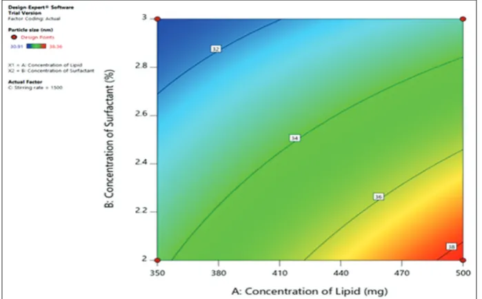
The particle size parameter shows that as the concentration of surfactant increases, the particle size decreases. The particle size of
all the batches ranges from 30.91±0.30 to 38.36±0.13 nm. Batch 7 was found to have the lowest particle size.
Effect of concentration of lipid and surfactant on particle size
Increased quantity of cocoa butter (lipid) caused an increase in particle size. The fact that the size of lipid nanoparticles is highly reliant on lipid concentration can be described in terms of the propensity of lipid to coalesce or unite at high lipid concentration. According to Stoke’s law, this kind of behavior can be described by the difference in density between internal and external phases. Increase in particle size of SLNs because of a reduction in the diffusion rate of the solute molecules in the outer phase as a result of viscosity increases in the lipid-solvent phase. Moreover, the increase in particle size might be due to increased quantity of lipid which provides additional space for drug molecules to get entrapped [22].
On increasing the concentration of tween 80, the particle size was found to get decreased. This might be due to the surfactant-induced decrease in surface tension between the aqueous phase and organic phase. Besides, the surfactant helps to stabilize the fresh generated surfaces and avoids particle aggregation. Higher concentrations of surfactants permit improved stabilization of the smaller droplets of lipids and thus prevent them from aggregating into larger droplets [22] (Figs. 1 and 2).
Response 2: Entrapment efficiency (% entrapment)
Final equation in terms of coded factors
Entrapment efficiency=80.55 + 2.08* A + 4.50*B − 1.50* C − 2.43* AB − 3.11* AC−0.7662*BC
Entrapment efficiency parameter of all the eight formulation ranges from 73.99±0.48 to 89.99±0.87. Batch 7 was found to have highest entrapment efficiency.
Effect of concentration of lipid and surfactant on entrapment efficiency
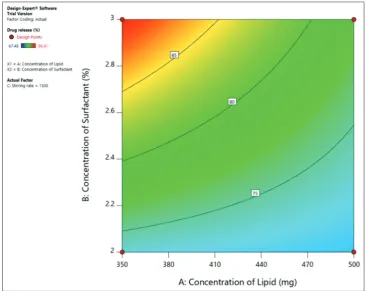
The concentration of lipids affects entrapment efficiency. Higher the concentration of lipids greater will be the entrapment of active material into the lipid. Low concentration of lipids can lead to the lower entrapment of active material into the lipid often leading to nanoparticles with larger particle size. With increasing the amount of lipid, % EE was found to increase because lipids act as solubilizing agents for the highly lipophilic drug. As the concentration of the surfactant increased, there is an increase in entrapment efficiency. This might be due to the increased solubility of the drug in the lipid by increasing the concentration of the surfactant [23,24] (Figs. 3 and 4).
Effect of stirring rate on entrapment efficiency
The entrapment efficiency is directly proportional to stirring speed. Higher the stirring speed, there is a rapid division of nanoparticles. This may have lesser chances of particle aggregation. Hence, as the stirring speed increases, the particle size decreases.
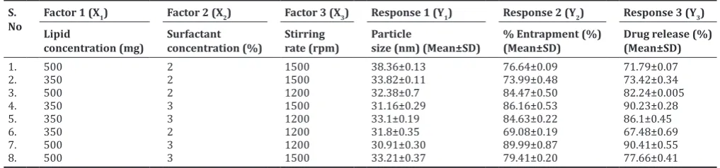
Table 3: 23 factorial design for optimization of lavender oil loaded SLNs
S.
No Factor 1 (XLipid 1) Factor 2 (X2) Factor 3 (X3) Response 1 (Y1) Response 2 (Y2) Response 3 (Y3) concentration (mg) Surfactant concentration (%) Stirring rate (rpm) Particle size (nm) (Mean±SD) % Entrapment (%)(Mean±SD) Drug release (%)(Mean±SD)
1. 500 2 1500 38.36±0.13 76.64±0.09 71.79±0.07
2. 350 2 1500 33.82±0.11 73.99±0.48 73.42±0.34
3. 500 2 1200 32.38±0.7 84.47±0.50 82.24±0.005
4. 350 3 1500 31.16±0.29 86.16±0.53 90.23±0.28
5. 350 3 1200 33.1±0.19 84.63±0.22 86.1±0.45
6. 350 2 1200 31.8±0.35 69.08±0.19 67.48±0.69
7. 500 3 1200 30.91±0.30 89.99±0.87 90.41±0.55
8. 500 3 1500 33.21±0.37 79.41±0.20 77.66±0.41
Fig. 3: Three dimensional plot for effect of concentration of lipids and concentration of surfactant on entrapment efficiency Fig. 1: Three dimensional plot for effect of concentration of lipids and concentration of surfactant on particle size
Fig. 5: Three dimensional plot for the effect of concentration of lipids and concentration of surfactant on drug release Fig. 4: Contour plot for the effect of concentration of lipids and concentration of surfactant on entrapment efficiency Response 3: In vitro drug release studies
Final equation in terms of coded factors
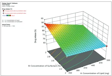
Drug release=79.92+0.6087*A + 6.18*B − 1.64*C − 2.76*AB −4.16*AC −0.5137*BC
Drug release parameter shows that drug release of all the eight formulation ranges from 67.48±0.69 to 90.41±0.55. Batch 7 was found to have the highest drug release.
Effect of concentration of lipid and surfactant on drug release
As the lipid concentration increases, the drug release rate decreases; this might be due to the higher concentration of drug present in the inner core of the vesicle. Drug release might also get decreased due to differences in the partition coefficient of the active material and lipophilicity of lipids. This lipophilicity decreased the diffusion of the
drug from the cocoa butter to the aqueous medium of dissolution. As the surfactant concentration increases, the drug release rate increases due to the increased solubility of drugs in the external phase. This might be due to the high affinity of surfactants toward aqueous environments [22] (Figs. 5 and 6).
From the above observation, batch 7 was found to be optimized. Hence, it was further evaluated for zeta potential and was then formulated into the gel. Image of SLNs of batch 7 is shown in Fig. 7 which was observed in digital microscope.
pH
Zeta potential
Zeta potential can be either positive or negative depending on the chemistry of the nanoparticles, which is an electric potential that is created due to the presence of a charge on the particle surface. Zeta potential indicates the degree of repulsion between equally charged particles in the formulation. Repulsive forces prevent aggregation of the particle during storage. Zeta potential is thus indicative of possible physical stability of a formulation. The ideal value of zeta potential is
−30 mV to +30 mV. The zeta potential of the formulation was found
to be 22.1 mV. Hence, the formulation was found to have moderate stability.
SLNs covered by a non-ionic surfactant like Tween 80 tend to remain stable regardless of having a lower zeta potential. Greater steric stabilization and less electrostatic stabilization are responsible for this kind of behavior. Surface encapsulation of the SLNs lessens the electrophoretic mobility of the particles and thus lowers the zeta potential. Hence, zeta potential measurement was not taken into consideration as a primary parameter in the selection of the optimal formulation [25].
Stability study
The stability studies were studied over different storage conditions of 5°C and 25°C as per the International Conference on Harmonization (ICH) guidelines. Both physical and chemical changes were studied after 3 months. Physical stability was checked in terms of appearance and particle size, whereas chemical studies were checked in terms of entrapment efficiency and drug release profile. The results show that there was no significant change in particle size, entrapment efficiency and drug release of the SLN formulation stored at 5°C and 25°C after 3 months (Table 4).
Characterization of gel
Physical evaluation
The SLN based gel of LEO was found to be homogenous, smooth, and consistent.
Fig. 7: Image of batch 7 (lavender essential oil loaded solid lipid nanoparticles) captured in a digital microscope at ×100
Table 4: Stability study of SLN (batch 7)
Evaluation parameter Initial After 3 months (°C)
5 25
Particle size (nm) 30.91 31.87 31.23
Entrapment efficiency (%) 89.99 87.34 86.98
% Drug release 90.41 88.83 88.25
SLN: Solid lipid nanoparticles
Fig. 6: Contour plot for the effect of concentration of lipids and concentration of surfactant on drug release
pH measurement
The pH of the formulation was found to be 6.8. It was found to be compatible with the skin pH [26].
Viscosity study
showed with non-Newtonian flow. This behavior might be due to its low flow resistance when applied at the high shear condition.
The results show that as the rpm increases, there was decrease in viscosity of the gel (Table 5).
Spreadability
Spreadability plays an important role in patient compliance and helps in the uniform application of the gel to the skin. A good gel spreads easily and takes less time to spread on the skin. The spreadability of the gel was found to be 6.28±0.4 cm.
In vitro drug release of optimized SLN based gel
The drug release of the gel after 8 h was found to be 88.73% (Table 6 and Fig. 8).
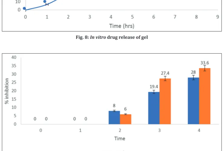
Ex vivo study
The % inhibition of carrageenan-induced edema method against 1% gel LEO loaded SLN based gel is shown in Table 7 and Fig. 9. Ex vivo studies were studied on Wistar rats. Percentage inhibition of the gel was found to be 28±0.1%. The anti-inflammatory activity was also studied in Voveran gel of diclofenac which was found to be 33.6±0.05% (Table 7).
Stability studies
The stability studies were studied over different storage conditions of 5°C and 25°C as the per the ICH guidelines. Physical and chemical
Fig. 8: In vitro drug release of gel
Fig. 9: Ex vivo studies of optimized solid lipid nanoparticles gel formulation
Table 5: Viscosity study of lavender oil loaded SLN based gel S.
No. R.P.M Viscosity at 0.2% concentration (cps)
1. 5 2658
2. 10 2585
3. 20 2514
4. 50 2403
5. 100 2311
R.P.M: Revolutions per minute, SLN: Solid lipid nanoparticles
Table 6:In vitrodrug release of optimized lavender oil loaded SLN based gel
S. No. Time % Drug release (Mean±SD)
1. 0 0
2. 1 6.85±0.4
3. 2 20.87±0.6
4. 3 38.6±0.7
5. 4 46.63±0.9
6. 5 54.8±1.2
7. 6 62.26±0.5
8. 7 74.9±1.1
9. 8 88.73±0.1
changes were studied after 3 months. Physical studies were checked in terms of visual examination, whereas the chemical changes were checked in terms of pH, spreadability, and drug release and viscosity. The results show that there was no significant change found in stability stored at 5°C and 25°C after 3 months. The results are stated in Table 8.
CONCLUSION
In the present work, lavender oil loaded SLNs were successfully prepared by the hot homogenization technique. The various physicochemical properties, particle size, and drug release entrapment efficiency were affected and can be controlled using the optimization technique. Alteration in the concentration of lipid, concentration of surfactant and stirring time can overcome the problems associated. 23 factorial design was used for the experiment and 8 formulations were developed. F7 formulation was found to be optimized. Its particle size was smaller as desired; entrapment efficiency and drug release were found to be maximum. SLN based gel was also developed successfully. Stability studies were conducted after 3 months, which did not show any remarkable change. Hence, the desired aim and objective could be achieved properly by lavender oil loaded SLN based gel.
REFERENCES
1. Vashishtha V, Sharma G, Gaur M, Bairwa R. A review on some plants having anti-inflammatory activity. J Phytopharm 2014;3:214-21. 2. Abdulkhaleq LA, Assi MA, Abdullah R, Zamri-Saad M, Taufiq-Yap YH,
Hezmee MNM, et al. The crucial roles of inflammatory mediators in inflammation: A review. Vet World 2018;11:627-35.
3. Ward PA, Lentsch AB. The acute inflammatory response and its regulation. Arch Surg 1999;134:666-9.
4. Verma S, Makkar D. Solid lipid nanoparticles: A comprehensive review. J Chem Pharm Res 2016;8:102-14.
5. Ekambaram P, Sathali A. Solid lipid nanoparticles: A review. Sci Rev Chem Commun 2012;2:20-102.
6. Cardia GFE, Silva-Filho SE, Silva EL, Uchida NS, Cavalcante HAO, Cassarotti LL, et al. Effect of lavender (Lavandula angustifolia) essential oil on acute inflammatory response. Evid Based Complement Alternat Med 2018;2018:1413940.
7. Vijayan V, Shaikh A, Sakthivel S, Reddy R. Formulation and characterization of solid lipid nanoparticles loaded neem oil for topical
delivery. J Acute Dis 2013;2:282-6.
8. Nasseri M, Golmohammadzadeh S, Arouiee H, Jaafari MR, Neamati H. Antifungal activity of Zataria multiflora essential oil-loaded solid lipid nanoparticles in-vitro condition. Iran J Basic Med Sci 2016;19:1231-7. 9. Wencui Z, Qi Z, Ying W, Di W. Preparation of solid lipid nanoparticles
loaded with garlic oil and evaluation of their in vitro and in vivo characteristics. Eur Rev Med Pharmacol Sci 2015;19:3742-50. 10. Shi F, Zhao JH, Liu Y, Wang Z, Zhang YT, Feng NP, et al. Preparation
and characterization of solid lipid nanoparticles loaded with frankincense and myrrh oil. Int J Nanomedicine 2012;7:2033-43. 11. Aland R, Ganesan M, Rao R. In vivo evaluation of tazarotene solid
lipid nanoparticles gel for topical delivery. Int J Pharm Sci Drug Res 2019;11:45-50.
12. Dianzani C, Foglietta F, Ferrara B, Rosa AC, Muntoni E, Gasco P, et al. Solid lipid nanoparticles delivering anti-inflammatory drugs to treat inflammatory bowel disease: Effects in an in vivo model. World J Gastroenterol 2017;23:4200-10.
13. Khare A, Singh I, Pawar P, Grover K. Design and evaluation of voriconazole loaded solid lipid nanoparticles for ophthalmic application. J Drug Deliv 2016;2016:6590361.
14. Jourghanian P, Ghaffari S, Ardjmand M, Haghighat S,
Mohammadnejad M. Sustained release curcumin loaded solid lipid nanoparticles. Adv Pharm Bull 2016;6:17-21.
15. Shilpa N, Chakravarthi N. Moxifloxacin loaded solid lipid nanoparticles preparation and characterization. Asian J Pharm Res 2012;2:105-12. 16. Madan JR, Khude PA, Dua K. Development and evaluation of solid
lipid nanoparticles of mometasone furoate for topical delivery. Int J Pharm Investig 2014;4:60-4.
17. Khalil R. Solid Lipid Nanoparticles for Topical Delivery of Meloxicam. Portugal: Annual International Interdisciplinary Conference 2013. p. 24-6.
18. Kushwaha AK, Vuddanda PR, Karunanidhi P, Singh SK, Singh S. Development and evaluation of solid lipid nanoparticles of raloxifene hydrochloride for enhanced bioavailability. Biomed Res Int 2013;2013:584549.
19. Gupta R, Gupta G. Formulation, development and evaluation of anti-inflammatory potential of cordia obliqua topical gel on animal model. Pharmacogn J 2017;9:s93-8.
20. Kuchekar M, Mohite M, Phadtare G. Formulation and evaluation test for voriconazole gel. Eur J Pharm Med Res 2016;3:466-70.
21. Damor B, Dashora A, Parra S. Evaluation of analgesic and anti-inflammatory activity of methanolic extract of Guazuma ulmifolia. J Appl Pharm Sci Res 2018;1:23-9.
22. Shah D, Gupta A, Shah Y. Effect of lipid and surfactant concentration on cefpodoxime proxetil solid lipid nanoparticles. Eur J Biomed Pharm Sci 2017;4:817-23.
23. Ekambaram P, Abdul HS. Formulation and evaluation of solid lipid nanoparticles of ramipril. J Young Pharm 2011;3:216-20.
24. Bhalekar M, Upadhyay P, Madgulkar A. Formulation and characterization of solid lipid nanoparticles for anti-retroviral drug darunavir. Appl Nanosci 2017;7:47-57.
25. Shah R, Eldridge D, Palombo E, Harding I. Optimization and stability assessment of solid lipid nanoparticles using particle size and zeta potential. J Phys Sci 2014;25:59-75.
26. Kasar P, Kale K, Phadtare D. Formulation and evaluation of topical antifungal gel containing itraconazole. Int J Curr Pharm Res 2018;10:71-4.
Table 8: Stability study of optimized lavender oil loaded SLN based gel (Viscosity)
S. No. R.P.M Viscosity (cps)
(Before 3 months) Viscosity (cps)(After 3 months)
1. 5 2658 2789
2. 10 2585 2677
3. 20 2514 2603
4. 50 2403 2561
5. 100 2311 2453
R.P.M: Revolutions per minute, SLN: Solid lipid nanoparticles
Table 7: Stability study of lavender oil loaded SLN based gel
Months Visual examination pH Spreadability (Mean±SD) In vitro drug release (Mean±SD)
Initial Transparent, smooth, homogenous 6.8 6.28±0.4 cm 88.73±0.1
After 3 months Transparent, smooth, homogenous 6.7 6.05±0.2 cm 86.41±0.5