Mouse models to study inner ear development
and hereditary hearing loss
LILACH M. FRIEDMAN, AMIEL A. DROR and KAREN B. AVRAHAM*
Department of Human Molecular Genetics and Biochemistry, Sackler School of Medicine, Tel Aviv University, Tel Aviv, Israel
ABSTRACT Hereditary sensorineural hearing loss, derived from inner ear defects, is the most common hereditary disability with a prevalence of 1 in 1000 children, although it can be present in up to 15% of births in isolated communities. The mouse serves as an ideal animal model to identify new deafness-related genes and to study their roles in vivo. This review describes mouse models for genes that have been linked with hearing impairment (HI) in humans. Mutations in several groups of genes have been linked with HI in both mice and humans. Mutant mice have been instrumental in elucidating the function and mechanisms of the inner ear. For example, the roles of collagens and tectorins in the tectorial membrane, as well as the necessity of intact links between the hair cell projections, stereocilia and kinocilia, have been discovered in mice. Accurate endolymph composition and the proteins which participate in its production were found to be crucial for inner ear function, as well as several motor proteins such as prestin and myosins. Two systematic projects, KOMP and EUCOMM, which are currently being carried out to create knock-out and conditional mutants for every gene in the mouse genome, promise that many additional deafness-related genes will be identified in the next years, providing models for all forms of human deafness.
KEY WORDS: mouse, inner ear development, deafness gene, hearing
Introduction
Hereditary hearing loss (HHL) in humans
Hearing impairment (HI) is traditionally classified as conduc-tive and sensorineural, based on the defecconduc-tive part of the hearing organ. While conductive HI results from defects in the external or middle ear, sensorineural HI results from a defect located along the auditory pathway, from the cochlea to the auditory cerebral cortex. A conductive defect yields a mild to moderate HI and in most cases may be medically solved. In contrast, a sensorineural defect yields a mild to profound HI and, thus far, sensorineurally hearing impaired persons may be aided with cochlear implants or hearing aids, but their problem cannot be completely solved [recently reviewed in (Petit, 2006)]. Therefore, further study is required to enable development of better therapies for senso-rineural HI.
At least 60% of persons with early-onset HI have hereditary hearing loss (HHL) due to genetic mutations. In most of these cases, a single mutation in a single gene is responsible for the hearing loss. About 70% of HHL cases in human are isolated or
*Address correspondence to: Karen B. Avraham. Department of Human Molecular Genetics and Biochemistry, Sackler School of Medicine, Tel Aviv University, Tel Aviv, Israel. Fax: +972-3-640-9360. e-mail: [email protected]
Electronic Supplementary Material (Table S1 - genes that have been linked both with inner ear defects in mice and HHL in humans) is available for this paper at: http://www.ijdb.ehu.es/web/paper.php?doi=072365lf
0214-6282/2007/$30.00 © UBC Press
Printed in Spain www.intjdevbiol.com
Abbreviations used in this paper: ABR, auditory brainstem response; BM, basilar membrane; ENU, N-ethyl-N-nitrosourea; EP, endocochlear potential; HHL, hereditary hearing loss; HI, hearing impairment; IHC, inner hair cells; NSHL, non-syndromic hearing loss; OHC, outer hair cells; PBM, PDZ binding motif; RP, retinitis pigmentosa; SHL, syndromic hearing loss; TM, tectorial membrane; WT, wild type.
include genes that encode for extracellular matrix components, gap junction and adhesion proteins, ion channels and transport-ers, other cell surface proteins and receptors, as well as myosins (molecular motors), cytoskeletal proteins, transcription factors and other proteins that interact with them to create hearing-related networks.
If mutations in a given gene lead to defective development of the inner ear and to early-onset HHL, the affected gene may be considered as having a role in inner ear development or function. A better understanding of inner ear development is required to understand the mechanisms by which specific mutations lead to HHL.
Mouse models
The study of sensorineural HHL in humans is limited by the inability to follow inner ear development. Genetic linkage analysis of HHL in humans is possible only in large families that contain several hearing impaired members. In addition, the search for the responsible gene in HHL patients may be more complicated than analysis of other inherited characters, since hearing-impaired persons from different families tend to marry each other and
marriages between hearing children of hearing-impaired parents are also not rare. As a result, one family may carry two or more deafness-related mutations (Petit, 2006). Moreover, due to the absence of a satisfactory human cell line with similar character-istics to the developing inner ear, only primary cultures or model animals may be used to study the interactions between proteins expressed in the inner ear, their spatial and temporal expression patterns, their functions and for any other biological study.
Mutant mouse models that exhibit HHL due to inner ear defects may help to identify genes that have a role in the development or function of the inner ear. When a gene is suspected as respon-sible for HHL in humans, similar mutations may be engineered in mice to verify this hypothesis. Gene-targeted mutagenesis, or ‘knockout’ mice, may also be used to uncover the gene’s role by comparison with wild type mice. Knockout mice have also been made for genes suspected as essential for hearing due to known interactions of their products with proteins encoded by other known deafness-related genes, or due to the expression of their products in the inner ear. In addition, mouse models are used to identify new genes that have a role in inner ear development and normal hearing. Many strains of hearing-impaired mice have
arisen spontaneously during the last century. Moreover, coinci-dental mutagenesis of mouse chromosomes, by chemicals (mainly by ENU, N-ethyl-N-nitrosourea), by X-ray radiation or by coinci-dental insertion of an extrinsic sequence («gene trap») has been used to create new hearing-impaired mouse strains. Identification of the responsible gene in such strains is much easier than genetic linkage analysis in humans. In fact, many deafness-related genes were identified in humans only after their identifica-tion in hearing-impaired mice (Supplementary Table S1).
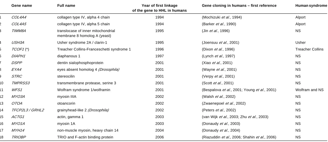
Mutations in more than 172 different genes have been reported as responsible for inner ear malformations or dysfunction in mice (most of them are listed in the Jackson Laboratory’s Hereditary Hearing Impairment in Mice database: http://www.jax.org/hmr/ master_table.html). Only 44 of them have already been linked to human HHL (these genes are listed in Supplementary Table S1). In addition, two genes that were linked with human HHL were found as not crucial for inner ear development and function in knockout mice (Table 1). Figure 1 illustrates the spatial expres-sion of some of the proteins encoded by genes associated with HHL.
Following the identification of the mutated gene, the mutant
mice may be used to follow defective inner ear development and to identify specific roles of the gene products. Examples of assays that have been used to evaluate the outcome of deafness-related mutations are shown in Figures 2 and 3. Inner ear development and defects may be followed using bright field light microcopy (Figure 2, A-C), transmission (TEM; Figure 2, D-F) or scanning electron microscopy (SEM; Figure 2, G-J), as well as by paintfill analysis (Figure 2, K-L). Physiological assays may be used to measure ion currents and voltage potentials. The patch clamp assay may be used to measure currents or membrane potentials in a single cell. Length change in individual cells may be used to measure electromotility of outer hair cells (Figure 2, M-N). Tem-poral and spatial expression patterns of specific mRNAs or proteins in the cells may be observed by in situ hybridization (ISH; Figure 2, O-P) and immunofluorescence (Figure 2, Q-V), respec-tively. Measurement of auditory brainstem response (ABR) to sound signals by scalp electrodes is the most widely used assay for evaluating hearing in mice (Figure 3, A-B). Vestibular defects may be assessed by swimming or other behavioral tests and may induce a characteristic circling behavior (Figure 3, C-E).
This review will describe mutant mouse models for some
B
C
D
E
F
G
B
C
D
E
F
H
I
J
A
O
K
L
P
Q
R
S
T
M
N
U
V
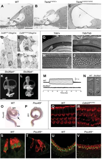
Fig. 2. Examples of assays which have been used to assess roles of specific genes in the mouse inner ear.(A-C) Light microscopic analy-sis (toluidine blue staining) of 1 mm thick sec-tions of the cochlear duct from wild type(A),
TectaY1870C/+(B) and TectaY1870C/Y1870C(C) mice.
Abnormal and detached tectorial membranes are observed when Tecta is mutated. Abbrevia-tions: LZ – limbal zone, MB – marginal band, HS – Hensen’s stripe. Arrows: an arrowhead in (B) – Kimura’s membrane, an arrow in (C) – tectorial membrane. Scale bars, 50 µm. Reprinted with permission from (Legan et al., 2005). (D-F) Trans-mission electron microscopy (TEM) of the organ of Corti at P30 from cochlea that expresses wild type connexin 26 (D), compared to cochlea in which the Cx26 gene was deleted (E-F). When Cx26 is absent, damaged Deiters’ cells do not stick to OHC, leading to hair cell degeneration. Abbreviations: D – Deiters’ cells, P – outer pillar cells. Arrows: disruption of the reticular lamina. Scale bars, 2.3 µm in (D), 1.25 µm in (E) and 0.8
µm in (F). Reprinted with permission from (Cohen-Salmon et al., 2002). (G-J) Scanning electron microscopy (SEM) of the organ of Corti basal portions at P0 from yellow submarine (Ysb) heterozygous (G-H)and homozygous (I-J)
mice. Ysb is a mutant allele of Sox2. Abnormal patches of hair cells are found in Ysb/Ysb mice. Three arrows in (H) indicate the OHC. Scale bars, 100 µm in (G, I), 20 µm in (H, J). Reprinted with permission from(Kiernan et al., 2005). (K-L) The paint-fill assay is used to present the endolymph labyrinth of the inner ear, in order to identify malformations within the inner ear cavi-ties (Bissonnette and Fekete, 1996). These fig-ures present lateral view of paint-filled P1 inner ears from heterozygous (K) and homozygous
(L)mice for the knocked-out allele of Slc26a4/ Pds (encoding pendrin). Slc26a4-/- mice exhibit
dilated cochleae and endolymphatic ducts and sacs. Scale bar, 500 µm. Reprinted with permis-sion from(Everett et al., 2001). (M-N) In vitro
analysis of OHC electromotility in wild type and mutant (Slc26a5/Prestin knockout) mice. (M)
Length changes of OHC in response to voltage steps (-120-60 mV in 20 mV steps) in whole-cell, voltage-clamp recordings. (N) Micrographs of OHC isolated from apical turns of cochleae. OHC that do not express prestin are shorter than wild type OHC and do not exhibit electromotility. Arrows in (N): open arrow -nucleus; filled arrow - stereocilia. Scale bar, 5
µm in (N). Reprinted with permission from (Liberman et al., 2002). (O-P) Whole mount in situ hybridizations (ISH) detect expression of Pou4f3(O) and Gfi1 (P) mRNAs in E18.5 cochleae from wild type (O) and dreidel (P) littermate mice. Dreidel mice, which do not express functional Pou4f3 protein, do not express Gfi1 mRNA. Arrow – Pou4f3
mRNA expression is detected as a blue band along the lateral wall of the cochlea. Reprinted with permission from (Hertzano et al., 2004). (Q-R) Whole mount immunohistochemistry detects spatial expression pattern of F-actin (shown in red, stained with rhodamine phalloidin) in stereocilia in the middle turn of wild type (Q) and waltzer V6J(R) organ of Corti at P7. The V6J allele was reported to be a functional null allele of Cdh23 (Di Palma et al., 2001a;
Di Palma et al., 2001b). Waltzer mice exhibit disorganized stereocilia (Lagziel et al., 2005). Scale bars, 5 µm. Figures from Ayala Lagziel and Thomas B. Friedman. (S-V) Immunohistochemistry of paraffin sections of E18.5 wild-type (S and U) and Pou4f3-/- (T and V) mouse inner ears. Expression of
Lhx3 (green) and myosin VI (red) was detected in the cochlea (S-T) and the vestibular system utricle (U-V). Whereas Lhx3 is expressed in the nuclei of all hair cells in the wild-type inner ears, Lhx3 expression could be detected only in the vestibular system of the Pou4f3 -/-mice but not in any of the
representative genes that are crucial for normal development and function of the mammalian inner ear. We will focus mainly on genes for extracellular and integral inner ear proteins that were found to be involved both in human HHL and mouse inner ear development. Nonetheless, some examples for genes encoding for intracellular proteins will be also mentioned.
Extracellular matrix components: cartilage and
tecto-rial membrane defects (collagen genes and Tecta)
The mammalian hearing organ, the organ of Corti, sits in the snail-shaped cochlea on a strand of connective tissue, the basilar membrane (BM). The collagen-based BM is graded in stiffness along the cochlea and vibrates in response to sound-induced movements of the cochlear fluids. These vibrations are detected by two types of hair cells, included in the sensory epithelium of the organ of Corti, the inner and outer hair cells (IHC and OHC, respectively). The mechanosensory hair bundles of the OHC project up from the reticular lamina, the apical surface of the sensory epithelium and are embedded in the overlying tectorial membrane (TM) [reviewed in (Raphael and Altschuler, 2003)]. A cross section of the organ of Corti is illustrated in Figure 1A. The mammalian TM has a unique and highly organized ultrastructure. It contains two main groups of components: collagen fibrils that are organized in heavy bundles and run radially across the TM and glycoproteins that compose the unusual striated-sheet matrix surrounding the fibrils (schematically illustrated in Figure 1B) (Hasko and Richardson, 1988). Collagens types II, IX and XI compose the radial fibrils (Slepecky et al., 1992; Thalmann, 1993), while two glycoproteins, alpha and beta tectorins (encoded by Tecta and Tectb), are the major components of the TM matrix (Legan et al., 1997).
Seven collagen proteins were linked with human HHL: COL2A1, COL4A3, COL4A4, COL4A5, COL9A1, COL11A1 and COL11A2 (Van Camp and Smith, 2006). Only five of these have mouse models (Supplementary Table S1). A mutation in COL11A2 was linked with autosomal dominant NSHL in humans (DFNA13 locus), but also with Stickler syndrome. The other collagen genes were only linked with SHL in humans, mainly Stickler (COL2A1, COL9A1 and COL11A1) and Alport (COL4A3-5) syndromes.
Alport syndrome-related collagens (chains alpha-3, 4 and 5 of collagen type IV) are included in basement membranes of the inner ear and the kidney’s glomeruli. In the cochlea, they are expressed in the BM, parts of the spiral ligament and stria vascularis. As a result, Alport syndrome (Alport, 1927) combines sensorineural HHL and progressive nephritis, often progressing
up to renal failure [reviewed in (Hudson et al., 2003)]. Following the identification of mutations in the human COL4A3 gene as responsible for Alport syndrome (Mochizuki et al., 1994), Col4a3 was knocked out in mice (Cosgrove et al., 1996). Homozygotes died at about 14 weeks of age due to renal failure. Defective basement membranes were found in the renal glomeruli and cochlear membranous labyrinth, similar to the human disease. The renal phenotype included progressive glomerulonephritis with proteinuria and microhematuria, focal multilaminated thick-ening and thinning of the glomerular basement membranes, as well as fibrotic glomeruli with collapsed capillaries. In the cochlear membranous labyrinth, both Col4a3 and Col4a4 chains were completely absent. Basement membranes of specific parts of the membranous labyrinth were significant thinner, thicker or unde-tectable compared to wild type cochleae and nearby capillaries were collapsed. Both renal and cochlear defects were progres-sive and HI was detected only after 6 weeks of age (Cosgrove et al., 1996; Cosgrove et al., 1998).
Stickler syndrome (Stickler et al., 1965) includes, in addition to a progressive sensorineural HHL, premature degenerative changes in various joints with abnormal epiphyseal development, vertebral abnormalities, osteoarthritis and sometimes also un-usual face and cleft palate. There are three types of Stickler syndrome: type 1 includes also progressive myopathy and blind-ness due to vitreoretinal degeneration and retinal detachment, while type 2 displays different vitreous defects with no retinal detachment [reviewed in (Snead and Yates, 1999)]. Type 3 is milder, with neither myopathy nor eye involvement (Vikkula et al., 1995). The Stickler syndrome-related collagens Col2a1, Col11a1 and Col11a2 are important components not only of the cochlear TM but also of the cartilage (Col2a1 is expressed also in the eye’s vitreous). Since the inner ear has a cartilage cover, which has an important role in its embryogenesis, mutated collagens types II and XI affect the inner ear size, structure and development.
COL2A1 was found to be involved in sensorineural deafness that accompanies several similar hereditary syndromes in hu-mans, such as Stickler syndrome, spondyloepiphyseal dysplasia congenita (SEDC) and chondrodysplasia. Dmm (autosomal semi-dominant disproportionate micromelia), a mouse with a mutated Col2a1 gene produced in 1966, is an offspring of a male whose spermatogonia had been irradiated. The Dmm mutation is a three-nucleotide deletion in the region encoding the C-propeptide globular domain of Col2a1. The deletion leads to the replacement of two amino acids, Lys and Thr, by a single amino acid, Asn, in the mutated protein (Pace et al., 1997). Dmm mice expressed a reduced level of collagen II and suffered from cartilage defects
Gene name Full name Main role of gene product Human reference* Human syndrome Mouse reference& Mouse strain
(mutagenesis method§)
Coch Coagulation factor Unknown. Secreted protein, (Robertson et al., 1998) NS (Makishima et al., 2005) Coch-/- (KO)
C homolog, cochlin most abundant protein in cochlea.
Myh9 non-muscle myosin Actin-binding motor protein (Lalwani et al., 2000) NS (autosomal dominant) (Parker et al., 2006) Myh9+/- (GT) Homozygotes
heavy polypeptide 9 died during gestation
HEREDITARY HEARING LOSS (HHL)- LINKED GENES IN HUMANS,
WHICH ARE NOT AS CRUCIAL FOR INNER EAR DEVELOPMENT AND FUNCTION IN MICE TABLE 1
that affect inner ear development as well. The homozygotes were dwarf with disproportionate short limbs (micromelia), had a cleft palate (Brown et al., 1981; Seegmiller et al., 1988) and died at birth due to lung hypoplasia (Foster et al., 1994). Inner ears of homozygous Dmm embryos had less collagen fibrils and pre-sented irregular cytodifferentiation of chondrocytes in the extra-cellular matrix, compared to wild type embryos (Berggren et al., 1997). As a result, dysmorphogenesis of the otic capsule and perilymphatic spaces during embryogenesis led to the develop-ment of malformed inner ears with a bulky cartilaginous capsule and a lack or reduction of defined perilymphatic spaces (Van de Water and Galinovic-Schwartz, 1987). More recently, a missense mutation in the mouse Col2a1 gene was produced spontaneously (R1417C). These mice were named sedc, since their phenotype was similar to human spondyloepiphyseal dysplasia congenita. Homozygous sedc adult mice had shortened noses, dysplastic vertebrae, femora and tibias, retinoschisis and hearing loss (Donahue et al., 2003). Gene targeted mutagenesis was used to create Col2a1 G574S mice, developed as a model for chondrod-ysplasia, following a parallel mutation that was found in humans. In addition to skeletal malformations, the mice were hearing impaired due to the development of a misshapen otic capsule. While the normal otic capsule is rounded, the transgenic otic capsule was flattened and elongated. The authors suggested that the weaker cartilage of the optic capsule could not resist the mechanical pressures from the developing brain and face and was squashed (Maddox et al., 1998). Heterozygote Col2a1 mu-tated mice displayed a milder but not normal phenotype.
Col9a1 is an example to a gene that was linked to HHL in mice before its mapping to a deafness-related locus in humans. Col9a1-knockout mice were raised as soon as 1994, but their inner ears were not studied and the observed phenotype was mainly non-inflammatory joint disease resembling human osteoarthritis (Fassler et al., 1994). Only 11 years later, following the re-finding that Col9a1 is highly expressed in the human inner ear (Abe et al., 2003) [collagens IX were found to be a major component of the TM also previously (Richardson et al., 1987)], the inner ears and hearing of Col9a1 knockout mice were studied (Asamura et al.,
2005). Indeed, these mice displayed a progressive hearing loss, most probably due to a disturbed organization of collagen fibrils in the TM, leading to an abnormal shape of this membrane. TM of Col9a1 knockout mice contained neither collagens IX nor col-lagens II. Therefore, it was suggested that colcol-lagens IX and II may interact in the TM to determine its three-dimensional structure (Asamura et al., 2005; Suzuki et al., 2005). A year later, a mutation in COL9A1 was linked to an autosomal recessive Stickler syn-drome in humans (Van Camp et al., 2006).
Cho mice arose spontaneously in 1971 (Seegmiller et al., 1971). Homozygotes had a cleft palate and died soon after birth due to lethal chondrodysplasia. The cho mutation is a 1-nt deletion in the Col11a1 gene that causes a frameshift and a premature termination codon, resulting in a truncated gene prod-uct that cannot assemble with other collagen molecules. Thus, cho is actually a functional null allele of Col11a1 (Li et al., 1995). Homozygotes were severely hearing impaired at birth due to underdevelopment of the organ of Corti in the lower turn of the cochlea, with no hair cells, supporting cells, nerve endings and pillar cells (Cho et al., 1991). Since heterozygous cho mice, which expressed both wild type and cho alleles of Col11a1, suffered from age-dependent osteoarthritis, it was suggested that the cho allele may have a destructive effect on connective tissues. How-ever, heterozygous mice were well hearing during their first two months of life and developed a moderate and progressive hearing loss later (age-related) that was not significantly differ from wild type mice (Szymko-Bennett et al., 2003). In contrast to findings in mice, human COL11A1-linked SHL is expressed also in heterozy-gotes: a point mutation in COL11A1 (G97V) was linked with an autosomal dominant Stickler syndrome (Richards et al., 1996) and a splice-donor-site mutation in this gene was linked with the similar autosomal dominant Marshall syndrome (Griffith et al., 1998).
Col11a2 was knocked out in mice by insertion of a neomycin-resistance cassette in the reverse orientation in place of exons 27 and 28. The inserted sequence included a premature termination codon. Thus, the full length protein was not expressed. The phenotype was much milder compared to cho (functional null
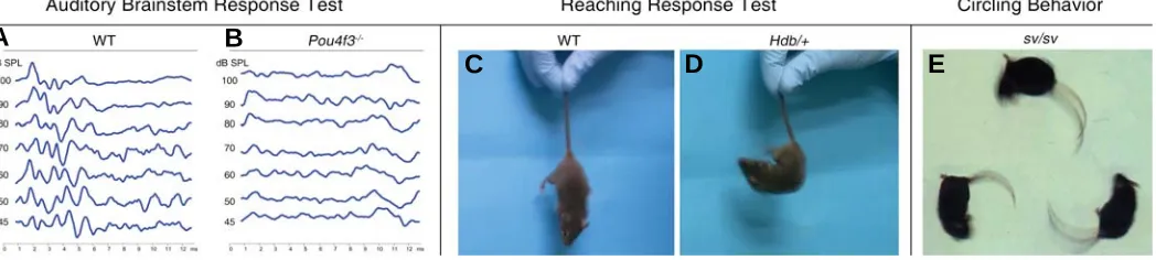
Fig. 3. Common behavioral tests for measuring and observing auditory and vestibular dysfunctions in mouse models. (A-B) Comparison between wild type and Pou4f3-/- auditory brainstem response (ABR) tests. WT mice show typical graphs with peaks in response to various intensities
(45-100 dB) of sound stimulations (A). Flattened graphs are observed in Pou4f3-/- mice even at the highest sound level (100 dB), indicating the profound
hearing loss of Pou4f3-/- mice at the measured frequency of 16 KHz (B). (C-D) Vestibular apparatus defects can be determined by reaching response.
By holding their tails, normal mice will stretch out their legs to make contact with the underneath surface (C). Hdb (headbanger) is a dominant mutated allele of Myo7a, induced by ENU mutagenesis (Rhodes et al., 2004). Mice heterozygous for this mutation (Hdb/+) failed in recapitulate their three dimensional position, curling up towards their tails (D). Figures from Amiel Dror. (E) A Snell’s waltzer mouse, which is homozygous for the sv allele of
Myo6 (spontaneous mutation), exhibits circling behavior, which is another strong indicator for an abnormal balance system.
B
C
D
E
Col11a1) mice. Homozygous mice had a smaller body size due to shorter long bones, receding snouts due to shorter nasal bones and hearing loss. The only morphologic abnormality observed in the inner ear was a larger and less compact TM with disorganized collagen fibrils (McGuirt et al., 1999; Li et al., 2001). The mild phenotype in homozygous mice correlates to the mild phenotype in humans (compared to mutations in other collagens): mutations in COL11A2 are responsible for a milder type of Stickler syndrome (type 3) with no eye abnormalities (Vikkula et al., 1995) and point missense mutations in this gene are responsible for NSHL (McGuirt et al., 1999).
Two mouse models with targeted mutations in Tecta (alpha tectorin) have been developed by the same group. Both mutations induced defective TM and HI. The first mutation was a targeted deletion in Tecta (named Tecta∆ENT). The only defect in the
homozygous mice, which did not express alpha tectorin (null mutation), was observed in the TM, which lacked all non-collageneous matrix and was completely detached from the organ of Corti and spiral limbus. Their inner ears were less sensitive to sound stimulation, supporting the hypothesis that the TM ampli-fies the hair cell response to low level signals (Legan et al., 2000; Lukashkin et al., 2004). Examination of homozygous Tecta∆ENT/
∆ENT mice, together with studying of the motion of the TM and BM
of the organ of Corti [e.g. (Hemmert et al., 2000)], helped to uncover the roles of these membranes [For details, see (Legan et al., 2005)].
The second mouse model carried a missense mutation in Tecta (Legan et al., 2005), identical to the Y1870C mutation that had been found in hearing impaired humans (Verhoeven et al., 1998). Homozygous TectaY1870C/Y1870C mice presented a
de-tached TM with no tectorins, similar to the Tecta∆ENT/∆ENT mice.
Heterozygous TectaY1870C/+ mice displayed a disrupted and
par-tially thinner TM that expressed tectorins and was still parpar-tially attached to the organ of Corti (Figure 2, A-C). Although the interactions between the heterozygote TM and OHC seemed normal, with an almost normal transport of feedback from OHC to BM, the sensitivity for sound signals was reduced due to an elevation in the neural activation thresholds. The space between the TM and the IHC was enlarged in heterozygotes and IHC and reticular lamina movements were specifically reduced at the characteristic frequency. Thus, the heterozygous TectaY1870C/+
mice helped to suggest a second role for the TM: although IHC hair bundles are not imbedded directly in the TM, the TM has still a role in transmitting the BM vibrations to the IHC in the charac-teristic frequencies. In other words, the TM adjusts the BM vibrations to optimally stimulate IHC at their best frequencies (Legan et al., 2005). This hypothesis was supported recently by a physiological study, suggesting that the hair bundles of the IHC are moved in response to fluid movements in the narrow space between the IHC and the TM. These fluid movements result from the TM vibration and movements of the OHC hair bundles (Nowotny and Gummer, 2006).
While mutations in TECTA have already been linked to NSHL in humans (Hughes et al., 1998; Verhoeven et al., 1998; Mustapha et al., 1999), TECTB mutations have not been found yet in hearing impaired persons. However, knockout mice for beta-tectorin were recently reported. Although the TM matrix in homozygous mice was disrupted, their inner ears were less sensitive only for low frequency tones, while in high frequencies the frequency
resolu-tion was sharpened with little or no loss of sensitivity (sharpness cochlear tuning). These results suggest a third role for the TM: to affect cochlear frequency resolution (Russell et al., 2007).
Intra-hair bundle link proteins: Cdh23, Pcdh15, Vlgr1
and Ush2a
Usher syndrome is the most common etiology for a combina-tion of hereditary deafness and blindness. This disease combines congenital sensorineural hearing loss and a progressive loss of the visual field due to retinitis pigmentosa (RP) that leads to a progressive retinal degeneration. Three clinical subtypes of Usher syndrome have been described. These types differ by the onset time and character of the hearing loss, onset time of the RP and involvement of vestibular dysfunction [recently reviewed in (Nikolopoulos et al., 2006)]. Thus far, mutations in nine genes have been linked to Usher syndrome in humans. Five of these genes have been also linked to NSHL in humans: MYOVIIA, USH1C/Harmonin, CDH23, PCDH15 and VLGR1/MASS1 (Van Camp and Smith, 2006). Mouse mutants are currently available for eight of the Usher-linked genes. The proteins encoded by Usher genes belong to different classes and have different functions. However, all these proteins have a role in the molecular function, development and/or maintenance of the hair cell hair bundle. Recently it was established that all the Usher-related proteins are bound (directly or indirectly) to each other through the harmonin’s PDZ sites and form a multi-protein unit that may be shuttled (via the motor myosins myosin VIIa and/or myosin XVa) along the hair cell’s actin filaments to their site of action within the stereocilia (Figure 1F) [recently reviewed in (Kremer et al., 2006; Reiners et al., 2006)]. Four Usher-related genes encode for adhesion proteins (cadherin 23, CDH23; protocadherin 15, PCDH15; Very Large G-protein coupled Receptor-1, VLGR1/ MASS1; and usherin, USH2A). The other Usher-related genes encode for intracellular hair cell proteins (Supplementary Table S1). The roles of Usher-related proteins in the eye have recently been reviewed (Reiners et al., 2006).
harmonin, the Usher-related cadherins are linked to cytoskeletal actin filaments and are part of the Usher-related multi-protein unit (Siemens et al., 2002; Adato et al., 2005b)].
In wild type mouse inner ears, cadherin 23 was localized to the hair cell stereocilia and Reissner’s membrane (Wilson et al., 2001; Boeda et al., 2002; Lagziel et al., 2005). In the mouse hair bundle, cadherin 23 was localized along the length of growing stereocilia and to the tips of mature stereocilia. More precisely, cadherin 23 was localized to links between stereocilia in the hair bundle (Boeda et al., 2002; Siemens et al., 2002; Siemens et al., 2004; Lagziel et al., 2005; Michel et al., 2005; Rzadzinska et al., 2005). Two splice variants of Cdh23 were found in the mouse inner ear. Both have PDZ-binding motifs that can bind harmonin. A truncated cadherin 23 that lacks the extracellular domain was also reported [reviewed in (Reiners et al., 2006)]. Protocadherin 15 is widely expressed in many tissues in mice (Alagramam et al., 2001a; Murcia and Woychik, 2001) and humans (Alagramam et al., 2001b), including the brain, cochlea and vestibule, from early development through adulthood. In the developing cochlea, protocadherin 15 was localized to the apical surface of hair cells, supporting cells, outer sulcus cells and spiral ganglion cells, while mature cochleae express protocadherin 15 only in hair cell stereocilia (Alagramam et al., 2001b).
Many mutant mice for Cdh23 are available. Four different mutations in Cdh23 arose spontaneously in mice: waltzer (Deol, 1956; Di Palma et al., 2001a; Wilson et al., 2001; Lagziel et al., 2005), waltzer niigata (Wada et al., 2001), modifier of deafwaddler – mdfw (Bryda et al., 2001) and age-related hearing loss – Ahl (Noben-Trauth et al., 2003). Injections of chemicals to male mice were also used to generate Cdh23 mutated offspring. Both chlorambucil, that induces deletion mutations [Albany-waltzer (Bryda et al., 1997)] and ENU, that induces point mutations (three types of waltzer-Jackson alleles; reported only in the Mouse Genome Database: http://www.informatics.jax.org) gave rise to Cdh23 mutated mice. Seven of these Cdh23-mutated mouse strains (except Ahl) displayed a similar phenotype: NSHL with circling behavior, head tossing and erratic movements that ap-pear in homozygotes from birth. Heterozygotes apap-peared normal at birth, but had a tendency to develop a progressive hearing loss at older ages and had a higher sensitivity for noise-induced hearing loss (Holme and Steel, 2004). The Ahl allele is a naturally-occurred Cdh23G753A dimorphism that appears in many common
laboratory inbred mouse strains. The replacement of guanosine 753 by adenosine causes in-frame skipping of exon 7, resulting in the tendency to develop a progressive hearing loss during aging and a higher sensitivity for noise-induced hearing loss (Davis et al., 2001; Noben-Trauth et al., 2003).
Waltzer mouse mutants exhibit a progressive disorganization of the hair bundle, which is first observed at the beginning of the bundle formation at embryonic day 18.5 (E18.5) and becomes more pronounced as the hair cells mature (Figure 2, Q-R). In addition, the kinocilium is misplaced. At older age, stereocilia seem thicker and fused, leading to hair cell degeneration (Di Palma et al., 2001a; Wada et al., 2001; Holme and Steel, 2002). C57BL/6J mice, which are homozygous for the Ahl allele, display hair cell degeneration in old age, more pronounced in the apical part of the cochlea. OHC are affected more than the IHC. Degeneration of the efferent nerve fibers was also observed (Mizuta et al., 1993). In the developing mouse inner ear hair cell,
cadherin 23 was located both in kinocilial and transient lateral links (Boeda et al., 2002; Lagziel et al., 2005; Michel et al., 2005), but waltzer mutated cadherin 23 was absent only from lateral links. Cadherin 23 was observed along kinocilia of mature vesti-bular hair cells as well (Lagziel et al., 2005). Hair cells of Cdh23-deficient zebrafish mutants lacked tip links and these fish had balance and hearing defects (Sollner et al., 2004). Two groups reported that cadherin 23 in mice is also a component of the tip links between stereocilia of the cochlear and vestibule hair bundles. Moreover, cadherin 23 has biochemical properties similar to those of the tip link. Therefore, it was suggested that cadherin 23 composes the tip link that regulates the mechanically gated ion channels in hair cells stereocilia (Goodyear and Richardson, 2003; Siemens et al., 2004).
The first mouse model for a mutated Pcdh15 was Ames-waltzer (av). Originally, Ames-Ames-waltzer mice were reported in 1956 as carrying a recessive spontaneous mutation causing deafness, circling behavior, head-tossing and hyperactivity, similar to the waltzer (v) phenotype (Schaible, 1956). In the following years, several mutations in the same locus arose independently, result-ing in similar phenotypes. The mutated gene was found to be Pcdh15 in an Ames-waltzer allele that was raised in transgenic mice following insertional mutagenesis (Alagramam et al., 1999). Circling behavior and a reduced AM1-43 dye uptake, that had been shown to correlate with normal transduction function in hair cells, preceded structural defects in the vestibule that could be observed by light or scanning electron microscopy. The functional defect led to disorganization of stereocilia in the cochlea and saccule, which resulted in hair cell dysfunction and progressive degeneration. While inner ears of P10 homozygotes displayed only abnormal stereocilia in the cochlea, saccular stereocilia began to be disorganized only at P30 and inner ears of adult homozygous mice (P50 or older) presented an almost complete degeneration of the cochlea’s organ of Corti and vestibular saccular macula (both supporting and hair cells were absent). In the cochlea, a secondary degeneration of the spiral ganglion neurons was also observed. The neuroepithelia of the utricle and the semicircular canals cristae appeared normal, but the utricular otoconia were large and malformed (Alagramam et al., 1999; Alagramam et al., 2001a; Alagramam et al., 2005). In another spontaneous Pcdh15 mutant, resulting from an insertion of a cytosine residue which led to a frame-shift and premature stop codon, the phenotype was very similar, although the mice were not completely deaf but only hearing impaired. Disorganization of cochlear stereocilia was observed in newborns (P0) (Hampton et al., 2003). An ENU-induced Pcdh15 mutated mouse presented a similar phenotype as well, with cochlear stereocilia disorganiza-tion not before the age of P2. In the cochlea, IHC were less affected compared to OHC (Washington et al., 2005). The three models described above are homozygous for functional null alleles. Milder phenotypes were reported in mice homozygous to less severe mutations in Pcdh15 (Pawlowski et al., 2006).
The Vlgr1/Mass1 gene in mice is transcribed to several splicing variants that encode integral and secreted proteins. The longest isoform, Vlgr1b, which is approximately 19 kb is size, is translated to the largest known cell surface protein (approximately 6300 amino acids), containing a large extracellular domain. Its intrac-ellular domain contains a PBM motif that may interact with harmonin’s PDZ domain. Although the Vlgr1b protein has a typical structure of a G-protein coupled receptor with seven transmem-brane domains, its function is unknown (McMillan et al., 2002; Yagi et al., 2005). Mass1 is a smaller (approximately 9400 bases) splice variant of Vlgr1. Vlgr1 receptors are expressed predomi-nantly in the neuroepithelium of the mouse developing brain (Yagi et al., 2005) and Vlgr1 mutations, in particular mutated Vlgr1b and Mass1 transcripts, have been associated with audiogenic sei-zures in mice (Skradski et al., 2001; McMillan and White, 2004; Yagi et al., 2005) and seizures in humans (Nakayama et al., 2002). The extracellular domains of Vlgr1 receptors contain multiple repeated units of CalX-β modules that bind Ca2+ cations and may have a role in Ca2+-dependent intercellular adhesion. It was also proposed that these modules may monitor the extracel-lular Ca2+ level and participate in intra- and extra-cellular Ca2+ trafficking (Nikkila et al., 2000; Weston et al., 2004). Additional motifs in the extracellular domains of Vlgr1 proteins were sug-gested to interact with other Usher-related proteins [reviewed in (Reiners et al., 2006)].
The first mouse model for mutant Vlgr1 was Mass1Frings, which
arose spontaneously in 1951 (Frings et al., 1951), serves as a mouse model for epilepsy due to its susceptibility to loud noise-induced seizures. The BUB/BnJ inbred mouse strain is homozy-gous for the Mass1Frings mutation and displays both audiogenic
seizures and progressive hearing loss that begins postnatally and progresses to complete deafness (Zheng et al., 1999; Skradski et al., 2001). BUB/BnJ mice are also homozygous for the Ahl allele of Cdh23, but this fact does not explain the deafness of all these mice, since in other strains homozygous to Ahl the probability and severity of hearing loss are much lower. The association of VLGR1 mutations with HHL included in Usher type II syndrome in humans (Weston et al., 2004) raised the possibility that the Mass1Frings mutation underlies hearing loss in BUB/BnJ mice.
Indeed, it was shown that the co-mutation of Cdh23 and Vlgr1 is responsible for most of the severe hearing loss in BUB/BnJ mice. In young BUB/BnJ mice, the cochlear stereocilia developed abnormally and remained immature. Stereocilia were discon-nected and detached, sometimes found outside their unit and the most severely affected bundles lost their polarity and graded height. At older ages, hair cells and spiral ganglion cells were degenerated (Johnson et al., 2005).
Wild type Vlgr1 receptors expression in the inner ear was found to be limited to the synapse region and the hair cell stereocilia, both in the vestibule and cochlea. In hair cells, Vlgr1 receptors were expressed only at the base of developing stereocilia in the same location and timing as ankle links: their expression is maximal at the perinatal period and diminished during hair cell development. A monoclonal antibody that is used to identify ankle links in chickens was found to bind the avian ortholog of Vlgr1b. Two mouse models with mutant Vlgr1 were developed: (a) knock-out mice that express no Vlgr1 proteins (Yagi et al., 2005) and (b) Vlgr1/del7TM mice, in which a targeted deletion was used to delete the transmembrane domain of Vlgr1 (McGee et al., 2006).
In both models, Vlgr1 receptors deficiency resulted in similar cochlear abnormalities. Homozygous mice did not display ankle links between the hair cell stereocilia. Although the hair bundles seemed normal at birth, they became disorganized thereafter. Mice homozygous for mutant Vlgr1 developed profound deafness by the third week of life and from this age displayed disorganized hair bundles, including displaced kinocilia, resulting in distorted stereocilia development. Thus, the Vlgr1 receptor is proposed to be a crucial member in the ankle link complex. Surprisingly, although developing vestibular hair bundles have ankle links and express Vlgr1 as well, only cochlear hair cells were damaged in homozygous mice. Vestibular cells were not degenerated and a vestibular phenotype was not observed, corresponding with a lack of vestibular symptoms in Usher II patients (McGee et al., 2006; Yagi et al., 2007).
Another integral Usher-related protein, usherin (encoded by the long transcript of Ush2a), was also suggested to be a compo-nent of the ankle links in developing stereocilia (Adato et al., 2005a). In humans, USH2A mutations are responsible for the most common genetic form of Usher syndrome (Eudy et al., 1998). Similarly, while knockout Ush2a-/- mice exhibited a
pro-gressive degeneration of photoreceptor cells, their hearing was only moderately affected, presenting moderate and non-progres-sive HI at higher frequencies. Although usherin was predicted as part of the ankle protein and was detected mainly in the base of developing stereocilia in both inner and outer hair cells (from E20) along the entire cochlea, Ush2a-/- mice presented normal hair
bundles and lost only OHC in the basal turn of the cochlea (Adato et al., 2005a; Liu et al., 2007).
The studies reviewed above suggest that the expression of link molecules early in the development of the hair bundle is essential for its correct formation and maturation. Correct maturation of the hair bundle is crucial for hair cell survival.
Genes responsible for endolymph production
junctions and are released from the epithelial basal cells to the extracellular space of the stria vascularis. Then, stria vascularis marginal cells take up the K+ ions and release them back into the endolymph. Stria vascularis marginal cells and Deiters’ cells, as an example for supporting cells, are illustrated in Figures 1C and 1D, respectively. A similar recycling pathway exists in the vesti-bule. This description is somewhat simplistic, since some of the K+ leakage from the endolymph is through outer sulcus cells and Reissner’s membrane [reviewed and illustrated in (Kikuchi et al., 2000; Wangemann, 2002)].
Several genes that account for HHL in humans encode pro-teins that participate in K+ circulation in the cochlea. Mutated mouse models were developed for the following genes: (a) Gjb2/ Cx26, Gjb6/Cx30 and Cldn14 that encode intercellular adhesion proteins: Gjb2 and Gjb6 genes encode the gap junction proteins connexin 26 (Cx26) and connexin 30 (Cx30), while Cldn14 encodes a tight junction protein; (b) Kcne1, Kcnq1 and Kcnq4 that encode potassium ion channels; and (c) Slc26a4 that encodes an anion transporter.
Gap junctions are channels interconnecting two cells and allow a rapid transport of wide variety of ions and small molecules (including nucleotides, siRNAs and inositol phosphates) between the connected cells. Gap junctions are composed of closely aggregated intramembranous channel particles (connexons), which in turn are hexameric assemblies of connexin proteins. The inner ear hair cells do not contain gap junctions. Two distinct networks of gap junctions exist in the cochlea: between connec-tive tissue cells and between non-sensory epithelial cells. Cx26 and Cx30 are part of both cochlear gap junction systems and can co-assemble to form hybrid (heteromeric) gap junctions. How-ever, the predominant connexin isoform expressed in cochlear supporting cells is Cx26 (Ahmad et al., 2003; Forge et al., 2003; Buniello et al., 2004).
In the human genome, GJB2 and GJB6 genes are located in the same chromosomal locus (DFNB1, 13q11-12). Mutations in this locus account for a high proportion of congenital hereditary NSHL with variability depending on the population [approximately 30-60%; e.g. (Zelante et al., 1997)]. GJB2 mutations are the most prevalent inherited source of deafness in humans (30-50% of prelingual hereditary NSHL cases). In most of these cases, the responsible mutations are small deletions in the GJB2 gene and the inheritance type is autosomal recessive. However, few cases of dominant inherited SHL due to GJB2 mutations were also reported. Thus far, more than a hundred deafness-related differ-ent mutations in GJB2 have been iddiffer-entified in humans. Large deletions in the GJB6 gene can also cause deafness in homozy-gotes. In addition, a combination of a large deletion in GJB6 and a point mutation in GJB2 can induce NSHL in heterozygotes [Connexins and Deafness Homepage; http://davinci.crg.es/deaf-ness/ (Ballana et al., 2007)].
Two different approaches, targeted mutagenesis (Gabriel et al., 1998) and ENU-induced mutagenesis (Coghill et al., 2002), were used to knock out the Gjb2/Cx26 gene in mice. Both approaches led to birth of well hearing heterozygous offspring only, while homozygous embryos died in utero due to placental defects. Two additional strategies were taken to generate mutant Gjb2 mouse models that will be both hearing impaired and viable. Gjb2 was specifically knocked out in the cochlear epithelial network (supporting and flanking epithelial cells), using the
con-ditional cre-loxP system to generate mice that are homozygous for Gjb2-loxP and carry Cre after an Otog promoter, which is expressed only in cochlear epithelial cells (Figure 2, D-F) (Cohen-Salmon et al., 2002). In a second approach, targeted point mutagenesis was used to replicate the Cx26 R75W mutation (Kudo et al., 2003) that is responsible for autosomal dominant SHL (HHL and skin disease) in human heterozygotes (Richard et al., 1998). The dominant inheritance was explained by the ability of the mutant Cx26 to inhibit the function of gap junctions that co-assemble wild type and mutant Cx26 molecules (Richard et al., 1998).
Both Gjb2 knockout homozygotes and Cx26R75W
heterozy-gotes exhibited similar HI in adults and histological phenotypes, although the second model displayed a more severe phenotype. In both models, the inner ear development was normal until postnatal day 14 (P14). Only after onset of hearing, at P15-P16, epithelial cells began to die due to apoptosis. The IHC-neighbor-ing supportIHC-neighbor-ing cells were first damaged. Thereafter, OHC and their supporting cells began to die. The tunnel of Corti was collapsed. Cx26R75W heterozygotes displayed degeneration of all
organ of Corti that began at P14 and led to a complete degenera-tion of both hair cells and supporting cells by seven weeks of age. In Gjb2 knockout mice, IHC died only in the more profoundly hearing impaired mice (but displayed immature synapses even when they survived) and some of the intradental cells of the spiral limbus were degenerated at older age (P60). The reticular lamina at the apical surface of the sensory epithelium, which is composed of tight junctions between hair cells and their supporting cells, was disrupted from an early stage in Gjb2 knockout mice (Figure 2, E-F). Therefore, Cx26 seems to be essential for survival and function of the organ of Corti, but is not required for its normal development. Differences between the models were observed in the maintenance of electric potential difference between the endolymphatic and perilymphatic compartments of the cochlea, represented by the endocochlear potential (EP). In Gjb2 knockout mice, endolymphatic K+ concentration and EP were much lower in homozygous mice, as expected, supporting the hypothesis that Cx26-based gap junctions are required for K+ recycling in the cochlea. Surprisingly, EPs of Cx26R75W heterozygotes were
nor-mal, suggesting that the reason for apoptosis of organ of Corti cells in the presence of a mutant Cx26 is an impaired K+ transport by supporting cells rather than affecting endolymph homeostasis, as originally hypothesized. Since Cx26 was not knocked out in the vestibule in the conditional model and its vestibular expression was normal in homozygous mice, these mice did not exhibit vestibular defects. However, no vestibular or other abnormalities were found in the second model as well. In addition, although the dominant mutant Cx26 R75W was expressed also in the cochlear connective tissue cell system, no obvious structural change was observed in the stria vascularis or spiral ligament (Cohen-Salmon et al., 2002; Kudo et al., 2003).
A Gjb6 knockout mouse model was also developed by inser-tion of a missense mutainser-tion. Homozygous mice were viable and fertile, but hearing impaired and lacked EP. Degeneration of the organ of Corti, due to apoptosis, was observed from the age of P18, similar to Gjb2 mutant mice (Teubner et al., 2003).
compensate for the lack of Cx26 in the conditional knockout model (Cohen-Salmon et al., 2002). Different connexins differ in size and ionic selectivity and have distinct voltage-gating sensi-tivities. As a result, connexons assembled from different connexins have different permeation and gating functions (Bruzzone and Cohen-Salmon, 2005; Zhao et al., 2006)]. Thus, characteristics of homomeric connexons, assembled from Cx30 only, may be different from those of heteromeric connexons assembled from both Cx26 and Cx30. Even if the permeation of small ions (like K+) is similar in different connexon types, the delivery of bigger secondary messenger molecules may be different, affecting K+ influxes indirectly. A recent paper offered that some Gjb2 muta-tions affect the gap junction permeability for inositol triphosphate rather than for K+. The failure to recycle K+ from the supporting cells back to the endolymph was suggested to be secondary to inositol triphosphate transport (Beltramello et al., 2005). None-theless, the failure of Cx30 to compensate for Cx26 lack may result from its low expression. In the opposite case, over-expres-sion of Cx26 in Gjb6 knockout mice completely restored hearing sensitivity and prevented hair cell degeneration. Thus, at least Cx26 can compensate for the absence of Cx30, suggesting that heteromeric gap junctions that contain both Cx26 and Cx30 are not essential for normal hearing and for organ of Corti survival in mice. Interestingly, Gjb6 knockout mice under-expressed Cx26 protein in the cochlea, suggesting an accelerated degradation of the homomeric gap junctions. Gjb6 knockout mice that also carried the gene for over-expression of Cx26, over-expressed Cx26 in the liver, but in the cochlea Cx26 levels were normal, suggesting that homomeric Cx26 gap junctions are less stable than heteromeric Cx26-Cx30 assemblies, but have a similar function (Ahmad et al., 2007).
Although connexin 29 (Cx29) is not involved in K+ ions recy-cling in the cochlea, it is worth mentioning, since mutations in the GJE1/Cx29 gene were found in NSHL patients recently (Yang et al., 2007). The cochlear distribution of Cx29 is very different from that of Cx26 and Cx30. Unlike Cx26 and Cx30, which are mostly expressed in cochlear supporting cells and fibrocytes, Cx29 is expressed mainly in Schwann cells of the spiral ganglion and at lower abundance in the stria vascularis (Eiberger et al., 2006; Tang et al., 2006b). The expression of Cx29 in brain and other organs is also mainly in myelinating cells. Two groups created knockout Gje1 mice. While one group reported no abnormalities in Cx29-deficient C57BL/6 mice, including normal myelin sheets (Eiberger et al., 2006), the other group reported hearing loss due to severe demyelination at the soma of spiral ganglion neurons (neuropathy), with a penetrance of ~50% and no damage to the inner ear neuroepithelium in BALB/c mice (Tang et al., 2006b).
Tight junctions, the most apical junctions in epithelial cells, serve as the major ion-selective barrier against paracellular transfer of fluids. In addition, they contribute to the maintenance of cellular polarity by forming an intramembrane barrier that restricts the lateral diffusion of apical and basolateral membrane components. Tight junctions are composed of at least three types of transmembrane proteins: occludin, claudins and members of the junction adhesion molecule (JAM) family. More than 20 claudins are known, each with a distinct permeability [recently reviewed in (Kondoh et al., 2006)]. In the cochlea, the essential separation of perilymph from endolymph is achieved by tight junctions that seal the spaces between the cells bordering the
fluid compartments. Following the identification of recessive mutations of human CLDN14 as responsible for profound NSHL in humans (Wilcox et al., 2001), Cldn14-null mice were created to explore the role of claudin 14 in the inner ear. Claudin 14 was detected in tight junctions of the cochlea’s reticular lamina (tight junctions between hair cells and supporting cells and between neighboring supporting cells). Cldn14-null mice had a normal EP, but were deaf. No vestibular phenotype was observed. Although the reticular lamina tight junctions seemed normal microscopi-cally in Cldn14-null mice, the hair cell stereocilia were lost or disorganized during the first 3 weeks of life, rapidly followed by hair cell degeneration. OHC were degenerated before IHC. Since claudin 14 has a higher permeability to K+ than Na+, it may be required to maintain the proper ionic composition of the perilym-phatic fluid surrounding the basolateral surface of OHC. The accurate ionic composition of this fluid may be essential for OHC survival (Ben-Yosef et al., 2003).
The genes Kcne1, Kcnq1 and Kcnq4 encode for subunits of slow voltage activated potassium channels, which are the major determinants of cellular repolarization in excitable cells. They open in response to depolarization and facilitate selective efflux of K+ across the plasma membrane. Each channel is composed of four alpha and some beta subunits. While the pore-forming alpha subunits are sufficient to form functional channels, beta subunits determine the channel’s unique properties, including its single-channel conductance, overall channel activity, voltage dependence, activation time dependence, temperature and pH sensitivity, as well as drug sensitivity [reviewed in (Wangemann, 2002)].
Stria vascularis marginal cells and vestibular dark cells secrete K+ into the endolymph only by K+ channels composed of Kcnq1 (alpha) and Kcne1 (beta) subunits. Therefore, Kcnq1/Kcne1 channels are responsible for endolymph formation (Marcus et al., 1997; Neyroud et al., 1997; Marcus et al., 1998; Nicolas et al., 2001). In cardiac myocytes, Kcnq1/Kcne1 K+ channels carry the slowly activating rectifier K+ current that plays a major role in the repolarization phase of the cardiac action potential. Therefore, mutations in KCNE1 or KCNQ1 in humans induce indistinguish-able SHL phenotypes (Jervell and Lange-Nielsen Syndrome) of HHL and cardiac symptoms, including prolonged QT intervals and arrhythmias followed by syncope or sudden death (Neyroud et al., 1997; Schulze-Bahr et al., 1997; Tyson et al., 1997).
birth, a collapse of the Reissner’s membrane and a decrease in the endolymphatic space volume began to be detected. A spon-taneous point mutation in Kcne1 also arose in mice (punk rocker mice; Kcne1pkr). Homozygous mice expressed a severely
trun-cated Kcne1 protein and a similar phenotype to that of Kcne1 knockout mice (Letts et al., 2000). Kcnq1 knockout mice also exhibited cardiac repolarization defects (Casimiro et al., 2001; Casimiro et al., 2004). While Kcnq1 is the channel core, it appears that Kcne1 is required for its trafficking to the plasma membrane, since vestibular dark cells in Kcne1 knockout mice expressed Kcnq1 in their cytoplasm rather than in their apical membranes (Nicolas et al., 2001). Thus, Kcne1 seems to be essential for Kcnq1 membrane targeting and/or stability of Kcnq1 in the mem-brane.
Kcnq4 is an alpha subunit of an M-type K+ channel. M-type channels are very slow voltage-dependent K+ channels. In neu-rons, M-channels can oppose sustained membrane depolariza-tion and repetitive firing of acdepolariza-tion potentials following a strong excitatory input, but they also can transiently elevate the neuron excitability following its exposure to modulatory neurotransmit-ters (Cooper and Jan, 2003). Accordingly, Kcnq4 channels were found in neurons of several nuclei of the central auditory pathway. However, Kcnq4 was also detected in the basolateral membrane of cochlear (Beisel et al., 2000) and vestibular (Rocha-Sanchez et al., 2007) mouse hair cells (both OHC and IHC). After the onset of hearing (P12–14), it localized exclusively to the basal pole. Therefore, it was suggested that Kcnq4 channels are responsible for the secretion of surplus K+ ions from the hair cell to the perilymph surrounding its basolateral membrane and for setting the hair cell resting membrane potential (Kharkovets et al., 2000; Boettger et al., 2002; Beisel et al., 2005; Rocha-Sanchez et al., 2007). In humans, KCNQ4 mutations induce autosomal dominant NSHL, suggesting that the mutated gene has a dominant negative effect when it is co-expressed with the wild type allele (Kubisch et al., 1999). Two mouse models with mutated Kcnq4 were devel-oped: a homozygous knockout mouse and a knock-in mouse with a point mutation that imitates the dominant negative mutation in humans. No vestibular symptoms were observed in both mouse models, although Kcnq4 is strongly expressed in WT vestibular hair cells. The mice had normal hearing at postnatal stages, but displayed a progressive hearing loss that was accompanied with a progressive degeneration of OHC. The progression of both deafness and OHC loss was faster in homozygous knockout and knock-in mice (weeks) compared to heterozygous knock-in mice (months). Using a selective inhibitor of Kcnq channels to isolate Kcnq-dependent K+ currents, no Kcnq-dependent K+ currents were detected in OHC from homozygous or dominant negative heterozygous mice, resulting in depolarized resting membrane potentials of the OHC. IHC were not significantly affected. There-fore, it was proposed that Kcnq4 mutations induce a progressive HHL due to chronic depolarization of OHC, leading to their degeneration (Kharkovets et al., 2006). Recently, Kcnq4 expres-sion in OHC was found to be regulated by thyroid hormones. The thyroid hormone receptor TRα directly affected Kcnq4 expression during OHC final differentiation. In TRα1 knockout mice, Kcnq4 was expressed but abnormally distributed along both the basal and lateral membranes of the OHC (Winter et al., 2006).
The SLC26 (solute carrier protein 26) family of anion exchang-ers includes integral proteins with 10-12 transmembrane domains
that can transport several anions, including chloride, iodide, sulfate, nitrate, bicarbonate, hydroxyl, oxalate and formate. Each member in this family has different affinity and specificity per each anion. Two members of the SLC26 have been linked with HHL in humans: SLC26A4/pendrin and SLC26A5/prestin. SLC26A4 mutations were associated with both SHL (Pendred syndrome) (Everett et al., 1997) and NSHL (Li et al., 1998; Usami et al., 1999), while SLC26A5 was associated only with NSHL (Liu et al., 2003).
Pendred Syndrome, first described in 1896 (Pendred, 1896), is characterized by sensorineural deafness and enlarged thyroid goiter with elevated iodine discharge after perchlorate adminis-tration. Most of the patients also displayradiologically detectable structural malformations of the innerear, the most common feature of which is an enlarged vestibularendolymphatic duct [reviewed in (Glaser, 2003)]. Enlarged endolymphatic ducts were also observed in some patients with NSHL due to mutations in SLC26A4 (Li et al., 1998; Usami et al., 1999). In heterologous expression systems, pendrin hasbeen shown to transport iodide, chloride, formate and nitrate(Scott et al., 1999; Scott and Karniski, 2000). Using mice and rats, pendrin was found to be expressed on apical membranes of thyroid, kidney and inner ear cells. The absence of pendrin was proposed as directly responsible for the defective organification of iodide in Pendred patients. However, Slc26a4 knockout mice lack thyroid symptoms (Everett et al., 2001) and the exact role of pendrin in the thyroid is still not clear. In the mouse inner ear, pendrin was detected on apical mem-branes of cells covered the endolymphatic cavities, which are considered to have a role in endolymph homeostasis (Everett et al., 1999; Royaux et al., 2003; Yoshino et al., 2004). In addition, the cochlear expression of pendrin included also supporting cells of the organ of Corti (Claudius and Deiters’ cells), as well as the spiral ligament and the spiral ganglion. Recently, a more sensitive approach (postembedding immunogold analysis under an elec-tron microscope) revealed some pendrin expression also in OHC and IHC, in particular in their apical membranes and stereocilia (Yoshino et al., 2006).
Slc26a4 knockout mice (Pds-/-) exhibited waltzer-like
vestibu-lar dysfunction and complete deafness. Their inner ears devel-oped normally only until E15, two days after the beginning of pendrin expression in wild type mice. Thereafter, a severe dilata-tion of endolymphatic cavities was developed, both in cochlea and vestibule (Figure 2, K-L). This dilatation was proposed to be secondary to an altered osmotic condition and an increased volume of the endolymphatic fluid. During the second postnatal week, hair cells began to degenerate. In the vestibule, the otoconia and otoconial membranes were also destructed (Everett et al., 2001). After weaning, the strial vascularis marginal cells of Pds-/- mice displayed irregular shapes and sizes, resulting in a
thinner stria vascularis. In adult Pds-/- mice, hyperpigmentation of
strial vascularis cells preceded their degeneration, suggesting free radical damage. Functional experiments revealed that Pds-/- mice
basal cell barrier of the stria vascularis. Thus, pendrin may serve a role in maintaining the EP without affecting K+ secretion from the stria vascularis marginal cells, but rather by affecting K+ fluxes in intermediate cells. Kcnj10 knockout mice did not generate an EP, but had a reduced endolymphatic volume and K+ concentration (Marcus et al., 2002). Therefore, pendrin deficiency may have additional outcomes. Another role of pendrin was recently re-vealed both in cochlea (Wangemann et al., 2007) and vestibule (Nakaya et al., 2007). Ca2+ channels (Trpv5 and Trpv6) in vesti-bular and cochlear epithelial cells reabsorb calcium ions from the endolymph and are inhibited by a low pH. In the cochlea, Trpv5 and Trpv6 are expressed in the strial vascularis marginal cells and sulcus epithelial cells, respectively. These channels maintain the low Ca2+ concentration of the normal endolymph. Pendrin-knock-out mice displayed lower pH and higher Ca2+ concentration in the endolymph, resulting in a reduced transepithelial potential in the utricle. The higher Ca2+ level in the endolymph may inhibit sensory transduction necessary for hearing and promote hair cell degeneration. Thus, in the inner ear, pendrin was proposed to function as a Cl-/HCO
3- that mediates secretion of alkaline HCO3 -ions to the endolymphatic space and one of its important roles may be to maintain the endolymph pH (Nakaya et al., 2007; Wangemann et al., 2007). The hyperpigmentation of stria vascularis in adult Pds-/- mice raised the hypothesis that an
inflammation process is involved in their degeneration. Indeed, this hyperpigmentation and marginal cell reorganization occurred concurrently with invasion of macrophages specifically to the stria vascularis and expression of macrophage and complement mark-ers (Jabba et al., 2006). The winged helix/forkhead gene Foxi1 (also known as Fkh10 ) was proposed to induce pendrin expres-sion, since Foxi1-null mice do not express pendrin and exhibit a similar phenotype to pendrin knockout mice (Hulander et al., 2003).
SLC26A5/prestin - the motor protein of outer hair cell
electromotility
The mammalian cochlea presents two mechanisms for ampli-fication of sound signals: (a) ampliampli-fication of stereocilia motions by mechano-electric transducer channels (exists in all known auditory organs); and (b) OHC somatic electromotility – a voltage-dependent rapid alteration of the length and stiffness of OHC (exists only in mammalian inner ears), termed also as the co-chlear amplifier. Electromotility includes shortening of depolar-ized OHC and lengthening of hyperpolardepolar-ized cells, independently on ATP or OHC Ca2+ level. Amplification by OHC electromotility is believed to amplify cochlear vibrations and enable the acute hearing sensitivity and frequency selectivity of the mammalian cochlea. This mechanism enables the cochlear response to low (<1 KHz) frequency signals [recently reviewed in (Frolenkov, 2006)].
Prestin is an integral protein that is expressed only in the cochlear OHC (an OHC is illustrated in Figure 1D). Prestin molecules, both as monomers and tetramers, are abundantly expressed along the OHC lateral membrane and for a lesser extent – in the basal membrane. Developmental expression of prestin coincides with the appearance of electromotility (Belyantseva et al., 2000; Zheng et al., 2000; Yu et al., 2006). Although prestin belongs to the SLC26 family of anion
exchang-ers and has a similar structure to other membexchang-ers of this family, clear evidence indicating that it functions as ion transporter has not reported yet. Moreover, a knock-out of prestin (Slc26a5) in mice did not affect whole-cell currents of OHC (Liberman et al., 2002). Instead, prestin is considered as the voltage-dependent motor protein responsible for OHC electromotility (Zheng et al., 2000), as Slc26a5 knockout mice displayed no OHC electromotility (Figure 2M) and frequency selectivity. These mice support the hypothesis that OHC electromotility enhances the inner ear sensitivity, since they exhibited 40-60 dB loss of cochlear sensi-tivity with no disruption of OHC hair bundles and mechano-electrical transduction. In addition, Slc26a5-null mice displayed shorter OHC (Figure 2N), which is not surprising, as prestin is very abundant in the lateral walls of these cells. At 4-9 weeks of age, a secondary apoptosis of OHC was observed in the cochlea’s basal quarter in Slc26a5-null mice, followed by IHC degeneration, although IHC do not express prestin. However, the HI preceded hair cells degeneration by at least two weeks, implying that lack of electromotility was the primary reason for hearing loss (Liberman et al., 2002; Cheatham et al., 2004; Wu et al., 2004). Recently, the typical distribution of prestin along the OHC lateral membrane
was found to depend on the thyroid hormone receptor TRβ
(Winter et al., 2006). Although the absolute magnitude of OHC electromotility in heterozygous mice was about half of normal (Liberman et al., 2002), cochlear function and appearance in mice with only one copy of the Slc26a5 gene were normal (Cheatham et al., 2005). It was suggested that prestin senses voltage by binding an intracellular Cl- ion in depolarized cells. As a result, its conformational is altered. Thus, prestin is a very efficient direct voltage-to-force converter. Its function is associated with a typical nonlinear capacitance, which may be measured [recently re-viewed in (Dallos et al., 2006)].
Unconventional myosins
Unconventional myosins are motor molecules that contain an actin-binding domain in their N-terminal motor or head domain. Using ATP as an energy source, they can move along actin filaments. Unconventional myosins also have binding sites for proteins on their C-terminal tails and thus, they may serve as «cars» that drag cargo proteins to their target sites in the cell. The mammalian inner ear expresses several unconventional myo-sins, each of which has a unique expression pattern and function in the inner ear. Mutations in five myosin genes (Myo1a, Myo3a, Myo6, Myo7a and Myo15a) have been associated with HHL in humans. The expression pattern of myosin 1A in the mouse inner ear has not been studied yet. Myo3a, Myo6, Myo7a and Myo15a are expressed within the mouse inner ear only in hair cells, and have a role in hair bundle organization [recently reviewed in (Hertzano and Avraham, 2005)]. Two of them, myosins VIIa and XV, can bind the PDZ sites on harmonin or whirlin and are part of the Usher-related network that is illustrated in Figure 1F [reviewed in (Reiners et al., 2006)]. Thus, myosin VIIa (Boeda et al., 2002; Senften et al., 2006) and myosin XVa (Belyantseva et al., 2005) actively transport harmonin and whirlin, together with attached proteins, to the proper sites in the stereocilia. Recently, myosin IIIa was also shown to be localized at stereocilia tips and required for their proper maintenance (Schneider et al., 2006).