Hydra, a model system for environmental studies
BRIAN QUINN
1,2,*, FRANÇOIS GAGNÉ
3and CHRISTIAN BLAISE
31Irish Centre for Environmental Toxicology, Galway-Mayo Institute of Technology, Galway, Ireland, 2Ryan Institute, National University of Ireland Galway, Galway, Ireland and
3Fluvial Ecosystem Research, Environment Canada, Montreal, Quebec, Canada
ABSTRACT Hydra have been extensively used for studying the teratogenic and toxic potential of numerous toxins throughout the years and are more recently growing in popularity to assess the impacts of environmental pollutants. Hydra are an appropriate bioindicator species for use in environmental assessment owing to their easily measurable physical (morphology), biochemical (xenobiotic biotransformation; oxidative stress), behavioural (feeding) and reproductive (sexual and asexual) endpoints. Hydra also possess an unparalleled ability to regenerate, allowing the as-sessment of teratogenic compounds and the impact of contaminants on stem cells. Importantly, Hydra are ubiquitous throughout freshwater environments and relatively easy to culture making them appropriate for use in small scale bioassay systems. Hydra have been used to assess the environmental impacts of numerous environmental pollutants including metals, organic toxicants (including pharmaceuticals and endocrine disrupting compounds), nanomaterials and industrial and municipal effluents. They have been found to be among the most sensitive animals tested for metals and certain effluents, comparing favourably with more standardised toxicity tests. Despite their lack of use in formalised monitoring programmes, Hydra have been extensively used and are regarded as a model organism in aquatic toxicology.
KEY WORDS:
Hydra, toxicity, bioassay, metal, regeneration
Introduction
Aquatic ecosystems are subjected to the release of numerous contaminants contributing multiple stresses on organisms living in this environment. The study of the effects of these anthropogenic inputs of contaminants as well as natural sources of chemicals on aquatic organisms represents an important endeavour in the protection of aquatic ecosystems. It is generally agreed that measuring only the chemical and physical attributes of water cannot provide the sole assessment of the health of an aquatic ecosystem (Ten Brink and Woudstra, 1991). The concept of bio-logical monitoring or biomonitoring is a product of the assumption that the measurement of the condition or health of biota can be used to assess the health of an ecosystem (Herricks and Cairns, 1982). The toxic effects of these stressors could be measured at all levels of biological organisation (from molecular to individuals to population and to communities). The cnidarian, Hydra spp, is an ancestral metazoan that has recently gained increased attention in aquatic toxicology as a sensitive and possible target species of the benthic community (Pascoe et al., 2003, Segner et al., 2003)
www.intjdevbiol.com
*Address correspondence to: Brian Quinn. Irish Centre for Environmental Toxicology, Galway-Mayo Institute of Technology, Dublin Road, Galway, Ireland.
Tel: +353-91-742-515. Fax: +353-91-742-500. e-mail: [email protected]
Final, author-corrected PDF published online: 14 June 2012.
ISSN: Online 1696-3547, Print 0214-6282 © 2012 UBC Press
Printed in Spain
Abbreviations used in this paper: AChE, acetylcholinesterase; EC50 , effective concen-tration needed to effect 50% of the exposed population; HO heme oxidase; LC50, lethal concentration needed to kill 50% of the exposed population; LOEC, lowest observable effect concentration; MAC, minimal effective concentration; MEC, minimal effect concentration; NOEC, no observable effect concentration; NM, nanomaterial; SOD, superoxide dismutase; TT, toxicity threshold; WEC, whole embryo culture bioassay.
History of Hydra in environmental studies
Toxicity tests using the Hydra have been increasingly used over the years. Since Hydra were first described in the scientific literature in the early 1700s (Campbell, 1989), they have been used to advance knowledge in many areas of biological research (Slobodkin and Bossert, 2001), particularly focusing on develop-mental studies. Hydra have the astonishing capacity to regenerate which makes them immortal organisms (Bosch, 2009). Hydra have a long history as model systems in developmental biology result-ing from the remarkable plasticity in their differentiation capacity and their ability to regenerate missing body parts (Bode, 2003; Holstein et al., 2003; Hemmrich et al., 2007). Hydra have also become an established and valuable indicator species for use in toxicity testing. Since the early eighties, Hydra have been used in developmental toxicity studies to detect the teratogenic potential of chemicals as they are the highest animal life capable of whole body regeneration (Johnson et al., 1982). Initially, this was done by the use of dissociated cells to make an artificial embryo (Johnson
et al., 1982) and later Wilby (1988) recommended the use of
dis-sected gastric sections to study regeneration. Toxicity was initially based on morphological changes that occurred in the animals as described by Johnson et al., (1980). These morphological changes were later used as the bases of a morphology and regenerative scale from 10 to 0 devised by Wilby (1988). This scale provides the most commonly used morphological and regenerative endpoints used in Hydra toxicity testing today. As Hydra reproduce asexually under normal conditions, population growth has also been used as a toxicity endpoint in the Hydra population reproduction toxic-ity test method (Stebbing and Pomroy, 1978). Testing protocols have been developed for survival and morphology (Blaise and Kusui, 1997, Trottier et al., 1997), population growth (Holdway, 2005), and teratogenicity (Johnson et al., 1982, Quinn et al., 2008b). Traditionally Hydra were used to assess the acute and regenerative toxicity of metals but organic compounds including more recently pharmaceuticals and nanomaterials have also been investigated (see section 4 below). Initially Hydra were used in toxicity tests on their own (Johnson et al., 1982; Blaise and Kusui, 1997; Beach and Pascoe, 1998; Pachura-Bouchet et al., 2006) and later as part of a bioassay battery of organisms (Arkhipchuk and Malinovskaya, 2002). The use of a battery of bioassays for the evaluation of complex environmental samples has been widely recommended as superior to a single bioassay, since it is unlikely that a single bioassay will be responsive to all possible toxicants (Clarke and Barrick, 1990). The sensitivity of Hydra morphology as an indicator of sub-lethal toxicity and the rapidity with which population growth rate effects can be observed make Hydra a uniquely useful toxicology test species.
Hydra anatomy
Hydra are one of the simplest multi-cellular organisms known
and consist of a tube made up of two connected epithelial cell layers (Steele, 2002). There is an opening or mouth (hypostome) at the top end of the tube, enclosed by tentacles that contain sting-ing cells (nematocysts), allowsting-ing the Hydra to catch prey (Steele, 2002). The mouth and tentacles are called the hydranth (Holdway, 2005). The rest of this organism is known as the column and has four distinctive sections: the gastric section located between the tentacles and the first (apical) bud; the budding section which produces the buds; the peduncle which is located between the
lowest bud and basal disc and the basal disc which is the foot-like formation (Holdway, 2005; see also in this issue Böttger and Has-sel, 2012). This structural complexity, simpler than vertebrates with central nervous system and specialized organs, but more complex than cultured cells, makes Hydra comparable to a living tissue whose cells and distant regions are physiologically connected (Galliot et al., 2006).
Like other cnidarians, Hydra are diploblastic organisms with 2 tissue layers: the outer ectoderm and inner endoderm, separated by an acellular mesoglea layer (Slobodkin and Bossert, 2001; Hoffmeister-Ullerich, 2007; see also in this issue Sarras, 2012). The endoderm lines the gastrovascular cavity, a water-filled sac, which acts both as a hydrostatic skeleton and the site for food digestion and nutrient absorption (Slobodkin and Bossert, 2001). Having a simple tubular body and being diploblastic, all of the epithelial cells of the Hydra are in constant contact with the aqueous environment, allowing toxic substances to be exposed to all body surfaces of the animal (Beach and Pascoe, 1998; Karntanut and Pascoe, 2000).
Hydra possess a simple nervous system which include a nerve
net that stretches throughout the body (Sakaguchi et al., 1996). Its structure and the simple anatomy of Hydra allow it to be a valuable and sensitive indicator of pollution or other pressures in the outside environment (Beach and Pascoe, 1998; Holdway et al., 2001).
Fast reproductive rate
Hydra have the ability to reproduce both sexually and
asexu-ally. Under favourable conditions and for the majority of the time,
Hydra typically reproduce asexually by budding which results in
the rapid production of a large numbers of genetically identical organisms (Pollino and Holdway, 1999). This has the advantage for toxicity testing of producing Hydra that are genetically similar (Beach and Pascoe, 1998). This lack of genetic variation allows experimental results to be reproduced more easily with decreased coefficient of variation (Beach and Pascoe, 1998). This method of reproduction produces a high reproductive rate allowing large numbers of Hydra to be cultured in a short period of time (Holdway, 2005). Hydra can also go through sexual reproduction, where they will make male and/or female gonads and stimulate a sexual cycle (Littlefield et al., 1991; Martin et al., 1997). They tend to reproduce sexually under stressful conditions such as variations in water temperature or other environmental stimuli that precede declines in population density (Brien, 1953; Ribi et al., 1985; Martin et al., 1997; Yoshida et al., 2006).
Ease of culture
Hydra are relatively easy organisms to culture and maintain
tests using H. littoralis and H. vulgaris have demonstrated that
Hydra may require a minimum of 6 mg/L of dissolved oxygen, a
maximum water hardness of 750 mg CaCO3/L, a pH range of 6 - 8, temperatures of 20 - 30°C and daily feeding of Artemia in order to
achieve logarithmic growth (Loomis, 1953; Fu et al., 1991). Stud-ies have shown that calcium ions and potassium ions are required for Hydra stock cultures, especially H. littoralis and H. viridissima (Lenhoff and Brown, 1970). Other ions which are often added to
Hydra culture medium include: chloride, magnesium, sodium and
bicarbonate (Muscatine and Lenhoff, 1965).
Widespread prevalence in freshwater ecosystems
Hydra are ubiquitous inhabitants of freshwater environments
generally found attached to natural submerged substrates such as sticks, rocks and plants (Slobodkin and Bossert, 2001) where they remain as sessile polyps or when food is scarce as floating polyps carried passively by water currents (Lomnicki and Slobodkin, 1966). The Green H. viridissima are usually found in clear waters while pink H. vulgaris are usually found in more turbid waters (Holdway et al., 2001). As Hydra occupy one of the lower trophic levels within freshwater foodwebs, changes in their population could have an indirect but significant effect on the rest of the freshwater community. Ecologically, Hydra play the role of both predators and prey in aquatic ecosystems (Slobodkin and Bossert, 2001). As predators, Hydra have been shown to ingest cladocerans (Schwartz and Hebert, 1989), copepods (Link and Keen, 1995),
rotifers (Walsh, 1995), and larval fish (Elliott et al., 1997), as well as their standard laboratory food, brine shrimp, Artemia sp. (Loomis and Lenhoff, 1956). They can be prey themselves for flatworms (Slobodkin and Bossert, 2001). Hydra are ecologically important and play an important role in structuring the planktonic make-up of ponds (Schwartz et al., 1983) and are therefore a valuable indicator species in ecotoxicology.
Endpoints
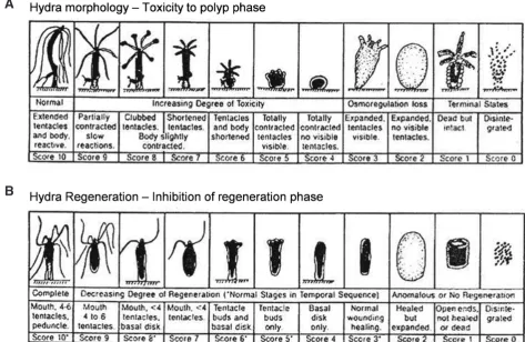
MorphologyToxicity in Hydra is typically measured by drastic changes in morphology assessed using a binocular microscope (Bossert and Galliot, 2012). As the amount of toxicant increases Hydra progres-sively exhibit morphological changes that were conventionally measured and expressed based on the observed changes in the animal with normal, bulbed or clubbed tentacles, shortened tentacles, tulip phase and disintegration as endpoints (Table 1). More recently, progressive changes in animal morphology have been measured and scored on a scale from 10 (normal, elongated tentacles and body), 8 (clubbed or bulbed tentacles), 6 (shortened tentacles), 5 (tulip phase), 2 (loss of osmoregulation) to score 0 (disintegrated) devised by Wilby (1988) (Table 1 and Fig. 1A). Scores 10–6 are reversible, sub-lethal indicators while the tulip phase (score 5 and below) is considered irreversible and used as the endpoint for lethality (Blaise and Kusui, 1997). The
progres-Fig. 1. Toxicity in Hydra based on morphological changes (A) and inhibition of regeneration (B) based on the scale from 10 to 0 devised by Wilby (1988) (reproduced with kind permission of Wilby, Tesh and Shore, 1989). Pictures of morphological changes in Hydra at various stages of the health index are shown in Bossert and Galliot (2012, this issue).
sive changes observed using the Wilby scale have the advantage over the conventional method of being more sensitive, reveal more detail on the pattern of response and provide a means of studying the ability of the animals to recover after exposure (Karntanut and Pascoe, 2000). Today this scale forms the basis of most Hydra toxicity tests and has been used extensively to assess toxicity of numerous compounds and effluents.
Regeneration
Hydra have an unsurpassed capability of regeneration and can
be considered a perpetual embryo as it permanently renews its in-ventory of differentiated cells (including nerve cells) from pluripotent stem cells (Müller, 1996). When Hydra polyps are cut into pieces they are able to regenerate the absent structures entirely (Bode, 2003; Holstein et al., 2003; Galliot et al., 2006; Hoffmeister-Ullerich, 2007). But regeneration can also occur after dissociation of the tissues, from cells that form reaggregates (Noda, 1971; Gierer et
al., 1972). In initial toxicological studies, regeneration was
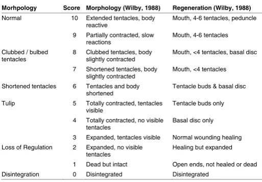
mea-sured by the use of dissociated cells to make an artificial embryo (Johnson et al., 1982) and later Wilby (1988) recommended the use of dissected gastric sections (located below the hypostome (mouth) and above the budding region) consisting of mitotically active multipotent stem cells to study regeneration. In both cases the developmental toxicity (D) was compared to adult survival toxicity (A) to produce a toxicity index (TI) based on the A/D ratio, that specifically evaluates the ability of a toxicant to alter devel-opment and unambiguously ranks substances according to their hazard potential (Johnson et al., 1982; Wilby, 1988; Quinn et al., 2008b). Morphological changes are observed using a binocular microscope and the degree of regeneration assessed. Today this is most commonly done using Wilby’s (1988) classification (Table 1 and Fig. 1B), with a score ≤5 corresponding to lethality (Pachura-Bouchet et al., 2006).
Hydra have been successfully used to examine the teratogenic
potential of several chemicals including effluents and water samples
(Fu et al., 1991, Fu et al., 1994) and various chemicals (Johnson
et al., 1986; Mayura et al., 1991; Yang et al., 1993; Bowden et al.,
1995,) including endocrine disrupting compounds (Pascoe et al., 2002; Pachura-Bouchet et al., 2006) and pharmaceuticals (Pascoe
et al., 2003; Quinn et al., 2008b; Quinn et al., 2009). Good
correla-tion was found between the in vitro Hydra regeneracorrela-tion assay and teratogenicity in vivo, as reported by Bowden et al., (1995) and Wilby and Tesh (1990), who proposed it as a screening tool for teratogenicity. Furthermore the combination of Hydra developmental hazard index (A/D ratio) and rat whole embryo culture test have been recommended for use together to facilitate the rapid detec-tion and ranking of hazardous chemicals associated with complex mixtures of chemical waste (Mayura et al., 1991; Yang et al., 1993). In many studies the regenerated animal’s ability to feed was also observed as a further sub-lethal behavioural endpoint (Pascoe et
al., 2003; Quinn et al., 2008b; Quinn et al., 2009).
Reproduction
As mentioned above Hydra have the ability to reproduce sexu-ally under stressful conditions or most commonly asexusexu-ally under favourable conditions. Hydra have the capacity to sense their envi-ronment such as food availability and temperatures, which determine the outcome of asexual reproduction and longevity (Schaible et
al., 2011). Indeed, a clear trade-off was found between asexual
reproduction and maintenance with food intake favouring budding, while starvation limits the budding process and maintains survival. This gives the opportunity to examine the outcomes of energy allocation upon various environmental stresses on the fitness of
Hydra. Under asexual reproduction Hydra that are well fed use the
excess of cells that are produced by forming buds in the middle of their body (Müller, 1996; Böttger and Hassel, 2012). The ability of
Hydra to regenerate is due to the constantly proliferating epithelial
and interstitial cells in its body column, in the absence of bisection this constant cell renewal allows the animal to bud at a rapid rate (Hoffmeister-Ullerich, 2007). This asexual mode of reproduction involves a tissue consisting of stem cells with continuous renewal potential. Hydra represent a key organism to study the effects of pollution on stem cell activity, integrity and capacity to differentiate into many other tissues. Indeed, the potential impacts of pollution on stem cell regeneration could compromise the future of offspring.
Hydra have a high asexual reproductive rate resulting in large
numbers being cultured in a short period of time (Holdway, 2005). This allows the reproductive effects of a possible toxicant to be determined (Mitchell and Holdway, 2000). The Hydra population reproduction toxicity test method determines the maximum con-centration at which a chemical or wastewater has no statistically significant effect over 7-days of exposure on the population growth as measured by changes in the number of intact hydroids (one hydroid equals one animal plus any attached buds) with the no observable effect concentration (NOEC) and lowest observable effect concentration (LOEC) determined (Stebbing and Pomroy, 1978; Holdway, 2005) . It measures the biological effects of low levels of contaminants using the rate of asexual reproduction of
Hydra. The mean relative population growth rate (K) is calculated
and is defined as:
K = ln (ny) – ln (nx)
T
Morhpology Score Morphology (Wilby, 1988) Regeneration (Wilby, 1988)
Normal 10 Extended tentacles, body
reactive Mouth, 4-6 tentacles, peduncle 9 Partially contracted, slow
reactions Mouth, 4-6 tentacles Clubbed / bulbed
tentacles 8 Clubbed tentacles, body slightly contracted Mouth, <4 tentacles, basal disc 7 Shortened tentacles, body
slightly contracted Mouth, <4 tentacles Shortened tentacles 6 Tentacles and body
shortened Tentacle buds & basal disc Tulip 5 Totally contracted, tentacles
visible Tentacle buds only
4 Totally contracted, no visible
tentacles Basal disc only 3 Expanded, tentacles visible Normal wounding healing Loss of Regulation 2 Expanded, no visible
tentacles Healing but expanded 1 Dead but intact Open ends, not healed or dead Disintegration 0 Disintegrated Disintegrated
TABLE 1
HYDRA TOXICITY CLASSIFICATION SCHEME
BASED ON MORPHOLOGY AND REGENERATION SCORES (10-1) TAKEN FROM WILBY (1988)
where nx is the number of Hydra at the beginning of the first day (tx), ny is the number of Hydra after y - x days (ty) and T is the length of the test period in days (ty – tx).
The Hydra reproduction toxicity test is a useful, cost effective and relatively easy test to assess the population reproductive tox-icity of both pure chemicals (Pollino and Holdway, 1999; Holdway
et al., 2001) and effluents and environmental samples (Mitchell
and Holdway, 2000; Rosenkrantz et al., 2008) in a relatively short time. This test method assesses population reproduction attributes which are extremely difficult and or expensive to do in many other tests or with many other types of test organisms. An alternative technique for measuring asexual reproduction is simply to count the number of hydranths in each well before and after a 96 hour exposure (Quinn et al., 2007). Although not as sensitive as the morphology or feeding tests, hydranth numbers potentially offer another end-point of interest to assess Hydra sub-lethality. Hydra can also undergo energy demanding sexual reproduction, where they will make male and/or female gonads and stimulate a sexual cycle. Hydra are generally dioecious (i.e. male and female organs are kept on separate individuals), but hermaphroditism and sex reversals can occur (Lenhoff, 1983). Moreover, the appearance of sexual dimorphism does stop asexual budding activity i.e., both processes occur at the same time. Sexual reproduction occurs more frequently in larger individuals that reached full adult size where growth is stopped. The size of individuals was related to bud numbers while no clear trend was found between either male or female gonad sizes. They tend to reproduce sexually under stressful conditions (Holdway, 2005). Temperature was found to regulate sexual reproduction in H. oligactis with sexual reproduc-tion occurring at lower temperatures (10-12 oC) (Littlefield et al.,
1991). However there is a cost for sexual reproduction. Sexually reproducing organisms undergo aging (Brien, 1953; Yoshida et al., 2006). The price of aging is the result of facilitating reproduction in the early life stage of organisms. Indeed, signs of aging were observed in sexually differentiated Hydra such as reduced food capture, contractile movements and reproduction with a higher rate of mortality in these populations. Moreover, the number of germ cells increased with a concomitant drop in the number of somatic cells. Interestingly, the Chlorella algae symbiosis with H. viridis was also shown to influence regeneration and sexual differentia-tion in polyps (Habetha et al., 2003; see in this issue Kovacevic, 2012). Under a low feeding regime, asexual growth was reduced in polyps lacking the algae, suggesting increased food assimilation. According to Habetha et al.,2002) in most cases, female gonads were produced only when symbiotic algae were present but had no effects of spermatogenesis. During oogenesis, symbionts were actively transferred from endodermal epithelial cells to the ectoder-mal oocytes indicating the involvement of green algae in the control of sexual differentiation in the green Hydra. The possible outcome for this association involves the potential effects of biotoxins and cyanobacterial blooms on the integrity of algae-Hydra symbiosis.
Feeding
Feeding behaviour is ecologically significant because of its direct effects on reproduction, population growth and abundance and composition of species it predates and is therefore a useful endpoint to study toxicity in aquatic ecosystems (Juchelka and Snell, 1994). Toxicant modification of feeding behaviour could eventually lead to reduced survival and reproduction, resulting in
adverse consequences at the population level (Halbach, 1984; Kooijman and Metz, 1984). Feeding is one of an organism’s most basic interactions with the environment and is a function of many physiological parameters (Lasker et al., 1982). Feeding rates are particularly important in regulating Hydra population densities. Laboratory studies have shown that Hydra population growth rates are directly related to frequency of feeding (Muscatine and Lenhoff, 1965; Otto and Campbell, 1977) and in the field, Hydra population densities were shown to closely follow increases in zooplankton abundance (Cuker and Mozley, 1981). The feeding process is divided into a series of discrete steps: capture of prey with nematocysts, transport of prey to mouth, mouth opening, ingestion, digestion and ejection of exoskeleton. The steps between capture and ingestion are termed feeding reaction or response (Lehhoff, 1961). Feeding behaviour in Hydra is initiated by the association of glutathione (GSH) with a putative external chemoreceptor (Bellis et al., 1992) and a wide variety of environmental parameters are know to affect this response (Lehhoff, 1961; see in this issue Pierobon, 2012). It is possible that the occurrence of oxidative stress and/or diminished amounts of GSH by its extensive conjugation to xenobiotics could potentially hamper feeding activity in Hydra (Quinn, 2004). Animal feeding behaviour has already been identified as a potential endpoint for the study of the more subtle effects of pharmaceuticals (Fent
et al., 2006). The feeding reaction has been successfully used in
several studies to examine the sub-lethal toxicity of several com-pounds including heavy metals (Beach and Pascoe, 1998; Quinn
et al., 2007) and pharmaceuticals (Pascoe et al., 2003; Quinn et al.,
2008a; Quinn et al., 2009) with the sub-lethal response investigated using feeding bioassay proved to be considerably more sensitive than that recorded in lethal studies (Beach and Pascoe, 1998).
Attachment
Attachment to a substrate is needed in order to feed, grow and reproduce and is therefore essential to Hydra. In a study by Quinn
et al., (2007) a clear relationship between increased toxicity to a
reference chemical (CdCl2) based on morphology and a decrease
in attachment was evident with Hydra attachment significantly re-duced by a concentration as low as 0.02 mg/L when compared to the control. To our knowledge this is the first time attachment has been reported as a toxicity endpoint. However an in vitro study by Lomnicki and Slobodkin, (1966) observed the secretion of a ‘bubble’ relating to food intake resulting in effectively turning the animal upside down and in Hydra detachment. Hydra attachment may be a sensitive toxicity endpoint that merits further study.
Biochemistry and biotransformation of xenobiotics
Metalsform the biochemical basis of their high sensitivity towards heavy metals (for more detail on metals see Section 4.1). Despite this, metals may be sequestered and expelled by Hydra following exposure, as has been observed with uranium accumulated in discharged nematocyst cells which are routinely discarded as new cells replace them (Hyne et al., 1992a). Hydra have some capacity to express heat shock proteins of 70 kDa family which can constitute a protection mechanism against heavy metals since there is a cross protection mechanism between thermo tolerance and metal tolerance (Brennecke et al., 1998). However, there were some reports on the inability of Hydra oligactis to acquire thermo tolerance due to the low levels of heat shock protein expression in this species (Gellner et al., 1992). The relative inability of
Hy-dra oligactis to protect against temperature increases and heavy
metal contamination makes this test species useful to study the interaction of temperature changes and metal pollution in the context of climate changes.
Organics
However both cellular stress responses and phase I and II detoxification enzymes have been identified and characterised in Hydra (Fig. 2). Hydra were found to posses glutathione S-transferase (GST) activity (Stenersen et al., 1987) an enzyme that plays an important role during phase II biotransformation in the detoxification and metabolism of many xenobiotic and endogenous compounds (Hoarau et al., 2004). Moreover, GST activity was induced in Hydra following exposure to the antiepileptic drug and common environmental pollutant carbamazapine, at environmen-tally relevant concentrations (Quinn, 2004), indicating a phase II biotransformation response. Carbamazepine exposure also led to the induction of oxidative metabolism as shown by a significant increase in both heme oxidase (HO) and lipid peroxidation, indi-cating that Hydra appear to possess both mixed function oxidase and conjugation capabilities that are inducible upon exposure to this xenobiotic (Quinn, 2004; Vernouillet et al., 2010). This was corroborated in a separate study where no significant bioaccumu-lation of carbamazepine by H. attenuata fed with carbamazepine contaminated T. platyurus was found, indicating that either uptake of the drug was weak or there is a high detoxification activity as revealed by the increased HO and cytochrome P450 3A4-like activity observed (Vernouillet et al., 2010).
Acetylcholinesterase
either the head or foot suggests that the cholinergic neural system is not involved in regeneration.
Oxidative stress
On the other hand, Hydra have the capacity to protect against oxidative stress since they contain two superoxide dismutase (SOD) genes (Dash et al., 2007). The SOD are of the type MnSOD which are principally found in the mitochondria and of the type CuZnSOD which is cytosolic in cells. Hydra subjected to thermal, starvation, metal and oxidative stresses responded by regulating both forms of SOD. The same group of investigators also identified a glutathione peroxidase family using phospholipid hydroperoxide as a co-substrate (Dash et al., 2006). As above, Hydra exposed to starvation, metal and oxidative stresses responded by regulat-ing upward their glutathione peroxidase transcripts, makregulat-ing them a useful test species to examine the impact of pollution-induced oxidative stress. For example, exposure of Hydra to increasing concentrations of carbamazepine, a persistent drug commonly found in municipal effluents, led to the induction of cytochrome P450-3A-like, heme oxidase activity and lipid peroxidation sug-gesting that biotransformation potentiates the toxicity of this drug by oxidative stress (Quinn, 2004).
DNA integrity
The genotoxicity potential of contaminants can also be in-vestigated using Hydra. In a study investigating the toxic effects of aspiron and metamizole sodium, the latter caused nucleolar structural damage in 90 % of the Hydra cells as early as 30 min of exposure (Arkhipchuk et al., 2004). Exposure of brown and green Hydra to increasing concentration of aluminium was genotoxic as determined by the Comet assay (Kovačević, 2007). The brown were more sensitive to aluminium than green Hydra, showing again a protective role of symbiosis to metals and the evolutionary advantage provided by symbiosis (see in this issue Kovacevic, 2012). Indeed, DNA tail length and intensity changes were stronger in brown than in green Hydra. However, behavioural responses to the presence of aluminium ions were observed more rapidly in green Hydra.
Toxins
MetalsHydra are generally sensitive to metal salts and other cations
(Holdway et al., 2001). For example, the reported 7-day toxicity
Toxin
e.g. Carbamazepine
Phase I
• Oxidation
• Reduction
• Hydrolysis
• Conjugation
- HO
- LPO
- MFO (CYP450)
- Glutathione peroxidase
Phase II
Phase I metabolite
Phase II metabolite
-GST
- Sulfotransferase?
Fig. 2. Evidence of biotransformation of xenobiotics in Hydra in a two-phase process. The first-phase reactions include oxidation, reduction and hydrolysis with the greatest importance ascribed to oxidation enzymes involved in the metabolism of the majority of xenobiotics, giving rise to more polar compounds. In second-phase reactions, the metabolites produced in the first phase are conjugated with products of the endogenous metabolism (e.g. GST) to give rise to polar compounds subsequently eliminated from the body.
threshold for the green Hydra was 0.56 and 250 mg/L for cadmium
and zinc respectively. This makes them excellent freshwater inver-tebrates for testing for the presence of dissolved metals based on their sensitivity and to rapidly determine population reproduction in the laboratory. Copper has been regularly found to be the most toxic heavy metal in comparative acute toxicity studies between
Hydra species (H. vulgaris, H. oligactis and H. viridissima),
fol-lowed by cadmium and zinc (Beach and Pascoe, 1998; Pollino and Holdway, 1999; Karntanut and Pascoe, 2000; Holdway et al., 2001; Karntanut and Pascoe, 2002; Karntanut and Pascoe, 2005). The green Hydra (H. viridissima) containing stable algal symbiotes has been routinely observed as the most sensitive Hydra species with a 96 hour LC50 (the lethal concentration need to kill 50% of the
exposed population) range of 8.5 - 28 mg/L for copper, 3 - 210 mg/L
for cadmium and 935 - 11,000 mg/L for zinc (Pollino and Holdway,
1999; Holdway et al., 2001; Karntanut and Pascoe, 2002; Karntanut and Pascoe, 2005). The increased acute sensitivity of this species to copper may result from copper affecting the zoochlorellae as it is a potent algaecide (Pollino and Holdway, 1999). However in an interesting study, Karntanut and Pascoe (2005) compared the toxicity of heavy metals to both symbiotic and aposymbiotic (free of their endosymbiotic algae) H. viridissima. Although the toxicity was similar for both groups, at the lower Cu concentrations the symbiotic
Hydra was better able to tolerate the toxicant. They hypothesized
that at low concentrations the copper taken up by symbiotic Hydra may be sequestered by the algae, providing a degree of protection for the polyp itself. However at the higher concentrations it is prob-able that any defence systems are overwhelmed by the toxicant effect and any slight benefit derived from the endosymbiotic algae is of little consequence. Indeed in some cases hormetic effects of metals on Hydra at lower concentrations have been observed (Pollino and Holdway, 1999; Karntanut and Pascoe, 2002).
Sensitivity to metals is thought to be partially due to the inability of Hydra to conjugate and expel metals owing to the lack of the metal binding protein metallothionein responsible for the uptake, transport and regulation of metals. This makes it a particularly sensitive species to metal contamination and a very effective bioindicator species for exposure of metals in the environment. Indeed toxicity endpoints for copper exposure were in the range of dissolved copper concentrations that one would expect to find in many contaminated environmental sites (Pollino and Holdway, 1999). Hydra have also been shown to accumulate metals, poten-tially leading to exposure to higher concentrations in the environ-ment. Deposits were observed in the discharged nematocysts of
H. viridissima after a 24 hour exposure to 200 - 3900 mg/L uranium
in a single compound mixture as well as in an effluent (Hyne et
al., 1992a) and were presumed to be responsible for the reduced
post-exposure ability of the Hydra to capture live Artemia sp. (Hyne
et al., 1992a). In this study aluminium, magnesium and zinc were
also found within the symbiotic algal cells of the Hydra (Hyne et
al., 1992a). In another study, copper, cadmium and zinc were
demonstrated to accumulate in H. vulgaris through both waterborne and food-borne exposure routes (Karntanut and Pascoe, 2007).
The acute effects of metals on Hydra are based on lethality established by the morphological effects on the animal e.g. > stage 5 on the Wilby (1988) scale (Table 1 and Fig 1A). However a reduction in population growth measured by reduced asexual budding is another sensitive and environmentally relevant end-point. Effects on population growth were observed at 8 - 16 mg/L
for copper (Stebbing and Pomroy, 1978; Pollino and Holdway, 1999; Karntanut and Pascoe, 2005), 0.8 mg/L for cadmium
(Hold-way et al., 2001), 75 mg/L for zinc (Holdway et al., 2001) and 50 mg/L for lead (Browne and Davis, 1977) on H. viridissima and 60 mg/L for nickel on H. littoralis (Santiago-Fandiño, 1983). However
as highlighted by Holdway et al., (2001) metal toxicity is greatly modified by the abiotic factors such as water hardness, pH and temperature used by the investigators rather than just species dif-ferences. High water hardness tends to increase complexation of metals as well as provide competing cations (Ca2+ and Mg2+) which
can decrease the effects of toxic divalent metals (Riethmuller et
al., 2001). The toxicity of uranium to H. viridissima, as measured
by population growth, was significantly reduced when water hard-ness was increased from 6.6 mg CaCO3/L to levels of both 165
and 330 mg CaCO3/L (Riethmuller et al., 2001). Lower pH values
may also increase the concentration of soluble and bioavailable free metal ions, which are more capable of causing internal toxic effects (Riethmuller et al., 2001, Walker et al., 2006). For example, based on population growth uranium was determined to be more toxic to H. viridissima at a pH of 6.6 than a pH of 8.6 (Hyne et
al., 1992b). The toxicity of magnesium sulfate (MgSO4), and the
influence of calcium (Ca), were assessed in very soft freshwater where Ca was shown to have an ameliorative effect on Mg toxicity (van Dam et al., 2010). It was concluded that magnesium can be toxic at concentrations approaching natural background levels, but toxicity is dependent on Ca concentrations, with exposure in very low ionic concentration, Ca-deficient waters posing the greatest risk to aquatic life (van Dam et al., 2010). Therefore, when com-paring the toxicity of metals or the potential impact of metals on the environment, exposure conditions and abiotic factors should also be taken into account.
Organic toxicants
Hydra reportedly have a lower sensitivity to organic compounds
than to metals and therefore a limited application for toxicity testing of organic toxicants. When selected organic toxicants were tested, the acute and sub-chronic toxicity endpoints for Hydra were higher than literature values for most species with toxicity endpoints be-yond the range that one would expect to find in the environment (Pollino and Holdway, 1999). Toxicity tests on H. oligactis using the polychlorinated biphenyls (PCBs) Aroclor 1016 and Aroclor 1254 resulted in a 72 hour LC50 range of 5,000 - 20,000 mg/L, although
sub-lethal inhibitory effects on reproduction and regeneration were seen at levels of 1,000 mg/L - 4,000 mg/L (Adams and Haileselassie,
1984). A low sensitivity for the reference toxicant 4-chlorophenol was reported by Mitchell and Holdway (2000) with a 96 hour LC50
of 34,000 mg/L similar to the value of 32,000 mg/L for H. vulgaris
reported by Pollino and Holdway (1999). These findings are con-sistent with those of previous studies, using toxicants such as the organochlorine pesticide lindane (Taylor et al., 1995), the chlori-nated hydrocarbon insecticide mirex (Lue and de la Cruz, 1978), ethylene dibromide (Herring et al., 1987), PCBs, atrazine, and DDT (Benson and Boush, 1983). In each case, Hydra species were less sensitive to the toxicant compared with other invertebrate species. Therefore, Hydra may have only limited application for the toxicity testing of organic toxicants and cannot be considered a sensitive model for invertebrates (Pollino and Holdway, 1999).
with a minimal affective concentration (MAC) range from 0.003 – 100,000 mg/L and toxicity correlated with increasing compound
hydrophobicity (Lum et al., 2003). When a suite of chlorophenols were tested with H. vulgaris, the 92 hour MAC range was 40 – 500,000 mg/L with the more chlorine-substituted compounds
generally being the most toxic (Mayura et al., 1991). Hydra have also been successfully used as part of a battery of test organisms to investigate the ecological risks for aquatic ecosystems posed by the toxicity of pesticides used in banana production (Castillo
et al., 2006) and to investigate the effect of insecticides (Dimiline
WP 25, Torak EC 24 and Gamacide 20) (Kalafatic et al., 1991).
Endocrine disrupting compounds (EDCs)
H. vulgaris (previously named H. attenuata) is reported to be one
of the most sensitive species to acute and chronic toxicity of the endocrine disrupting compound 4-nonylphenol compared to several freshwater invertebrates (Pachura-Bouchet et al., 2006). The toxic-ity recorded for Hydra with a 96 hour LC50 of 97.5 mg/L and a “no
observed effect concentration” (NOEC) for tentacle morphology of <25 mg/L, occurred at concentrations that are representative of
those at polluted sites (Pachura et al., 2005). The reproductive tox-icity of bisphenol A (BPA) on both asexual and sexual reproduction was investigated in H. oligactis (Fukuhori et al., 2005). Exposure to BPA at mg/L range (1-10 mg/L) had adverse effects on sexual reproduction where the asexual method was favoured. It was not clear whether the estrogenic properties of BPA accounted for the observed toxicity since the concentrations required were high com-pared to reported environmental concentrations and the low dose required for producing hormonal effects in fish. In another study, exposure of H. vulgaris to BPA and 17a-ethynylestradiol (EE2)
led to changes in the structure of polyps at concentrations above 58 and 42 mg/L BPA and EE2 respectively (Pascoe et al., 2002).
Regeneration was inhibited at 460 and 150 mg/L of BPA and EE2
respectively. The effect of exposure to EE2 was found to impair sexual reproduction in H. vulgaris, but at such high, environmentally unrealistic concentrations (500 mg/L) it was felt that the response
was probably the result of general toxicity rather than disruption of hormonal / signalling processes (Pascoe et al., 2002). This has led to the conclusion that these primitive cnidaria are not subject to disruption by estrogens or estrogen mimics (Pascoe et al., 2002). Notwithstanding this, the presence of indolamines, steroids and neuropeptides have been identified in cnidarian tissues, which indicates that endocrine disrupters might also disrupt the signal-ling pathways in Hydra (Tarrant, 2005). Evidence suggests that both classical fast (acetylcholine, glutamate, GABA, glycine) and slow (catecholamines and serotonin) transmitters as well as neu-ropeptides are involved in cnidarian neurotransmission, although cumulative data are incomplete (Kass-Simon and Pierobon, 2007). However, it remains unknown whether these compounds are involved in larger signal cascades comparable to the vertebrate hypothalamic-pituitary-gonadal axis. Regeneration of the apical, gastral and basal fragments was inhibited by dopamine synthesis inhibitors but did not produce morphological abnormalities (Os-troumova and Markova, 2002). In another study, head specific differentiation was influenced by specific protein kinase inhibitors (Cardenas et al., 2000). Head cellular proliferation was seemingly under the control of protein kinase C pathway while head cellular differentiation involved tyrosine protein-kinase Src signalling which is considered a proto-oncogene in vertebrates.
Pharmaceuticals
The addition of novel contaminants, particularly pharmaceuti-cal drugs into the environment, primarily by municipal effluents, is causing much environmental concern. Recent research has indicated that H. vulgaris are sensitive to pharmaceuticals typically found in wastewater effluents (Pascoe et al., 2003; Quinn et al., 2008a; Quinn et al., 2008b; Quinn et al., 2009) as reviewed by Blaise et al., (2006). Pharmaceuticals are continuously present at low concentrations so chronic effects are thought to be more relevant (Fent et al., 2006). Although lethality based on morpho-logical effects was observed at high environmentally unrealistic concentrations (mg/L) (Pascoe et al., 2003; Quinn et al., 2008a, Quinn et al., 2008b; Quinn et al., 2009), the toxicity threshold (TT) value (TT=(NOEC×LOEC)1/2) for ibuprofen was 320 mg/L,
(Quinn et al., 2008a) only a factor of 10 higher than the concen-tration found in Canadian effluents (22 mg/L (Brun et al., 2006).
When exposed as a mixture, pharmaceuticals were 2 to 3 orders of magnitude lower for the equivalent toxicity (EC50 and TT) for
the individual pharmaceutical, indicating they may act additively in a mixture, having sub-lethal effects at environmentally relevant (mg/L–ng/L) concentrations (Quinn et al., 2009). Similarly Pascoe
et al., (2003) found that Hydra regeneration was inhibited by 3 of
the 10 drugs examined (diazepam, digoxin and amlodipine) at 10
mg/L after an extended exposure time (17 days). When subjected
to tier two toxicity assessment under EU regulatory guidance using environmentally relevant concentrations, a MEC/PNEC value >1 was calculated for gemfibrozil, ibuprofen and naproxen indicating teratogenic potential (Quinn et al., 2008b). Biochemical biomarker responses have also been observed in Hydra following exposure to pharmaceuticals. Despite not being bioaccumulated (Vernouillet
et al., 2010) carbamazepine was found to affect global cytochrome
and cytochrome P450 3A-like activity, lipid peroxidation (LPO), heme oxidase (HO) and GST activity (Quinn, 2004; Vernouillet et
al., 2010). Oxidative metabolism with an increase in LPO was found
at a threshold concentration of 7.1 mg/L (Quinn, 2004), similar to
maximum concentration of 6.3 mg/L reported in WWTPs (Ternes,
1998). These results indicate the potential for chronic effects to occur at environmentally relevant concentrations and the useful-ness of Hydra to assess these potential effects.
Nanomaterials
We found only a handful of studies dealing with the toxicity of nanomaterials (NMs) to Hydra. The toxicity of 11 NMs were examined in a test battery of aquatic biotests (Blaise et al., 2008) where Hydra proved to be the most sensitive species with 96 hour EC50 in the 0.1-1 mg/L range for inorganic NMs: copper and zinc
iron oxides, indium tin oxides and holmium oxides nanoparticles.
Hydra were also seemingly sensitive to single-wall carbon
nano-tubes with an EC50 in the 1-10 mg/L range. Interestingly rod-shaped
nanocrystals proved excitatory to cnidarians (Malvindi et al., 2008). Exposure of Hydra to rod-shaped semiconductor quantum dots resulted in an unexpected tentacle-writhing behaviour suggesting the involvement of tentacle neurons and depends on Ca2+ ions.
(Tortiglione et al., 2009). At acidic pH, amino-pegylated coated cadmium quantum dots were actively internalized by tentacle and body ectodermal cells in Hydra vulgaris. Negatively charged quantum dots were not bioavailable to the Hydra. The uptake of positively charged nanoparticles involved annexin proteins in the cell membranes which are involved in pro-apoptotic mechanisms of cell death. In another study, the toxicity of uncapped titanium dioxide and zinc-capped titanium dioxide was also examined in the Hydra to determine the resulting effects of capping in the
Hy-dra (Yeo and Kang, 2010). Although there was no marked overall
toxicity of these nanoparticles as determined by the absence of tissue necrosis or apoptotic cells, some damage was found with zinc capped TiO2 and uncapped TiO2 under UV-A photo
condi-tions. However it was shown that it was the photo conditions that produced effects on morphology and cytotoxicity.
Industrial and municipal effluents
Hydra have also been used to test the environmental impact of
both municipal and industrial effluents, most commonly as a mem-ber of a battery of test organisms in bioassay studies. In a study of acute toxicity to ten industrial effluents on H. vulgaris, four were found to be lethal and eight sub-lethal with a 96 hour LC50 varying
from 18.8 - 100% effluent (Blaise and Kusui, 1997). From this study the simple and cost-effective Hydra microassay appears to be a valuable (sub)lethal toxicity screening tool for effluents. Another study investigating the developmental hazards of industrial waste-water samples tested on H. vulgaris resulted in a minimal effect concentration (MEC) (based on both adult morphology and artificial embryo toxicity) of 6-31% effluent (Fu et al., 1991). These authors also reported 72 and 96 hour LC50 ranging from 2.7 to >100% for
industrial wastewaters, which closely matched the LC50 values of
fathead minnow (Pimephales promelas) bioassays run in parallel (Fu et al., 1994). As part of a battery of bioassays, Hydra species have been used to assess the toxicity of vinasse (a byproduct of the sugar industry) (Ferreira et al., 2011), animal feed additives (Marroquin-Cardona et al., 2009) and pesticides (Castillo et al., 2006). When exposed to retention pond water containing gold mine effluent, population reproduction in H. viridissima was reduced by 80 to 100% by a treatment of 0.1% pond water (pH 6.5), in which copper and zinc were the most likely toxic components (Dam et al., 2008). A significant decrease in population growth, in H. viridissima, was also noted when exposed to 100% retention pond water (pH 7.5 - 8.0) from a uranium mine (Hyne et al., 1992a) although ad-ditional work showed the retention pond water to be toxic at 32% with a reduced pH of 6.6 (Hyne et al., 1992b).
Municipal effluents are an important and highly complex source of pollution into the environment. Due to this complexity biologi-cal assessment with the use of bioassays rather than chemibiologi-cal analysis is often favoured. In a toxicity test of municipal sewage on the morphology of H. vulgaris the 96 hour LC50 varied from
16.7 - >98% effluent and the 96 h EC50 for tentacle clubbing from
4.9 - >98% effluent making the Hydra assay a more sensitive indicator of toxicity than the Microtox® test (Pardos et al., 1999).
H. hexactinella has also been used to test the toxicity of urban
runoff water collecting in stormwater basins where only one out of the three basin samples tested were toxic to Hydra with a 96 hour LC50 of 61% stormwater (Rosenkrantz et al., 2008). However,
the basin water found to be toxic had the highest levels of copper, cadmium, lead, nickel and zinc (Rosenkrantz et al., 2008). This
supports the conclusion that, like the metal-laden mine effluents tested, effluents are most likely to be toxic to Hydra if they contain metals. The H. vulgaris bioassay was also included in a battery of tests used to appraise the detoxification capacity of the three strains of Pleurotus fungi on municipal effluent (Dellamatrice et
al., 2005) and the impact of sewage effluent on the deterioration
of wetlands (Oberholster et al., 2008).
Comparison of hydra sensitivity with other test
organ-isms
Hydra are often used as part of a battery of test organisms
representing different levels of biological organization used to assess the toxicity of a chemical or effluent. Bioassays with other invertebrates, such as daphnia (Daphnia magna) and Ceriodaphnia (C. affinis or C. dubia), are widely used to detect chronic toxicity (after 21-days exposure) of chemical solutions, environmental and waste waters (Viganò et al., 1996). In several studies the Hydra bioassay, particularly when chronic changes in morphology and reproduction rate were measured, was more sensitive than other bioassays undertaken with invertebrate, vertebrate and plant test organisms (Diaz-Baez et al., 2002; Ronco et al., 2002a; Arkhipchuk
et al., 2006b; Oberholster et al., 2008). In particular, sub-lethal effects
on Hydra morphology were found to be considerably more sensi-tive than lethal effects (Blaise and Kusui, 1997), closely matching the toxicity response of the fathead minnow (Fu et al., 1994) and are generally more sensitive than the Microtox test (Pardos et al., 1999). H. vulgaris is particularly sensitive to Cd and Cu, respond-ing at similar concentrations to G. pulex, an indicator species of freshwater quality and with 48 and 72 hours LC50 values showing
similar sensitivity to Daphnia magna (Beach and Pascoe, 1998).
H. vulgaris have also been shown to be more sensitive than G. pulex for variety of toxicants including benzene, trichloroethylene
and allylamine (Slooff et al., 1983).
Hydra was seen as a suitable animal model in biotesting
strategies designed for toxicity assessment of all types of water media and incorporating diverse toxicity including cytotoxicity and genotoxicity endpoints (Arkhipchuk and Malinovskaya, 2002; Arkhipchuk and Garanko, 2002). For specific objectives, the use of bioassays with Hydra and mammalian cell culture in tandem is recommended to facilitate the rapid detection and ranking of the developmental hazards of different toxins (Mayura et al., 1991, Yang et al., 1993). When used in parallel with the rat whole embryo culture (WEC) bioassay the Hydra regeneration assay was found to support the in vivo potential teratogenic hazard data (Yang et
al., 1993; Bowden et al., 1995). Hydra thus stands out as a useful
animal model for comprehensive and comparative assessment of different types of toxicity.
Use in monitoring
Hydra are a very popular model organism in aquatic toxicity and
Institute of the Supervising Scientist (eriss). In these monitoring programmes biological toxicity tests were developed using local aquatic species from different trophic levels and phyla, and with various endpoints. In assessing the toxicity of mine waste (Super-vising Scientist Report 110), the tests use two species of Hydra (H.
viridissima and H. vulgaris) using either survival or reproduction as
the endpoint, a water flea (Moinodaphnia macleayi) using either survival or reproduction as the endpoint, and a fish embryo/larva (Mogurnda mogurnda) with hatchability and survival as endpoints (Hyne et al., 1996). In the more recent ecotoxicological testing protocols for Australian tropical freshwater ecosystems (Super-vising Scientist Report, 173) the method is based on the Hydra population growth test as described by Hyne et al., (1996) using
H. viridissima (Riethmuller et al., 2003).
Hydra were also used in the WaterTox network, an
interna-tional network of laboratories from eight participating countries, that examined the applicability of a battery of simple, inexpensive bioassays in environmental management and the relevance of the test results in establishing the toxicological quality of water sources and drinking water. The core battery of tests consisted of 2 animal assays (48 hour acute toxicity to D. magna (Dutka, 1989) and 96 hour exposure lethality to H. vulgaris, (Trottier et al., 1997) and a vascular plant bioassay (L. sativa 120 hour exposure inhibition of root elongation (Dutka, 1989). This program initially established quality control mechanisms for the bioassays used (Ronco et al., 2002b) followed by examining the sensitivity, applicability and reproducibility of selected battery to screen drinking water and drinking water sources of the presence of toxicants (Diaz-Baez
et al., 2002). These bioassays have been subsequently used in
several other studies (Arkhipchuk and Garanko, 2002; Oberholster
et al., 2008) where Hydra was found to show good sensitivity.
Strengths and weaknesses of the Hydra model system
Standardised and relatively easy culture methods, consistent rapid asexual reproduction leading to a population of genetically identical clones, a diploblastic structure allowing exposure of cells directly to the environment, easily observable and quantifiable morphological changes and a widespread prevalence in freshwater ecosystems make Hydra a model test species for use in aquatic toxicology. For these reasons, Hydra have been used in numerous small scale tests and bioassays to study the toxicity of various pol-lutants. Hydra have been shown to regenerate into a healthy adult polyp from either a seriously injured intact individual (Loomis and Lenhoff, 1956), a ball of disassociated cells known as an artificial embryo (Johnson et al., 1982) or a dissected section of their gastric region (Quinn et al., 2008a). Due to this tremendous regenerative capacity, Hydra have been widely used in developmental biology and to assess the teratogenic effects of many environmental pol-lutants. Their rapid rate of asexual reproduction by budding allows the population reproduction effects of a potential toxicant to be determined in the laboratory, which is another enormous benefit of the Hydra model system.
However the main weakness of using Hydra as a toxicity test organism lies in its reported lack of sensitivity to organic toxicants. It has been reported that for organic toxicants acute and sub-chronic toxicity endpoints for Hydra were higher than literature values for most species and beyond the range that could be found in environ-ment (Pollino and Holdway, 1999). Despite Hydra having a large
potential for use in bioassays to test inorganic toxicants (particularly metals), it is felt that Hydra have a limited application for toxicity testing of organic toxicants. However as we have highlighted in section 4 there are several cases where Hydra were reported to have good sensitivity, particularly to the more novel contaminants nonylphenol and pharmaceuticals. Another apparent drawback for the use of Hydra in toxicity testing lies in its reported inability to metabolize xenobiotics (Fu et al., 1994) particularly the lack of metallothionein. However it appears that the more we investigate the biochemistry of these animals the more biochemical / biomarker stress responses are being expressed (Section 3 and 4). One important drawback to the use of Hydra is the ability of the expo-sure parameters (water hardness, pH, conductivity, temperature and feeding) to affect the toxicity endpoints potentially leading to both false positive and negative results. Nevertheless, this can be overcome by careful experimental planning and the measurement of these parameters during exposures involving Hydra.
Conclusion
Part of the success of the use of Hydra in aquatic toxicology and in bioassays for toxicity testing is due to the wide array of lethal and non-lethal endpoints that can be used in both acute and chronic studies. For lethality, morphology and regeneration are appropriate endpoints. However the non-lethal endpoints which also include morphology and regeneration along with reproduc-tion, feeding, various biochemical endpoints and attachment are particularly sensitive and have been found by several authors to be more sensitive than other bioassays undertaken with invertebrate, vertebrate and plant test organisms. Hydra are commonly used in acute toxicity tests with a 96 hour exposure time. However several studies have found increased sensitivity of the hydra bioassay us-ing the standard toxicity endpoints (morphology and reproductive capacity) following extended chronic exposure periods of up to 21 days. Arkhipchuk et al., (2006b) reported 3-5 times increased sensitivity for chronic lethal effects, and up to 10-16 times for chronic sub-lethal effects over acute exposure effects, indicating exposure time should be carefully chosen. Clearly, the inclusion of numerous effects-based endpoints (morphology, regeneration, reproduction [hydranth number], biomarker expression, prey ingestion and attachment) can contribute to a better overall un-derstanding of the chronic effects of contaminants, and Hydra, as a recognized animal model for aquatic studies, has a role to play in this respect. Appropriately designed, relatively simple and inexpensive laboratory toxicity tests using Hydra with a selection of acute and sub-lethal endpoints are generally adequate, with small application factors, for predicting the environmental risk of polluting chemicals to freshwater ecosystems.
Acknowledgements
This work has been partially funded by Environment Canada and the Environmental Protection Agency (Ireland) under the Science, Technol-ogy, Research and Innovation for the Environment (STRIVE) Programme 2007–2013.
References
ANDERSEN, R. A., WIGER, R., DAAE, H. L. and ERIKSEN, K. D. H. (1988). Is the metal binding protein metallothionein present in the coelenterate Hydra attenuata?
Comp Biochem Physiol C 91: 553-557.
ARKHIPCHUK, V. V., BLAISE, C. and MALINOVSKAYA, M. V. (2006a). Use of
Hy-dra for chronic toxicity assessment of waters intended for human consumption. Environ Pollut 142: 200-11.
ARKHIPCHUK, V. V., BLAISE, C. and MALINOVSKAYA, M. V. (2006b). Use of
Hy-dra for chronic toxicity assessment of waters intended for human consumption. Environ Pollut 142: 200-211.
ARKHIPCHUK, V. V. and GARANKO, N. N. (2002). A novel nucleolar biomarker in plant and animal cells for assessment of substance cytotoxicity. Environ Toxicol 17: 187-94.
ARKHIPCHUK, V. V., GONCHARUK, V. V., CHERNYKH, V. P., MALOSHTAN, L. N. and GRITSENKO, I. S. (2004). Use of a complex approach for assessment of metamizole sodium and acetylsalicylic acid toxicity, genotoxicity and cytotoxicity.
J Appl Toxicol 24: 401-7.
ARKHIPCHUK, V. V. and MALINOVSKAYA, M. V. (2002). Quality of water types in Ukraine evaluated by WaterTox bioassays. Environ Toxicol 17: 250-257. BEACH, M. J. and PASCOE, D. (1998). The role of Hydra vulgaris (Pallas) in
assess-ing the toxicity of freshwater pollutants. Water Res 32: 101-106.
BELLIS, S. L., KASS-SIMON, G. and RHOADS, D. E. (1992). Partial characteriza-tion and detergent solubilizacharacteriza-tion of the putative glutathione chemoreceptor from
Hydra. Biochemistry 31: 9838-9843.
BENSON, B. and BOUSH, G. M. (1983). Effect of pesticides and PCBs on budding rates of green Hydra. Bull. Environ Contam Toxicol 30: 344-350.
BERGMANN, A.S.H. (2010). Apoptosis, stem cells, and tissue regeneration.
Sci-ence Signal 3: 8.
BLAISE, C., GAGNE, F., EULLAFFROY, P. and FERARD, J. F. (2006). Ecotoxicity of selected phamraceuticals of urban origin discharged tothe Saint-Lawrance river (Quebec, Canada): A review. Brazil J Aquat Sci Technol 10: 29-51.
BLAISE, C., GAGNE, F., FERARD, J. F. and EULLAFFROY, P. (2008). Ecotoxicity of selected nano-materials to aquatic organisms. Environ Toxicol 23: 591-8. BLAISE, C. and KUSUI, T. (1997). Acute toxicity assessment of industrial effluents with
a microplate-based Hydra attenuata assay. Environ Toxicol Water Qual 12: 53-60. BRIEN, P. (1953). La pérennité somatique. Biol Rev 28: 308-349.
BODE, H. R. (2003). Head regeneration in Hydra. Dev Dynam 226: 225-236. BOSCH, T. C. G. (2009). Hydra and the evolution of stem cells. BioEssays 31: 478-486. BOSSERT, P., and GALLIOT, B. (2012). How to use Hydra as a model system to
teach biology in a classroom? Int J Dev Biol 56: 637-652.
BÖTTGER, A. and ALEXANDROVA, O. (2007). Programmed cell death in Hydra.
Semin Cancer Biol 17: 134-146.
BÖTTGER, A. and HASSEL, M. (2012). Hydra, a model system to trace the emergence of boundaries in eumetazoans. Int J Dev Biol 56: 583-591.
BOWDEN, H. C., WILBY, O. K., BOTHAM, C. A., ADAM, P. J. and ROSS, F. W. (1995). Assessment of the toxic and potential teratogenic effects of four glycol ethers and two derivatives using the Hydra regeneration assay and rat whole embryo culture. Toxicol In vitro 9: 773-781.
BRENNECKE, T., GELLNER, K. and BOSCH, T. C. G. (1998). The lack of a stress response in Hydra oligactis is due to reduced hsp70 mRNA stability. Eur J
Bio-chem 255: 703-709.
BROWNE, C. L. and DAVIS, L. E. (1977). Cellular mechanisms of stimulation of bud production in Hydra by low levels of inorganic lead compounds. Cell Tissue
Res 177: 555-570.
BRUN, G. L., BERNIER, M., LOSIER, R., DOE, K., JACKMAN, P. and LEE, H. B. (2006). Pharmaceutically active compounds in Atlantic Canadian sewage treat-ment plant effluents and receiving waters, and potential for environtreat-mental effects as measured by acute and chronic aquatic toxicity. Environ Toxicol Chem, 25: 2163-2176.
CAMPBELL, R. D. (1989). Taxonomy of the European Hydra (Gnidaria: Hydrozoa): a re-examination of its history with emphasis on the species H. vulgaris Pallas, H.
attenuata Pallas and H. circumcincta Schulze. Zool J Linnean Soc 95: 219-244.
CARDENAS, M., FABILA, Y. V., YUM, S., CERBON, J., BÖHMER, F. D., WETZKER, R., FUJISAWA, T., BOSCH, T. C. G. and SALGADO, L. M. (2000). Selective protein kinase inhibitors block head-specific differentiation in Hydra. Cell Signal 12: 649-658.
CASTILLO, L. E., MARTINEZ, E., RUEPERT, C., SAVAGE, C., GILEK, M., PIN-NOCK, M. and SOLIS, E. (2006). Water quality and macroinvertebrate community response following pesticide applications in a banana plantation, Limon, Costa Rica. Sci Total Environ 367: 418-32.
CLARKE, S. M. and BARRICK, C. W. (1990). A bioassessment battery for use in an industrial setting: a new management approach. Environ Toxicol water 10: 81-90. CUKER, B. E. and MOZLEY, S. C. (1981). Summer population fluctuations, feeding
and growth of Hydra in an arctic lake. Limnol Oceanogr 26: 697-708.
DAM, R. V., HOGAN, A., HARFORD, A. and MARKICH, S. (2008). Toxicity and metal speciation characterisation of waste water from an abandoned gold mine in tropical northern Australia. Chemosphere 73: 305-313.
DASH, B., METZ, R., HUEBNER, H. J., PORTER, W. and PHILLIPS, T. D. (2006). Molecular characterization of phospholipid hydroperoxide glutathione peroxidases from Hydra vulgaris. Gene 381: 1-12.
DASH, B., METZ, R., HUEBNER, H. J., PORTER, W. and PHILLIPS, T. D. (2007). Molecular characterization of two superoxide dismutases from Hydra vulgaris.
Gene 387: 93-108.
DELLAMATRICE, P., MONTEIRO, R., KAMIDA, H., NOGUEIRA, N., ROSSI, M. and BLAISE, C. (2005). Decolourization of Municipal Effluent and Sludge by Pleurotus
sajor-caju and Pleurotus ostreatus. World J Microbiol Biotechnol 21: 1363-1369.
DIAZ-BAEZ, M. C., SÁNCHEZ, W. A., DUTKA, B. J., RONCO, A., CASTILLO, G., PICA-GRANADOS, Y., CASTILLO, L. E., RIDAL, J., ARKHIPCHUK, V. and SRIVASTAVA, R. C. (2002). Overview of results from the WaterTox intercalibration and environmental testing phase II program: Part 2, ecotoxicological evaluation of drinking water supplies. Environ Toxicol 17: 241-249.
DUTKA, B. J. (1989). Short term root elongation toxicity assay. In: DUTKA, B. J. (ed.)
Methods for toxicological analysis of waters, wastewaters and sediments.
Burling-ton, Ontario, Canada: Environment Canada, National Water Research Institute. ELLIOTT, J. K., ELLIOTT, J. M. and LEGGETT, W. C. (1997). Predation by Hydra on
larval fish: field and laboratory experiments with Bluegill (Lepomis macrochirus).
Limnol Oceanog 42: 1416-1423.
FENT, K., WESTON, A. A. and CAMINADA, D. (2006). Ecotoxicology of human pharmaceuticals. Aquat Toxicol 76: 122-159.
FERREIRA, L. F., AGUIAR, M. M., MESSIAS, T. G., POMPEU, G. B., LOPEZ, A. M., SILVA, D. P. and MONTEIRO, R. T. (2011). Evaluation of sugar-cane vinasse treated with Pleurotus sajor-caju utilizing aquatic organisms as toxicological indicators. Ecotoxicol Environ Saf 74: 132-7.
FU, L.-J., STAPLES, R. E. and STAHL, R. G. (1994). Assessing acute toxicities of pre- and post-treatment industrial wastewaters with Hydra attenuata: A comparative study of acute toxicity with the fathead minnow, Pimephales promelas. Environ
Toxicol Chem 13: 563-569.
FU, L. J., STAPLES, R. E. and STAHL, R. G., JR. (1991). Application of the Hydra
attenuata assay for identifying developmental hazards among natural waters and
wastewaters. Ecotoxicol Environ Saf 22: 309-19.
FUKUHORI, N., KITANO, M. and KIMURA, H. (2005). Toxic effects of bisphenol A on sexual and asexual reproduction in Hydra oligactis. Arch Environ Contam
Toxicol 48: 495-500.
GALLIOT, B., MILJKOVIC-LICINA, M., DE ROSA, R. and CHERA, S. (2006). Hydra, a niche for cell and developmental plasticity. Semin Cell Dev Biol 17: 492-502. GELLNER, K., PRAETZEL, G. and BOSCH, T. C. G. (1992). Cloning and expression
of a heat-inducible hsp70 gene in two species of Hydra which differ in their stress response. Eur J Biochem 210: 683-691.
GIERER, A., BERKING, S., BODE, H., DAVID, C.N., FLICK, K., HANSMANN, G., SCHALLER, H. and TRENKNER, E. (1972). Regeneration of hydra from reag-gregated cells. Nat New Biol 239: 98-101.
HABETHA, M., ANTON-ERXLEBEN, F., NEUMANN, K. and BOSCH, T. C. G. (2003). The Hydra viridis / Chlorella symbiosis. Growth and sexual differentiation in polyps without symbionts. Zoology 106: 101-108.
HALBACH, U. (1984). Population dynamics of rotifers and its consequences for ecotoxicology. Hydrobiologia 109: 79-96.
HEMMRICH, G., ANOKHIN, B., ZACHARIAS, H. and BOSCH, T. C. G. (2007). Mo-lecular phylogenetics in Hydra, a classical model in evolutionary developmental biology. Mol Phylogenet Evol 44: 281-290.