_____________________________________________________________________________________________________
*Corresponding author: E-mail: [email protected];
(Past name: British Journal of Medicine and Medical Research, Past ISSN: 2231-0614, NLM ID: 101570965)
A Unique Presentation of Intestinal Obstruction:
A Case Report of Multiple Jejunal Diverticulae
Mahmoud A. Abd El Mohsen
1and Doaa M. Hasan
2*1
Department of Surgery, Kasr Al Ainy Faculty of Medicine, Cairo University, Egypt. 2
Kasr Al Ainy Faculty of Medicine, Cairo University, Egypt.
Authors’ contributions
This work was carried out in collaboration between both authors. Author MAAEM wrote the case presentation which he performed. Author DMH managed the literature searches and wrote the first draft of the manuscript. Both authors read and approved the final manuscript.
Article Information
DOI: 10.9734/JAMMR/2018/44119
Editor(s):
(1) Dr. Masahiro Hasegawa, Department of Orthopaedic Surgery, Mie University Graduate School of Medicine, Japan.
Reviewers:
(1) Ashrarur Rahman Mitul, Dhaka Shishu Hospital & Bangladesh Institute of Child Health, Bangladesh. (2)Einar Arnbjörnsson, Lund University, Sweden. Complete Peer review History:http://www.sciencedomain.org/review-history/26240
Received 15 June 2018 Accepted 06 September 2018 Published 14 September 2018
ABSTRACT
Small intestinal diverticulosis refers to the presence of multiple saclike mucosal herniations through weak points in the intestinal wall this is a rare presentation. Small intestinal diverticulae are far less common than colonic diverticulae. The prevalence of small intestinal diverticulae on autopsy ranges from 0.06% to 1.3%. Although there is no consensus on the management of asymptomatic jejunal diverticular disease, some complications are potentially life threatening and require early surgical treatment.
We report a case of multiple jejunal diverticulae presented as intestinal obstruction in Kasr Alainy hospital. This is the first case to be documented in Kasr Alainy hospital.
1. INTRODUCTION
Small intestinal diverticulosis refers to the presence of multiple saclike mucosal herniations through weak points in the intestinal wall this is rare presentation. Small intestinal diverticulae are far less common than colonic diverticulae. The prevalence of small intestinal diverticulae on autopsy vary from 0.06% to 1.3% [1-3] and increases with age, as a result of which most of the cases present during the 6th or 7th decades. The etiology of this condition is unknown. It is assumed to develop as the result of abnormalities in peristalsis, intestinal dyskinesis, and high segmental intra-luminal pressures. Pathologically, they are pseudodiverticula of the pulsion type, resulting from increased intra-luminal pressure and weakening of the bowel wall [4].
The majority of jejunal diverticulosis cases are discovered accidentally during radiological investigations [5]. Symptomatic presentations are rare and generally problematica. Jejunal and jejuno-ileal localization is approximately three times less frequent than duodenal, but roughly four times more likely to develop complications [3]. Surgical exploration is practically the treatment of choice in case of acute complicated presentation.
Although there is no agreement on the management of asymptomatic jejunal diverticular
disease, some complications are possibly life threatening and require urgent surgical intervention.
We report a case of multiple jejunal diverticulae presented as intestinal obstruction in Kasr Alainy hospital.
2. CASE PRESENTATION
A 62- year old male patient not known to be diabetic or hypertensive reported to the emergency room with generalized abdominal pain and gave a history of absolute constipation for 4 days with episodes of vomiting.
The patient underwent Open cholecystectomy and Nephrolithotomy 22 years ago and repair of recurrent paraumblical hernia with mesh 18 years ago.
On physical examination Heart rate was 116/min, Blood pressure was 118/76 mmHg and the respiratory rate was 14 breaths/min. On abdominal examination distention was present, Bowel sounds were absent. Routine blood tests showed normal white blood cells count and elevated serum creatinine (2.96 mg/dl).
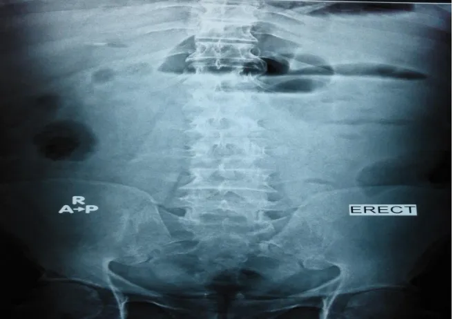
Abdominal x-ray revealed air fluid levels (Fig. 1).
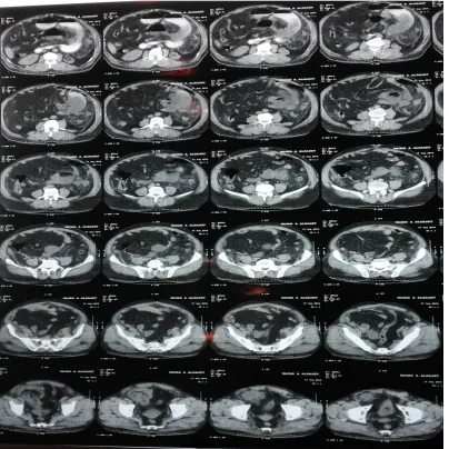
Computed tomography of the abdomen (Fig. 2) showed intestinal obstruction with filled defects with contrast as diverticulae of small bowel.
Fig. 2. CT abdomen
A decision was made that urgent Laparotomy was required on the basis of the general condition of the patient and the investigations showing signs of bowel obstruction. Upon exploration we found markedly dilated jejunal loop with fibrinous adhesions around, the rest of the small bowel was collapsed. The large intestine was collapsed too.
An adhesive band was found 1 m from the duodenojejunal junction occluding the mentioned jejunal loop and other adhesions were found.
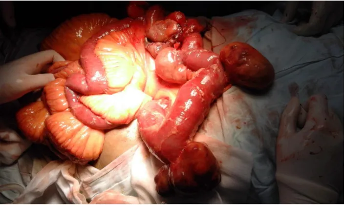
Multiple large jejunal diverticulae was found. The 1st was 25 cm from duodenojejunal. The last was 1 m from duodenojejunal (7 diverticulae). One of them was perforated (Figs. 3, 4).
Resection of the affected loop (20 cm from duodenojejunal junction –1m length) followed by 1ry jeujuno-jejunal End-to-end anastomosis in 2 layers (1st interrupted extra mucosal and the 2nd interrupted seromuscular).
Histopathology report confirmed an ulcerative jejunal diverticulitis with some areas of perforation.
Fig. 3. Intraoperative findings. Multiple giant diverticula
3. DISCUSSION
Diverticulosis of the colon is a very common condition. Described as early as the 17th century, most of the knowledge we now have is based on work during the 20th century. Age, sex, race, and geography all play a specific role in the development of diverticula [6]. Contrariwise Jejuno-ileal diverticulosis is a rare condition first described in 1794 by Sommering and later in 1807 by Sir Astley Cooper and is characterized by herniation of mucosa and submucosa through the muscular layer of the bowel wall (false diverticula) on the mesenteric border of the
bowel. Eighty percent occur in the jejunum, 15% in the ileum and 5% in both of them [7-9].
To our knowledge, this is the first case we have encountered and documented in Kasr Alainy. Usually jejunal diverticulae are asymptomatic and commonly discovered accidentally on radiological study or during autopsy [10]. Nonetheless, it could be presented with complication as obstruction, hemorrhage and diverticulitis perforation in 10-30% of the cases as in our case where one of the diverticulae was found perforated.
Once the jejunal diverticulosis is diagnosed, conservative management should be instituted to relieve symptoms and reduce the risk of complications associated with diverticular disease.
Diagnosis is often challenging and is performed mostly by imaging studies. A late diagnosis can be lethal, because perforation is associated with a high mortality in up to 40% of patients[11]. The differential diagnosis includes neoplasms (with or without perforation), foreign body perforation, traumatic haematoma, medication-induced ulceration (non-steroidal anti-inflammatory drug), and Crohn’s disease[12]. There is no consensus on therapeutic strategy and conservative management of symptomatic jejunal diverticular disease. If the inflammation is mild, medical management may be attempted (bowel rest and antibiotics). Surgery is the preferred treatment option. The resection must be limited because: 1/even when the entire segment of small bowel containing diverticula is resected, the diverticula can recur. 2/ in order to reduce the risk of short bowel syndrome. The surgical techniques used: 1/suturing the perforation (with omental patch closure) 2/invaginating the diverticulum (with a suture) 3/segmental small bowel resection and primary anastomosis. The first two techniques should be avoided due to a high mortality rate. In the presence of complications, surgical resection with reestablishment of the bowel continuity is the preferred treatment option [7].
4. CONCLUSION
In summary, Jejunal diverticulosis is a rare cause of intestinal obstruction and poses a dilemma even for an experienced surgeon. We should keep in mind that it may cause intestinal obstruction as it is usually overlooked in our differential diagnosis. Surgical intervention is indicated for acute abdomen intestinal obstruction.
CONSENT
Written informed consent was obtained from the patient for publication of this case report and accompanying images.
ETHICAL APPROVAL
As per international standards or university standards written ethical approval has been collected and preserved by the authors.
COMPETING INTERESTS
Authors have declared that no competing interests exist.
REFERENCES
1. Jeong J, Hong SS, Hwang J, Kim HJ, Chang YW. Acute diverticulitis of the terminal ileum: Ultrasonography and CT findings. Ultrasonography. 2014;34(1):74– 77.
DOI: 10.14366/usg.14041
2. Kassahun WT, Fangmann J, Harms J, Bartels M, Hauss J. Complicated small-bowel diverticulosis: A case report and review of the literature. World Journal of Gastroenterology. 2007;13(15):2240–2. Available:http://www.ncbi.nlm.nih.gov/pub med/17465510
3. Kassir R, Boueil-Bourlier A, Baccot S,
Abboud K, Dubois J, Petcu CA, Tiffet O. Jejuno–ileal diverticulitis:
Etiopathogenicity, diagnosis and management. International Journal of Surgery Case Reports. 2015;10:151–153. DOI: 10.1016/j.ijscr.2015.03.044
4. Lempinen M, Salmela K, Kemppainen E. Jejunal diverticulosis: A potentially dangerous entity. Scandinavian Journal of Gastroenterology. 2004;39(9):905–909. DOI: 10.1080/00365520410006288 5. Lin CH. Diverticulosis of the jejunum with
intestinal obstruction: A case report. World Journal of Gastroenterology. 2005;11(34): 5416.
DOI: 10.3748/wjg.v11.i34.5416
6. Makris K, Tsiotos GG, Stafyla V, Sakorafas GH. Small intestinal nonmeckelian diverticulosis. Journal of Clinical Gastroenterology. 2009;43(3):201–207. DOI: 10.1097/MCG.0b013e3181919261 7. Martel J, Raskin JB. History, incidence,
and epidemiology of diverticulosis. Journal of Clinical Gastroenterology. 2008;42(10): 1125–1127.
DOI: 10.1097/MCG.0b013e3181865f18 8. Patel VA, Jefferis H, Spiegelberg B, Iqbal
Q, Prabhudesai A, Harris S. Jejunal diverticulosis is not always a silent spectator: A report of 4 cases and review of the literature. World Journal of Gastroenterology. 2008;14(38):5916. DOI: 10.3748/wjg.14.5916
disorder. Journal of Medicine and Life. 2012;5(3):308–10.
Available:http://www.ncbi.nlm.nih.gov/pub med/23049633
10. Singh S, Aggarwal V, Sandhu HS. Perforated jejunal diverticulum: A rare complication. Saudi Journal of Gastroenterology. 2011;17(5):367. DOI: 10.4103/1319-3767.84502
11. Staszewicz W, Christodoulou M, Proietti S, Demartines N. Acute ulcerative jejunal
diverticulitis: Case report of an uncommon entity. World Journal of Gastroenterology. 2008;14(40):6265.
DOI: 10.3748/wjg.14.6265
12. Tayeb M, Khan FM, Rauf F, Khan MM. Phytobezoar in a jejunal diverticulum as a cause of small bowel obstruction: A case report. Journal of Medical Case Reports. 2011;5(1):482.
DOI: 10.1186/1752-1947-5-482
_________________________________________________________________________________
© 2018 El Mohsen and Hasan; This is an Open Access article distributed under the terms of the Creative Commons Attribution License (http://creativecommons.org/licenses/by/4.0), which permits unrestricted use, distribution, and reproduction in any medium, provided the original work is properly cited.
Peer-review history: