Ashalatha*, Karthik G. Vaidya and Pooja B.
Department of Biotechnology, Acharya Institute of Technology, Affiliated to Visvesvaraya Technological University (VTU), Dr. Sarvepalli Radhakrishnan Road, Soladevanahalli, Bengaluru, Karnataka-560107, India.
*Corresponding author’s E-mail:ashalatha@acharya.ac.in
Received: 03-02-2020; Revised: 23-04-2020; Accepted: 30-04-2020. ABSTRACT
Wound healing is a progressive and sequential series of events for the ultimate restoration of distressed tissue. Inflammation, tissue proliferation and remodulation are the three main phases involved in wound healing which is however preceded by a hemostasis phase which involves vasoconstriction, platelet aggregation and thrombus formation. The proliferation phase marks the formation of new cells and blood vessels and the overall process of wound healing is terminated by the remodulation phase, which involves the formation of new tissue made up of collagen fibers. Wounds are further classified as acute and chronic based on the duration taken for the healing process and the technologies involved in wound treatment intend to reduce the time taken to heal. To treat wounds, various wound care products have been developed such as negative pressure wound devices (NPWD), hyperbaric oxygen therapy, bioengineered skin substitutes, silk-based biomaterials and mesenchymal stem cells (MSCs)-based therapy. This review highlights the use of bioengineered skin substitutes, silk-based biomaterials and mesenchymal stem cells (MSCs) for treating wounds.
Keywords: Wound Healing, Wound Care, Skin Substitutes, Silk-based Biomaterials, Mesenchymal Stem Cells (MSCs)-based Therapy.
INTRODUCTION
he process of wound healing has been extensively
studied for many years now. There have been
advances in understanding the exact mechanisms
involved in this process and the field of wound care is
dependent on the understanding of the process of wound
repair. Wounds are classified, based on how long they take
to heal, as acute, wounds that heal within 10-12 weeks,
and chronic wounds, which take more than 12 weeks to
heal and it is based on this classification that the wound
care technologies are developed. Wound care has
improved greatly over the years and now, the field of
wound care has a tremendous number of treatment
options. New wound care products stimulate the synthesis
of collagen, promote angiogenesis and increase the rate of
re-epithelialization. Latest technologies are based on
manipulating the wound environment. While the
well-established techniques are still followed, new technologies
have developed and many new products have been
introduced into the market. There is a continuous
advancement in the production of better and more
efficient technologies to treat wounds.
This review will discuss the overall mechanism involved in
the process of wound healing and will highlight three new
prominent technologies involved in wound care.
Bioengineered skin substitutes, silk-based biomaterials
and mesenchymal stem cells have gained attention over
the years due to their great potential to treat wounds and
have henceforth been elaborated in this article.
WOUND HEALING MECHANISM
Wound healing is a forced response including a cascade of
events to orchestrate smooth progression and restoration
of tissue. Three significant phases of wound healing are
Inflammation, tissue proliferation and remodulation.
These events are not time constricted; as they overlap and
include systematic series of processes such as clotting,
inflammation,
granulation,
tissue
formation,
epithelialization, neovascularization, collagen formation
and wound contraction.
1,2When cells experience damage
associated or pathogen-specific molecular patterns, the
primary sensory neurons facilitate immediate response by
transmigrating mononuclear cells to the site to secrete
inflammatory cytokines.
3,4Hemostasis is a partial
modulatory phase before inflammation; which facilitate
vasoconstriction, platelet aggregation and thrombus
formation for the infiltration of cells.
5,6Neutrophils
eliminate the pathogens in the site while chemoattractants
like Transforming growth factor-
β
(
TGF-
β) and
monocyte
chemoattractant protein-1
(
MCP-1) attract monocytes to
the wound site, and cumulating its conversion to
macrophages for the further proliferation phase.
Proliferative phase involves the formation of new cells and
angiogenesis by endothelial cells, fibroblasts and
keratinocytes. Reactive oxygen species (ROS) sequester
the supply of blood to healing areas and supply phagocytic
and
bacteriostatic
effects
to
the
surrounding
environment.
7,8Macrophages and platelets deliver
essential growth factors and pro-inflammatory cytokines
and all these, lead to the production of extracellular matrix
and collagen fibers by fibroblasts and myofibroblast.
Angiogenesis enhances tissue granulation and
re-Advances in Wound Healing and Wound Care Technologies
–
A Review
T
epithelialization, which contributes to 80% wound closure
in humans.
9,10The remodulation is the longest and demarcation phase
which take even years to complete. This phase starts after
2-3 weeks, resulting in the restoration of wounded original
tissue with a collagenous scar without any epidermal
appendages. The ultimate aim of remodulation is wound
closure by the formation of a densely packed, less vascular
neotissue made of parallel collagen fibers.
11-13This is
characterized by an increased amount of type I collagen,
produced
by
myofibroblasts
and
formation
of
keratinocytes as layers. The active cells present during
proliferative phase undergo either withdrawal, or
apoptosis,
or
degraded
by
the
plasmatic
metalloproteinases.
14-17This accounts for the initial
redness of scars and the hypopigmentation upon
maturation. The various stages of the wound healing
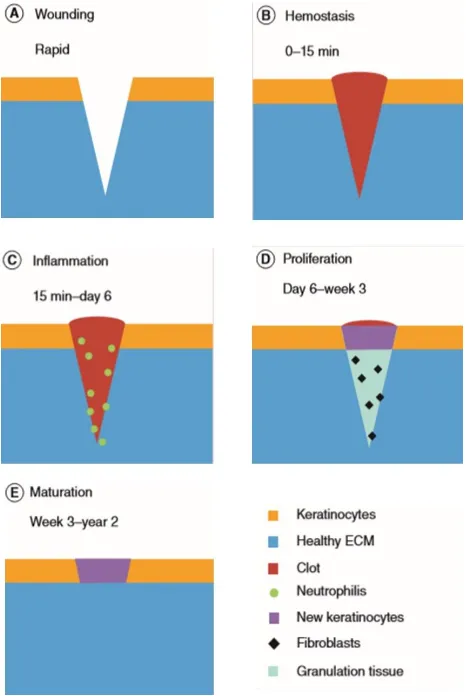
processcan be visualized in figure 1.
Figure 1: Successive stages of Wound healing. a)
Wounding: occurrence of a wound in healthy tissue. b)
Hemostasis: clot formation. c) Inflammation: Elimination
of pathogens in the injured site by neutrophils. d)
Proliferation: Occurrence of granulation tissue and
keratinocytes, infiltration of fibroblasts. e) Maturation:
The final phase, fibroblasts disappear and formation of
healthy ECM.
18ADVANCES IN WOUND CARE TECHNOLOGIES
Wound care is one of the emerging and affluent sectors in
the medical field. The wound care
armamentarium started
back in 69 BC when people used silver to treat infections.
19This domain has changed immensely over past decades
due to the furtherance in technology and evolved in the
usage of personalized medicine according to its
idiosyncratic phenotype.
Negative Pressure Wound Devices (NPWD) have been used
from the onset of the 21
stcentury and is still prevalent in
populations.
19This can be used for soft tissue injuries,
pathogen-infected wounds, debridement wounds as well
as enterocutaneous fistulas.
Hyperbaric oxygen therapy has been widely used in
recalcitrant wounds, mainly DFU. This can assist
angiogenesis, growth factor signaling, cellular mobility,
fibroblast proliferation and enhance leukocyte function.
20-22This therapy also inhibits the spreading of infectious
necrosis due to high level oxygen.
22Manipulation of Reactive Oxygen Species (ROS) in wound
therapy is another approach where they are dispensed to
stimulate angiogenesis, cellular migration as well as to
provide bacteriostatic effect to wound microenvironment.
But this technique is far more useful in acute wounds and
its critical concentration in treatment hinders its wide
usage.
7,8An establishing method in chronic wound treatment,
mainly DFU, is Cell-based therapy; application of healthy,
donor-derived mesenchymal stem cells (MSC) into the
circulation. The stem cells of diabetics or chronic wound
patients are defective so that the administration of these
MSC could help in normal restorative healing of
wounds.
23,24The use of bioengineered skin substitutes, silk-based
biomaterials and mesenchymal stem cells in wound
treatment are described below.
Bioengineered Skin Substitutes
Bioengineered skin substitutes may be produced either as
cellularized engineered skin grafts or as acellular dermal
regeneration templates (DRTs). These have been
developed to take care of two main problems affecting the
wound repair mechanism in cases of deeper wounds,
which are, reducing the quantity of healthy skin removed
from the patient and restoring the skin’s physiological
conditions avoiding scar formation.
25-34DRTs are made up
of porous and fibrous materials and are nothing but a 3D
scaffold which mimic the 3D architecture of human tissue
to support cell growth. The DRTs must be
non-immunogenic, biocompatible, stiff and flexible and should
support epidermal growth and promote the influx of blood
vessels once implanted.
35-37form a vascular network.
25Some of the commercially
available acellular dermal substitutes (Table 1) are
described below.
Alloderm, Dermacell and Dermamatrix are
non-crosslinked, decellularized dermis, obtained from
cadavers, which can be inserted in the wound bed.
28,30-33,37-40Alloderm is being used for burn victims since 1992 and it
has also been used for the treatment of severe soft tissue
defects.
41The structure of Alloderm can be seen in figure
2. Dermacell and Dermamatrix are intended for soft tissue
reconstruction (abdomen, nasal reconstruction, facial
defects, etc.) and for reconstructing breast.
25Integra is
made up of bi-layered extracellular matrix fibers which are
of cross-linked bovine collagen and chondroitin-6-sulphate
with a transient epithelium made using a silicone
membrane. The structure of Integra can be seen in figure
2. The silicone sheet is taken off only after the formation
of neodermis.
33,41,42The vascularization is faster in
Matriderm than in Integra. This is because of the presence
of elastin in Matriderm, which attracts more vascular cells
than Integra.
25Some of the commercially available
products are listed along with their composition and
applications in table 1.
Cellularized Dermal Substitutes:
Cellularized skin
substitutes can be categorized into three: (1) Epithelial
sheets: formed by seeding epithelial cells on polymeric
membranes. (2) Dermis equivalents: composed of
fibroblasts contained generally in either 3D porous
matrices or hydrogels. (3) Full-thickness (or composite)
equivalents: composed of an epidermis and dermis
equivalent. A few commercially available cellularized
dermal substitutes (Table 2) are described below.
Dermagraft is made up of a bio-degradable PLGA mesh
which is seeded with cryopreserved neonatal allogenic
fibroblasts. This material is advised to be used in patients
with sufficient blood supply.
44The structure of Dermagraft
can be seen in figure 2. TransCyte consists of a mesh (made
up of nylon) coated with bovine collagen which is seeded
with neonatal allogenic human fibroblasts which
synthesize extracellular matrix components along with
growth factors.
45The structure of TransCyte can be seen in
figure 2. OrCel is a bi-layered skin substitute composed of
human fibroblasts in a bovine collagen sponge. OrCel, after
being placed in the wound site, dissolves and gets replaced
by the patient’s skin.
27,45,46-49Apligraft is made up of
neonatal fibroblast cells which are seeded on a bovine type
I collagen gel with neonatal keratinocytes being cultured
on top of this dermal layer.
47,50,51The structure of Apligraft
can be seen in figure 2. Some of the commercially available
products are listed along with their composition and
applications in table 2.
Figure 2: Bioengineered Skin Substitutes. (a) Acellular: (i)
Alloderm, (ii) Integra; (b) Cellularized: (i) TransCyte, (ii)
Dermagraft, (iii) Apligraft.
52Table 1: Acellular Dermal Substitutes
25,43Product
Graft
Composition
Application
Alloderm
Allograft
Acellular Human Dermis, Non-crosslinked
Soft Tissue Reconstruction
Dermacell
Allograft
Acellular Human Dermis, Non-crosslinked
Chronic Non-healing Wounds
Dermamatrix
Allograft
Acellular Human Dermis, Non-crosslinked
Soft
Tissue
Replacement,
Breast
Reconstruction
Integra
Xenograft
Acellular Bovine Type I Collagen and
Chondroitin-6-sulphate Copolymer coated with
a Thin Silicone Elastomer, Crosslinked
Deep Partial Thickness and Full
Thickness Burns
Matriderm
Xenograft
Bovine Non-crosslinked Lyophilized Dermis
coated with Elastin Hydrolysate
Table 2: Cellularized Dermal Substitutes
25,43Product
Composition
Application
Dermagraft
Human Cultured Neonatal Fibroblasts Seeded on Polyglactin Scaffold
Diabetic Foot Ulcers
Transcyte
Nylon Mesh Coated with Bovine Collagen and Seeded with Allogenic
Neonatal
Human Foreskin Fibroblasts
Full and Partial Thickness Burns
Denovoderm
Autologous Fibroblasts in Collagen Hydrogel
Deep Defect of Skin
Orcel
Type I Bovine Collagen Matrix Seeded with Allogenic Neonatal Foreskin
Fibroblasts and Keratinocyte
Epidermolysis Bullosa
Apligraft
Bovine Collagen Matrix Seeded with Neonatal Foreskin Fibroblasts and
Keratinocytes
Diabetic and Venous Ulcers,
Epidermolysis Bullosa
Silk-based Biomaterials
Scaffolds that are derived from silk are said to be
biodegradable, biocompatible and also believed to mimic
the extracellular matrix of the skin.
49,54Their porosity and
good mechanical strength, along with the cellular and
molecular
modulators
which
promote
tissue
redevelopment,
help
them
create
an
excellent
microenvironment for wound healing.
55Among most of the
biomaterials reported, silk fibroin from
Bombyx mori
(mulberry silkworm) has been widely accepted as a material
for applications in the biomedical field due to its
biodegradability, low immunogenicity, biocompatibility,
cost-effectiveness,
tensile
properties
and
easy
processing.
56,57Silk is made up of two proteins, a central
protein called fibroin and a glue-like coating called sericin.
58The structure of silk fiber can be seen in
figure 3.
Biocompatibility problems have been reported to be caused
by sericin and sericin-free silk fibroin is reported to show
excellent
biocompatibility.
54,58-61Electrospinning
biocompatible polymers (natural and synthetic) hold great
potential in the field of wound healing as it closely mimics
the structure of the natural ECM and also allows to
incorporate bioactive molecules easily.
56,62-64Figure 3: Structure of Silk Fiber
56Silk Fibroin-based Biomaterials:
Silk fibroin-based
biomaterials were reported to have great mechanical
properties.
53,54,59,65,66Studies have stated that silk fibroin
blends show good biocompatibility
53,54,59,65and also that silk
fibroin when used in vivo, doesn’t induce any significant
immune response.
54,59,67,68Also it has been shown that
there was no trace of thrombus formation when sutures
coated with fibroin were introduced into animal connective
fibroin-blended materials are ideal biomaterials as they also
have high porosity.
53,54,59,70-73The disadvantages of silk fibroin-based biomaterials have
also been stated by many. Dry state pure silk fibroin films
cannot be used for wound care as they have poor
mechanical properties.
23,65Electrospinning silk fibroin
nanofibers can increase the porosity of the material but,
this also provides bacteria with a suitable growth
environment.
19,31,59,75When using a regenerated silk fibroin,
it is difficult to produce a uniformly thick material as its
brittleness
causes
it
to
fragment
easily.
24,32,53,64Antimicrobial effect is absent in silk
65,76and hence, many
studies blend another polymer, which is enriched with
antimicrobials or antibiotics such as an antimicrobial
peptide
59and vancomycin
73, with silk.
Silk Sericin-based Biomaterials:
Two silk fibroins are joined
together by a natural polymer called silk sericin to form silk
yarn. Silk sericin is highly hydrophilic and it can be
cross-linked with other polymers due to its organization, solubility
and structural composition
77. It has been reported that silk
sericin has antioxidant properties
78-80, good moisturizing
effect
78, improved cell attachment
78,81and enhanced of cell
proliferation
76,78.
Silk
sericin
also
shows
low
immunogenicity
82.
One of the biggest disadvantages of using silk sericin-based
polymers is that it causes hypersensitivity reactions.
58,77A
study reported that silk sericin caused type I
hypersensitivity
reaction
and
also
a
delayed
hypersensitivity reaction as it increased the production of
IgE, causing asthmatic reactions in a few patients.
67,83Skin
prick tests and skin patch tests confirm the same.
83-85Escherichia coli
were observed to grow better in agar
plates containing silk fibroin than those containing silk
sericin.
75It is also shown that silk sericin does not have
anti-bacterial properties.
86Mesenchymal Stem Cells-based Method
enhance
re-epithelialization,
increase
angiogenesis,
promote the formation of granulation tissue, modulate
inflammation and regulate the remodeling of ECM. This has
led to positive effects on wound healing and regeneration
of skin (figure 8).
89,90A study by Walter et al.
91demonstrated that MSCs derived
from human bone marrow increased the in vitro migration
of keratinocytes and fibroblasts which in turn increased the
rate of wound closure. Jeon et al.
92showed that the
migratory ability of human skin fibroblasts was elevated
significantly when cultured with MSCs derived from human
umbilical cord blood.
Studies have also shown that MSCs increase vascular
endothelial growth factor (VEGF) which promotes
angiogenesis.
93-96Rat wounds implanted with MSCs derived
from rat adipose tissue showed vasculogenesis by the direct
differentiation into vascular endothelial cells. MSCs also
secreted elevated levels of VEGF and hepatocyte growth
factor (HGF)
94. Various ways in which MSCs enhance wound
healing are summarized in figure 4.
MSCs have the ability to modulate the inflammatory
responses in a manner that favors wound healing. Wounds
transplanted with MSCs have shown lower number of
inflammatory cells and pro-inflammatory cytokines like
interleukin (IL)-1 and tumor necrosis factor-alpha
(TNF-α
).
93Jeon et al.
92reported that there was significant
increase in the levels of superoxide dismutase (SOD) and
glutathione peroxidase (GPx) when fibroblasts were
exposed to human umbilical cord MSC-conditioned
media.
97ECM remodeling enhancing ability has also been exhibited
by MSCs.
89,92,98Media conditioned using human umbilical
cord MSCs has been demonstrated to inhibit matrix
metalloproteinase (MMP)-1 expression. This suggests that
MSCs decrease the degradation of the collagenous matrix.
This preserves the matrix and enhances fibroblast
regeneration.
92A few therapeutic effects of MSCs are tabulated in table 3
below.
Figure 4: Enhanced Wound Healing by Mesenchymal Stem
Cells
87.
(VEGF – Vascular Endothelial Growth Factor, HGF – Hepatocyte Growth
Factor, IL-1 – Interleukin-1, TNF-α – Tumor Necrosis Factor-alpha, ICAM1 –
Intercellular Adhesion Molecule 1, SOD – Superoxide Dismutase, GPx –
Glutathione Peroxidase, MMP-1 – Matrix Metalloproteinase-1, HaCaT –
Immortalized Human Keratinocyte)
Table 3: Therapeutic effects of MSCs
87Therapeutic Effect
Source of MSC
Wound
Healing
Model
Reference(s)
Increased Cell Migration
Mouse Bone Marrow
Human Amniotic Fluid
Mouse
Mouse
Human
Mouse
99
100
101
Increased Immuno-modulation
Human Gingival Tissue
Mouse Bone Marrow
Human Bone Marrow
Mouse
Mouse
Mouse
102
103
104
Increased Angiogenesis
Mouse Bone Marrow
Dog Adipose Tissue
Mouse
Human Adipose Tissue
Mouse
Mouse
Mouse
Mouse
105
106
107
108
Increased Wound Healing Efficacy
Human Amniotic Fluid
Human Adipose Tissue
Human Bone Marrow
Mouse
Mouse
Mouse
CONCLUSION
The process of wound healing has been well understood
due to the extensive studies performed. Wound treatments
or wound care technologies have been improving
continuously and the advances in these technologies
continue to expand. The use of bioengineered skin
substitutes, silk-based biomaterials and mesenchymal stem
cells (MSCs) for wound care are a relatively recent
technology. Acellular and cellular skin substitutes have
been introduced into the market and have shown great
results. These come with drawbacks of their own and thus
there is scope of improvement in the field of bioengineering
skin substitutes. Silk-based biomaterials and MSCs-based
wound treatments are still under the scientific research and
testing phase and have not been commercialised yet. Both
these methods show great promise in accelerating the
process of wound healing.
There is a lot of scope for advances in the field of wound
care which continues to expand. New technologies and
ideas are being proposed frequently to treat wounds better
and to accelerate the healing process. The products
currently available in the market increase the various
techniques and methods that a medical practitioner can use
to provide wound treatment based on the type of wound.
REFERENCES
1. Cañedo-Dorantes, L., &Cañedo-Ayala, M., Skin Acute Wound
Healing: A Comprehensive Review, International Journal of
Inflammation, 2019, 1–15. doi:10.1155/2019/3706315, PMID:
31275545.
2. Nejati, R., Kovacic, D., &Slominski, A., Neuro-immune-endocrine
functions of the skin: an overview, Expert Review of Dermatology,
8(6), 2013, 581–583. doi:10.1586/17469872.2013.856690, PMID:
24587812.
3. Takeuchi, O., & Akira, S., Pattern Recognition Receptors and
Inflammation, Cell, 140(6), 2010, 805–820.
doi:10.1016/j.cell.2010.01.022.
4. Sinno, H., &Prakash, S., Complements and the Wound Healing
Cascade: An Updated Review, Plastic Surgery International, 2013,
1–7. doi:10.1155/2013/146764, PMID: 23984063.
5. Cho, H., Blatchley, M. R., Duh, E. J., &Gerecht, S., Acellular and
cellular approaches to improve diabetic wound healing, Advanced
drug delivery reviews, 146, 2019, 267-288.
doi:10.1016/j.addr.2018.07.019.
6. Rajendran, N. K., Kumar, S. S. D., Houreld, N. N., &Abrahamse, H., A
review on nanoparticle based treatment for wound healing, Journal
of Drug Delivery Science and Technology, 44, 2018, 421–430.
doi:10.1016/j.jddst.2018.01.009.
7. Dunnill, C., Patton, T., Brennan, J., Barrett, J., Dryden, M., Cooke, J.,
Georgopoulos, N. T., Reactive oxygen species (ROS) and wound healing: the functional role of ROS and emerging ROS-modulating technologies for augmentation of the healing process, 2015,
International Wound Journal, 14(1), 89–96. doi:10.1111/iwj.12557.
8. Bryan, N., Ahswin, H., Smart, N., Bayon, Y., Wohlert, S., & Hunt, J.
A., Reactive oxygen species (ROS)–a family of fate deciding
molecules pivotal in constructive inflammation and wound healing,
Eur Cell Mater, 24(249), 2015, p.e65. doi:10.22203/ecm.v024a18, PMID:23007910.
Concepts, European Surgical Research, 58(1-2), 2016, 81–94.
doi:10.1159/000454919.
10. Teller, P., & White, T. K., The Physiology of Wound Healing: Injury
Through Maturation, Surgical Clinics of North America, 89(3), 2009,
599–610. doi:10.1016/j.suc.2009.03.006.
11. Gonzalez, A. C. de O., Costa, T. F., Andrade, Z. de A., &Medrado, A.
R. A. P., Wound healing - A literature review, AnaisBrasileiros de
Dermatologia, 91(5), 2016, 614–620.
doi:10.1590/abd1806-4841.20164741, PMID: 27828635.
12. Szmyt, K., Krokowicz, Ł., Bobkiewicz, A., Cybułka, B., Ledwosiński,
W., Gordon, M., &Drews, M., Comparison of the effectiveness of the treatment using standard methods and negative pressure wound therapy (NPWT) in patients treated with open abdomen technique, Polish Journal of Surgery, 87(1), 2015, 22-30. doi: 10.1515/pjs-2015-0013, PMID: 25803065.
13. Volk, S. W., &Bohling, M. W., Comparative wound healing- Are the
small animal veterinarian’s clinical patients an improved translational model for human wound healing research? Wound
Repair and Regeneration, 21(3), 2013, 372–381.
doi:10.1111/wrr.12049.
14. Olczyk, P., Mencner, Ł., &Komosinska-Vassev, K., The Role of the Extracellular Matrix Components in Cutaneous Wound Healing,
BioMed Research International, 2014, 1–8.
doi:10.1155/2014/747584, PMID: 24772435.
15. Landén, N. X., Li, D., &Ståhle, M., Transition from inflammation to
proliferation: a critical step during wound healing, Cellular and
Molecular Life Sciences, 73(20), 2016, 3861–3885.
doi:10.1007/s00018-016-2268-0, PMID: 27180275.
16. Minutti, C. M., Knipper, J. A., Allen, J. E., &Zaiss, D. M. W.,
Tissue-specific contribution of macrophages to wound healing, Seminars
in Cell & Developmental Biology, 61, 2017, 3–11.
doi:10.1016/j.semcdb.2016.08.006.
17. Han, G., &Ceilley, R., Chronic Wound Healing: A Review of Current
Management and Treatments, Advances in Therapy, 34(3), 2017,
599–610. doi:10.1007/s12325-017-0478-y, PMID: 28108895.
18. Hassiba, A. J., El Zowalaty, M. E., Nasrallah, G. K., Webster, T. J.,
Luyt, A. S., Abdullah, A. M., &Elzatahry, A. A., Review of recent research on biomedical applications of electrospun polymer nanofibers for improved wound healing, Nanomedicine, 2016,
11(6), 715–737. doi:10.2217/nnm.15.211.
19. Murphy, P. S., & Evans, G. R. D., Advances in Wound Healing: A
Review of Current Wound Healing Products, Plastic Surgery
International, 2012, 1–8. doi:10.1155/2012/190436, PMID:
22567251.
20. Emmerson, E., Campbell, L., Davies, F. C. J., Ross, N. L., Ashcroft, G.
S., Krust, A., Hardman, M. J., Insulin-Like Growth Factor-1 Promotes Wound Healing in Estrogen-Deprived Mice: New Insights into
Cutaneous IGF-1R/ERα Cross Talk, Journal of Investigative
Dermatology, 132(12), 2012, 2838–2848.
doi:10.1038/jid.2012.228.
21. Wu, S. C., Marston, W., & Armstrong, D. G., Wound care: The role
of advanced wound healing technologies, Journal of Vascular
Surgery, 52(3), 2010, 59S–66S. doi:10.1016/j.jvs.2010.06.009.
22. Huang, E., Heyboer III, M., &Savaser, D. J., Hyperbaric oxygen
therapy for the management of chronic wounds: patient selection and perspectives, Chronic Wound Care Management and Research,
Volume 6, 27–37. doi:10.2147/cwcmr.s175721.
23. Law, B., Fowlkes, V., Goldsmith, J. G., Carver, W., & Goldsmith, E. C.,
Diabetes induced alterations in the extracellular matrix and their
impact on myocardial function, Microscopy and Microanalysis : The
Official Journal of Microscopy Society of America, Microbeam
24. Wang, H., Li, D., Zhai, Z., Zhang, X., Huang, W., Chen, X., Li, W., Characterization and Therapeutic Application of Mesenchymal Stem Cells with Neuromesodermal Origin from Human Pluripotent
Stem Cells, Theranostics, 9(6), 2019, 1683–1697.
doi:10.7150/thno.30487, PMID: 31037131.
25. Debels, H., Hamdi, M., Abberton, K., & Morrison, W., Dermal
matrices and bioengineered skin substitutes: a critical review of
current options, Plastic and reconstructive surgery Global
open, 3(1), 2015, e284. doi:10.1097/GOX.0000000000000219,
PMID: 25674365.
26. Proksch, E., Brandner, J. M., & Jensen, J. M., The skin: an
indispensable barrier, Experimental dermatology, 17(12), 2008, 1063-1072.https://doi.org/10.1111/j.1600-0625.2008.00786.x
27. Zhang, Z., &Michniak-Kohn, B. B., Tissue Engineered Human Skin
Equivalents. Pharmaceutics, 4(1), 2012, 26–41.
doi:10.3390/pharmaceutics4010026, PMID: 24300178.
28. Heidi, D., & Wayne, M., (2019). Scaffolds for dermal tissue
engineering, Handbook of Tissue Engineering Scaffolds: Volume Two, 2019, 147-172. doi:10.1016/b978-0-08-102561-1.00006-3.
29. Heimbach, D., Luterman, A., Burke, J., Cram, A., Herndon, D., Hunt,
J., Zawacki, B., Artificial Dermis for Major Burns, Annals of Surgery,
208(3), 1988, 313–320. doi:10.1097/00000658-198809000-00008,
PMID: 3048216.
30. Shahrokhi, S., Arno, A., &Jeschke, M. G., The use of dermal
substitutes in burn surgery: Acute phase, Wound Repair and
Regeneration, 22(1), 2014, 14–22. doi:10.1111/wrr.12119, PMID:
24393152.
31. Sun, B. K., Siprashvili, Z., &Khavari, P. A., Advances in skin grafting
and treatment of cutaneous wounds, Science, 346(6212), 2014,
941–945. doi:10.1126/science.1253836.
32. Dhivya, S., Padma, V. V., &Santhini, E., Wound dressings – a review,
BioMedicine, 5(4), 2015, 24-28. doi:10.7603/s40681-015-0022-9, PMID: 26615539.
33. Van der Veen, V. C., van der Wal, M. B. A., van Leeuwen, M. C. E.,
Ulrich, M. M. W., &Middelkoop, E., Biological background of dermal
substitutes, Burns, 36(3), 2010, 305–321.
doi:10.1016/j.burns.2009.07.012.
34. Bloemen, M. C. T., van Leeuwen, M. C. E., van Vucht, N. E., van
Zuijlen, P. P. M., &Middelkoop, E., Dermal Substitution in Acute Burns and Reconstructive Surgery: A 12-Year Follow-Up, Plastic and
Reconstructive Surgery, 2010, 125(5), 1450–1459.
doi:10.1097/prs.0b013e3181d62b08.
35. Laschke, M. W., &Menger, M. D., Vascularization in Tissue
Engineering: Angiogenesis versus Inosculation, European Surgical
Research, 48(2), 2012, 85–92. doi:10.1159/000336876.
36. Mazio, C., Casale, C., Imparato, G., Urciuolo, F., Attanasio, C., De
Gregorio, M., Netti, P. A., Pre-vascularized dermis model for fast and functional anastomosis with host vasculature. Biomaterials,
192, 2019, 159–170. doi:10.1016/j.biomaterials.2018.11.018.
37. Ma, P. X., Scaffolds for tissue fabrication, Materials Today, 7(5),
2004, 30–40. doi:10.1016/s1369-7021(04)00233-0
38. Domaszewska-Szostek, A., Krzyzanowska, M., &Siemionow, M.,
Cell-Based Therapies for Chronic Wounds Tested in Clinical Studies, Annals of plastic surgery, 83(6), 2019, e96-e109. doi:10.1097/sap.0000000000001947.
39. De Angelis, B., Orlandi, F., Fernandes Lopes MoraisD’Autilio, M.,
Scioli, M. G., Orlandi, A., Cervelli, V., & Gentile, P., Long-term follow-up comparison of two different bi-layer dermal substitutes in tissue regeneration: Clinical outcomes and histological findings,
International Wound Journal, 15(5), 2018, 695-706.
doi:10.1111/iwj.12912.
40. Shukla, A.; Dey, N.; Nandi, P. Acellular Dermis as a Dermal Matrix of
Tissue Engineered Skin Substitute for Burns Treatment, Ann Public Health Res, 2(3), 2015, 1023. PMID: 29911847.
41. Wainwright, D. J., Use of an acellular allograft dermal matrix
(AlloDerm) in the management of full-thickness burns, Burns, 21(4),
1995, 243–248. doi:10.1016/0305-4179(95)93866-i.
42. Moiemen, N. S., Vlachou, E., Staiano, J. J., Thawy, Y., & Frame, J. D.,
Reconstructive Surgery with Integra Dermal Regeneration Template: Histologic Study, Clinical Evaluation, and Current Practice, Plastic and Reconstructive Surgery, 117(SUPPLEMENT),
2006, 160S–174S. doi:10.1097/01.prs.0000222609.40461.68.
43. Nyame, T. T., Chiang, H. A., Leavitt, T., Ozambela, M., &Orgill, D. P.,
Tissue-Engineered Skin Substitutes, Plastic and Reconstructive
Surgery, 136(6), 2015, 1379–1388.
doi:10.1097/prs.0000000000001748.
44. Urciuolo, Casale, Imparato, &Netti., Bioengineered Skin
Substitutes: the Role of Extracellular Matrix and Vascularization in the Healing of Deep Wounds, Journal of Clinical Medicine, 8(12), 2019, 2083. doi:10.3390/jcm8122083, PMID: 31805652.
45. Demling, R. H., &DeSanti, L., Management of partial thickness facial
burns (comparison of topical antibiotics and bio-engineered skin
substitutes), Burns, 25(3), 1999, 256–261.
doi:10.1016/s0305-4179(98)00165-x.
46. Boyce, S. T., &Lalley, A. L., Tissue engineering of skin and
regenerative medicine for wound care, Burns & Trauma, 6(1), 2018, 4. doi:10.1186/s41038-017-0103-y, PMID: 30009192.
47. Falanga, V., &Sabolinski, M., A bilayered living skin construct
(APLIGRAFR) accelerates complete closure of hard-to-heal venous
ulcers, Wound Repair and Regeneration, 7(4), 1999, 201–207.
doi:10.1046/j.1524-475x.1999.00201.x.
48. Efanov, J. I., Tchiloemba, B., Duong, A., Bélisle, A., Izadpanah, A.,
Coeugniet, E., &Danino, M. A., Use of bilaminar grafts as life-saving interventions for severe burns: A single-center experience, Burns,
44(5), 2018, 1336–1345. doi:10.1016/j.burns.2018.01.020.
49. Forbes-Duchart, L., Marshall, S., Strock, A., & Cooper, J. E.,
Determination of Inter-Rater Reliability in Pediatric Burn Scar Assessment Using a Modified Version of the Vancouver Scar Scale,
Journal of Burn Care & Research, 28(3), 2007, 460–467.
doi:10.1097/bcr.0b013e318053d3bb.
50. Griffiths, M., Ojeh, N., Livingstone, R., Price, R., &Navsaria, H.,
Survival of Apligraf in Acute Human Wounds, Tissue Engineering,
10(7-8), 2004, 1180–1195. doi:10.1089/ten.2004.10.1180.
51. Kirsner, R. S., Sabolinski, M. L., Parsons, N. B., Skornicki, M., &
Marston, W. A., Comparative effectiveness of a bioengineered living cellular construct vs. a dehydrated human amniotic membrane allograft for the treatment of diabetic foot ulcers in a real world
setting, Wound Repair and Regeneration, 23(5), 2015, 737–744.
doi:10.1111/wrr.12332.
52. Vig, K., Chaudhari, A., Tripathi, S., Dixit, S., Sahu, R., Pillai, S., Singh,
S., Advances in Skin Regeneration Using Tissue Engineering, International Journal of Molecular Sciences, 18(4), 2017, 789. doi:10.3390/ijms18040789, PMID: 28387714.
53. Oliveira Barud, H. G., Barud, H. da S., Cavicchioli, M., do Amaral, T.
S., Junior, O. B. de O., Santos, D. M., Ribeiro, S. J. L., Preparation and characterization of a bacterial cellulose/silk fibroin sponge scaffold
for tissue regeneration, Carbohydrate Polymers, 128, 2015, 41–51.
doi:10.1016/j.carbpol.2015.04.007.
54. Suganya, S., Venugopal, J., Ramakrishna, S., Lakshmi, B. S., &Dev, V.
R. G., Naturally derived biofunctionalnanofibrous scaffold for skin
tissue regeneration, International Journal of Biological
Macromolecules, 68, 2014, 135–143.
doi:10.1016/j.ijbiomac.2014.04.031
55. Mogoşanu, G. D., &Grumezescu, A. M., Natural and synthetic
polymers for wounds and burns dressing, International Journal of
Pharmaceutics, 463(2), 2014, 127–136.
56. Chouhan, D., Chakraborty, B., Nandi, S. K., &Mandal, B. B., Role of non-mulberry silk fibroin in deposition and regulation of extracellular matrix towards accelerated wound healing,
ActaBiomaterialia, 48, 2017, 157–174.
doi:10.1016/j.actbio.2016.10.019.
57. Sofia, S., McCarthy, M. B., Gronowicz, G., & Kaplan, D. L.,
Functionalized silk‐based biomaterials for bone formation, Journal of Biomedical Materials Research: An Official Journal of The Society for Biomaterials and The Japanese Society for Biomaterials, 54(1), 2001, 139-148. doi:10.1002/1097-4636(200101)54:1<139::aid-jbm17>3.0.co;2-7.
58. Sobajo C, Behzad F, Yuan XF, Bayat A. Silk: a potential medium for
tissue engineering, Eplasty, 8, 2008; e47. PMID: 18997857.
59. Song, D. W., Kim, S. H., Kim, H. H., Lee, K. H., Ki, C. S., & Park, Y. H.,
Multi-biofunction of antimicrobial peptide-immobilized silk fibroin
nanofiber membrane: Implications for wound healing,
ActaBiomaterialia, 39, 2016, 146–155.
doi:10.1016/j.actbio.2016.05.008.
60. Kasoju, N., & Bora, U., Silk Fibroin in Tissue Engineering, Advanced
Healthcare Materials, 1(4), 2012, 393–412.
doi:10.1002/adhm.201200097.
61. Vepari, C., & Kaplan, D. L., Silk as a biomaterial, Progress in Polymer
Science, 32(8-9), 2007, 991–1007.
doi:10.1016/j.progpolymsci.2007.05.013, PMID: 19543442.
62. Bhardwaj, N., &Kundu, S. C., Electrospinning: A fascinating fiber
fabrication technique, Biotechnology Advances, 28(3), 2010, 325–
347. doi:10.1016/j.biotechadv.2010.01.004.
63. Schneider, A., Wang, X. Y., Kaplan, D. L., Garlick, J. A., &Egles, C.,
Biofunctionalizedelectrospun silk mats as a topical bioactive dressing for accelerated wound healing, ActaBiomaterialia, 5(7),
2009, 2570–2578. doi:10.1016/j.actbio.2008.12.013, PMID:
19162575.
64. M.M. Stevens, J.H. George, Exploring and engineering the cell
surface interface, Science 310(5751), 2005, 1135–1138.
doi:10.1126/science.1106587, PMID: 16293749.
65. Li, S., Li, L., Guo, C., Qin, H., & Yu, X., A promising wound dressing
material with excellent cytocompatibility and proangiogenesis action for wound healing: Strontium loaded Silk fibroin/Sodium alginate (SF/SA) blend films, International Journal of Biological
Macromolecules, 104, 2017, 969–978.
doi:10.1016/j.ijbiomac.2017.07.020.
66. Marsano, E., Corsini, P., Canetti, M., &Freddi, G., Regenerated
cellulose-silk fibroin blends fibers, International Journal of
Biological Macromolecules, 43(2), 2008, 106–114.
doi:10.1016/j.ijbiomac.2008.03.009.
67. Altman, G. H., Diaz, F., Jakuba, C., Calabro, T., Horan, R. L., Chen, J.,
Kaplan, D. L., Silk-based biomaterials. Biomaterials, 24(3), 2003,
401–416. doi:10.1016/s0142-9612(02)00353-8.
68. Hardy, J. G., Römer, L. M., &Scheibel, T. R., Polymeric materials
based on silk proteins, Polymer, 49(20), 2008, 4309–4327.
doi:10.1016/j.polymer.2008.08.006.
69. Sakabe, H., Ito, H., Miyamoto, T., Noishiki, Y., & Ha, W. S., In vivo
blood compatibility of regenerated silk fibroin, Sen’iGakkaishi,
45(11), 1989, 487–490. doi:10.2115/fiber.45.11_487.
70. Shefa, A. A., Amirian, J., Kang, H. J., Bae, S. H., Jung, H.-I., Choi, H.,
Lee, B.-T., In vitro and in vivo evaluation of effectiveness of a novel TEMPO-oxidized cellulose nanofiber-silk fibroin scaffold in wound
healing, Carbohydrate Polymers, 177, 2017, 284–296.
doi:10.1016/j.carbpol.2017.08.130.
71. Min, B.-M., Lee, G., Kim, S. H., Nam, Y. S., Lee, T. S., & Park, W. H.,
Electrospinning of silk fibroin nanofibers and its effect on the
72. Ju, H. W., Lee, O. J., Lee, J. M., Moon, B. M., Park, H. J., Park, Y. R.,
Park, C. H., Wound healing effect of electrospun silk fibroin nanomatrixin burn-model, International Journal of Biological
Macromolecules, 85, 2016, 29–39.
doi:10.1016/j.ijbiomac.2015.12.055.
73. Zhang, X., Baughman, C. B., & Kaplan, D. L., In vitro evaluation of
electrospun silk fibroin scaffolds for vascular cell growth,
Biomaterials, 29(14), 2008, 2217–2227.
doi:10.1016/j.biomaterials.2008.01.022, PMID: 18279952.
74. Kaur, J., Rajkhowa, R., Afrin, T., Tsuzuki, T., & Wang, X., Facts and
myths of antibacterial properties of silk, Biopolymers, 101(3), 2013,
237–245. doi:10.1002/bip.22323.
75. Lan, Y., Li, W., Guo, R., Zhang, Y., Xue, W., & Zhang, Y., Preparation
and characterisation of vancomycin-impregnated gelatin
microspheres/silk fibroin scaffold, Journal of Biomaterials Science,
Polymer Edition, 25(1), 2013, 75–87.
doi:10.1080/09205063.2013.836951
76. Terada, S., Sasaki, M., Yanagihara, K., & Yamada, H., Preparation of
silk protein sericin as mitogenic factor for better mammalian cell culture, Journal of Bioscience and Bioengineering, 100(6), 2005,
667–671. doi:10.1263/jbb.100.667
77. Kamalathevan, P., Ooi, P. S., & Loo, Y. L., Silk-Based Biomaterials in
Cutaneous Wound Healing, Advances in Skin & Wound Care, 31(12),
2018, 565–573. doi:10.1097/01.asw.0000546233.35130.a9
78. Kunz, R. I., Brancalhão, R. M. C., Ribeiro, L. de F. C., &Natali, M. R.
M., Silkworm Sericin: Properties and Biomedical Applications,
BioMed Research International 2016, 2016, 1–19.
doi:10.1155/2016/8175701, PMID: 27965981.
79. Chlapanidas, T., Faragò, S., Lucconi, G., Perteghella, S., Galuzzi, M.,
Mantelli, M., Faustini, M., Sericins exhibit ROS-scavenging, anti-tyrosinase, anti-elastase, and in vitro immunomodulatory activities,
International Journal of Biological Macromolecules, 58, 2013, 47–
56. doi:10.1016/j.ijbiomac.2013.03.054.
80. Dash, B. C., Mandal, B. B., &Kundu, S. C., Silk gland sericin protein
membranes: Fabrication and characterization for potential biotechnological applications, Journal of Biotechnology, 144(4),
2009, 321–329. doi:10.1016/j.jbiotec.2009.09.019.
81. Tsubouchi, K., Igarashi, Y., Takasu, Y., & Yamada, H., Sericin
Enhances Attachment of Cultured Human Skin Fibroblasts,
Bioscience, Biotechnology, and Biochemistry, 69(2), 2005, 403–405.
doi:10.1271/bbb.69.403.
82. Lamboni, L., Gauthier, M., Yang, G., & Wang, Q., Silk sericin: A
versatile material for tissue engineering and drug delivery,
Biotechnology Advances, 33(8), 2015, 1855–1867.
doi:10.1016/j.biotechadv.2015.10.014.
83. Gowda, G., Shivalingaiah, A. H., Vijayeendra, A. M., Sarkar, N.,
Nagaraj, C., &Masthi, N. R. R., Sensitization to silk allergen among workers of silk filatures in India: a comparative study, Asia Pacific Allergy, 6(2), 2016, 90. doi:10.5415/apallergy.2016.6.2.90, PMID: 27141481.
84. Wen C, Ye S, Zhou L, Yu Y., Silk-induced asthma in children: a report
of 64 cases, Annals of Allergy, 65(5), 1990, 375-378. PMID: 2244708.
85. Dewair, M., Baur, X., & Ziegler, K., Use of immunoblot technique for
detection of human IgE and IgG antibodies to individual silk proteins, Journal of Allergy and Clinical Immunology, 76(4), 1985,
537–542. doi:10.1016/0091-6749(85)90772-9.
86. Kaur, J., Rajkhowa, R., Tsuzuki, T., Millington, K., Zhang, J., & Wang,
X., Photoprotection by Silk Cocoons, Biomacromolecules, 14(10),
2013, 3660–3667. doi:10.1021/bm401023h.
87. Lee, D. E., Ayoub, N., &Agrawal, D. K., Mesenchymal stem cells and
88. Ding, D.-C., Shyu, W.-C., & Lin, S.-Z., Mesenchymal Stem Cells, Cell
Transplantation, 20(1), 2011, 5–14. doi:10.3727/096368910x
89. Lee, S. H., Jin, S. Y., Song, J. S., Seo, K. K., & Cho, K. H., Paracrine
Effects of Adipose-Derived Stem Cells on Keratinocytes and Dermal Fibroblasts, Annals of Dermatology, 24(2), 2012, 136. doi:10.5021/ad.2012.24.2.136, PMID: 22577262.
90. Schlosser, S., Dennler, C., Schweizer, R., Eberli, D., Stein, J. V.,
Enzmann, V., Plock, J. A., Paracrine effects of mesenchymal stem cells enhance vascular regeneration in ischemic murine skin,
Microvascular Research, 83(3), 2012, 267–275.
doi:10.1016/j.mvr.2012.02.011.
91. Walter, M. N. M., Wright, K. T., Fuller, H. R., MacNeil, S., & Johnson,
W. E. B., Mesenchymal stem cell-conditioned medium accelerates skin wound healing: An in vitro study of fibroblast and keratinocyte
scratch assays, Experimental Cell Research, 316(7), 2010, 1271–
1281. doi:10.1016/j.yexcr.2010.02.026.
92. Jeon, Y. K., Jang, Y. H., Yoo, D. R., Kim, S. N., Lee, S. K., & Nam, M. J.,
Mesenchymal stem cells’ interaction with skin: Wound-healing effect on fibroblast cells and skin tissue, Wound Repair and
Regeneration, 18(6), 2010, 655–661.
doi:10.1111/j.1524-475x.2010.00636.x.
93. Liu, L., Yu, Y., Hou, Y., Chai, J., Duan, H., Chu, W., Du, J., Human
Umbilical Cord Mesenchymal Stem Cells Transplantation Promotes Cutaneous Wound Healing of Severe Burned Rats, PLoS ONE, 9(2),
2014, e88348. doi:10.1371/journal.pone.0088348, PMID:
24586314.
94. Nie, C., Yang, D., Xu, J., Si, Z., Jin, X., & Zhang, J., Locally Administered
Adipose-Derived Stem Cells Accelerate Wound Healing through Differentiation and Vasculogenesis, Cell Transplantation, 20(2),
2011, 205–216. doi:10.3727/096368910x520065.
95. Cao, Y., Sun, Z., Liao, L., Meng, Y., Han, Q., & Zhao, R. C., Human
adipose tissue-derived stem cells differentiate into endothelial cells in vitro and improve postnatal neovascularization in vivo, Biochemical and Biophysical Research Communications, 332(2),
2005, 370–379. doi:10.1016/j.bbrc.2005.04.135.
96. Lu, F., Mizuno, H., Uysal, C. A., Cai, X., Ogawa, R., &Hyakusoku, H.,
Improved Viability of Random Pattern Skin Flaps through the Use of Adipose-Derived Stem Cells, Plastic and Reconstructive Surgery,
121(1), 2008, 50–58. doi:10.1097/01.prs.0000293876.10700.b8.
97. Schafer, M., & Werner, S., Oxidative stress in normal and impaired
wound repair, Pharmacological Research, 58(2), 2008, 165–171.
doi:10.1016/j.phrs.2008.06.004.
98. Ojeh, N. O., &Navsaria, H. A., An invitroskin model to study the
effect of mesenchymal stem cells in wound healing and epidermal regeneration, Journal of Biomedical Materials Research Part A,
102(8), 2013, 2785–2792. doi:10.1002/jbm.a.34950.
99. Hu, C., Yong, X., Li, C., Lü, M., Liu, D., Chen, L., Liang, G.,
CXCL12/CXCR4 axis promotes mesenchymal stem cell mobilization to burn wounds and contributes to wound repair, Journal of Surgical
Research, 183(1), 2013, 427–434. doi:10.1016/j.jss.2013.01.019.
100. Roubelakis, M. G., Trohatou, O., Roubelakis, A., Mili, E.,
Kalaitzopoulos, I., Papazoglou, G., Anagnou, N. P., Platelet-Rich Plasma (PRP) Promotes Fetal Mesenchymal Stem/Stromal Cell Migration and Wound Healing Process, Stem Cell Reviews and
Reports, 10(3), 2014, 417–428. doi:10.1007/s12015-013-9494-8.
101. Furumoto, T., Ozawa, N., Inami, Y., Toyoshima, M., Fujita, K., Zaiki,
K., Maeda, A., Mallotusphilippinensis bark extracts promote preferential migration of mesenchymal stem cells and improve
wound healing in mice, Phytomedicine, 21(3), 2014, 247–253.
doi:10.1016/j.phymed.2013.09.003.
102. Zhang, Q.-Z., Su, W.-R., Shi, S.-H., Wilder-Smith, P., Xiang, A. P.,
Wong, A., Le, A. D., Human Gingiva-Derived Mesenchymal Stem Cells Elicit Polarization of M2 Macrophages and Enhance Cutaneous
Wound Healing, STEM CELLS, 28(10), 2010, 1856–1868.
doi:10.1002/stem.503, PMID: 20734355.
103. Xu, J., Wu, W., Zhang, L., Dorset-Martin, W., Morris, M. W., Mitchell,
M. E., &Liechty, K. W., The Role of MicroRNA-146a in the Pathogenesis of the Diabetic Wound-Healing Impairment: Correction With Mesenchymal Stem Cell Treatment, Diabetes,
61(11), 2012, 2906–2912. doi:10.2337/db12-0145, PMID:
22851573.
104. Qi, Y., Jiang, D., Sindrilaru, A., Stegemann, A., Schatz, S., Treiber, N.,
Scharffetter-Kochanek, K., TSG-6 Released from Intradermally Injected Mesenchymal Stem Cells Accelerates Wound Healing and Reduces Tissue Fibrosis in Murine Full-Thickness Skin Wounds,
Journal of Investigative Dermatology, 134(2), 2014, 526–537.
doi:10.1038/jid.2013.328.
105. Waterman, R. S., Tomchuck, S. L., Henkle, S. L., & Betancourt, A. M.,
A New Mesenchymal Stem Cell (MSC) Paradigm: Polarization into a Pro-Inflammatory MSC1 or an Immunosuppressive MSC2
Phenotype, PLoS ONE, 5(4), 2010, e10088.
doi:10.1371/journal.pone.0010088, PMID: 20436665.
106. Rustad, K. C., Wong, V. W., Sorkin, M., Glotzbach, J. P., Major, M. R.,
Rajadas, J., Gurtner, G. C., Enhancement of mesenchymal stem cell angiogenic capacity and stemness by a biomimetic hydrogel
scaffold, Biomaterials, 33(1), 2012, 80–90.
doi:10.1016/j.biomaterials.2011.09.041, PMID: 21963148.
107. Kim, H., Choi, K., Kweon, O.-K., & Kim, W. H., Enhanced wound
healing effect of canine adipose-derived mesenchymal stem cells with low-level laser therapy in athymic mice, Journal of
Dermatological Science, 68(3), 2012, 149–156.
doi:10.1016/j.jdermsci.2012.09.013.
108. Lu, Y., Tian, H., & Hong, S., Novel 14,21-dihydroxy-docosahexaenoic
acids: structures, formation pathways, and enhancement of wound
healing, Journal of Lipid Research, 51(5), 2009, 923–932.
doi:10.1194/jlr.m000059, PMID: 19965612.
109. Mangi, A. A., Noiseux, N., Kong, D., He, H., Rezvani, M., Ingwall, J.
S., &Dzau, V. J., Mesenchymal stem cells modified with Akt prevent remodeling and restore performance of infarcted hearts, Nature
Medicine, 9(9), 2003, 1195–1201. doi:10.1038/nm912
110. Jun, E., Zhang, Q., Yoon, B., Moon, J.-H., Lee, G., Park, G., You, S.,
Hypoxic Conditioned Medium from Human Amniotic Fluid-Derived Mesenchymal Stem Cells Accelerates Skin Wound Healing through TGF-β/SMAD2 and PI3K/Akt Pathways, International Journal of
Molecular Sciences, 15(1), 2014, 605–628.
doi:10.3390/ijms15010605, PMID: 24398984.
111. Lee, E. Y., Xia, Y., Kim, W.-S., Kim, M. H., Kim, T. H., Kim, K. J., Sung,
J. H., Hypoxia-enhanced wound-healing function of adipose-derived stem cells: Increase in stem cell proliferation and up-regulation of
VEGF and bFGF, Wound Repair and Regeneration, 17(4), 2009, 540–
547. doi:10.1111/j.1524-475x.2009.00499.x.