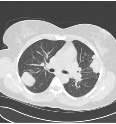
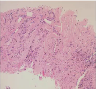
Non-neoplastic Pulmonary Hyalinising Granuloma
5
0
0
Full text
Figure
Related documents