R E S E A R C H
Open Access
Clinicopathologic features and surgical outcome
of solid pseudopapillary tumor of the pancreas:
analysis of 17 cases
Xiao-Guang Wang
1, Quan-Fa Ni
1, Jian-Guo Fei
1, Zheng-Xiang Zhong
1and Peng-Fei Yu
2*Abstract
Background:We summarize our experience of the diagnosis, surgical treatment, and prognosis of solid pseudopapillary tumors (SPTs).
Methods:We carried out a retrospective study of clinical data from a series of 17 patients with SPT managed in two hospitals between October 2001 and November 2011.
Results:All of the 17 patients were female and the average age at diagnosis was 26.6 years (range 11 years to 55 years). The tumor was located in the body or tail in ten patients, the head in five patients, and the neck in two patients. The median tumor size was 5.5 cm (range 2 cm to 10 cm). All 17 patients had curative resections, including seven distal pancreatectomies, five local resections, four pancreaticoduodenectomies, and one central pancreatectomy. Two patients required concomitant splenic vein resection due to local tumor invasion. All patients were alive and disease-free at a median follow-up of 48.2 months (range 2 to 90 months). There were no significant associations between
clinicopathologic factors and malignant potential of SPT. Ki-67 was detected in three patients with pancreatic parenchyma invasion.
Conclusions:The SPT is an infrequent tumor, typically affecting young women without notable symptoms. Surgical resection is justified even in the presence of local invasion or metastases, as patients demonstrate excellent long-term survival. Positive immunoreactivity for Ki-67 may predict the malignant potential of SPTs.
Background
The solid pseudopapillary tumor (SPT) of the pancreas was first reported by Frantz in 1959 [1]. It is a rare neoplasm of low malignant potential, and accounts for approximately 1% of pancreatic tumors [2]. This tumor primarily affects young women and is usually treated with surgical resection [3]. After resection and follow-up, there is generally a relatively favorable prognosis. Recently, the number of cases reported in the literature has been steadily rising; however, the pathogenesis and guidelines for SPT treatment remain unclear. In this study, we report our clinical experience with 17 cases of SPTs.
Methods
Between October 2001 and November 2011, 17 patients who underwent surgery for a pathologically confirmed SPT at the Department of Abdominal Surgery, Zhejiang Cancer Hospital and the Department of Surgery, Second Affiliated Hospital of JiaXing Medical College were reviewed retrospectively. Patients’ clinical presentation, radiological details, surgical data, pathological features, postoperative course, and long-term survival were col-lected and analyzed. Outpatient records combined with telephone interviews were used for follow-up.
Pathologically, SPT was defined as malignant if it demonstrated extrapancreatic invasion, distant metastases, pancreatic parenchymal invasion, or perineural or vascular invasion [4]. Univariate analyses of predictive features of malignancy were performed to compare clinicopathologic factors. All statistical analyses were performed with the computer program Statistical Package for Social Sciences (SPSS) 16.0 for Windows (Chicago, Illinois).
* Correspondence:yupengfei23@163.com 2
Department of Abdominal Surgery, Zhejiang Cancer Hospital, 38# Guangji Road, Zhejiang Province, Hangzhou 310022, China
Full list of author information is available at the end of the article
Results
Patient characteristics
All of the 17 patients were women, aged from 11 to 55 years (mean 26.6 years). The clinical presentation is unspe-cific, including abdominal pain (35.3%), abdominal discom-fort (27.3%), abdominal distension (27.3%), back pain (11.8%), and vomiting (9.1%). Three patients whose SPT was found during routine physical examinations were asymptomatic. The patients had a median symptom dur-ation of one month (range 5 days to 11 months). The tumors were 5.5 cm in diameter, on average (range 2 cm to 10 cm), and were located in the body or tail in ten patients, the head in five patients, and the neck in two patients. The clinical features of the 17 patients are listed in Table 1.
Preoperative examination and diagnosis
Radiological investigations were performed before oper-ation, including computed tomography (CT) in twelve patients, ultrasonography (US) in eight patients, mag-netic resonance imaging (MRI) in four patients, and US-guided fine needle aspiration cytology (FNAC) in two patients. Figures 1 and 2 show CT and US images of the SPT. The mass was described on cross-sectional imaging as heterogenous (solid and cystic) in ten patients and solid in seven patients. Calcifications were present in 4 of the 17 patients, while hemorrhage or ne-crosis was detected in 6 patients.
None of the patients had a definitive preoperative diagno-sis and a correct diagnodiagno-sis was made in six patients. Mis-diagnoses included pancreatic adenocarcinoma (n = 6), cystadenomas (n= 3), islet cell tumors (n= 1), and pancre-atic cyst (n= 1).
Surgical data
All 17 patients underwent surgical exploration. Seven patients with lesions in the pancreas body or tail underwent a distal pancreatectomy, including two spleen-preserving resections. Five patients underwent local resection and two of them had concomitant splenic vein resection due to local tumor invasion. The remaining five patients underwent pancreaticoduode-nectomy (Whipple, four cases) and central pancreatec-tomy (one case). Total surgery time ranged from 1.5 to 6.5 hours (mean 3.7 hours). Blood transfusion was needed in five patients during surgery; each patient received 2 units of blood.
All 17 patients had R0 resections and there were no sur-gical mortalities. Postsursur-gical complications occurred in five patients. One patient had pulmonary infection four days after surgery. Another patient had been found to have a pseudocyst in the first follow-up. Three patients had
pancreatic leakage. The median postsurgical stay was 10.3 days (range 7 to 17 days).
Pathological features
Grossly, the tumor is well encapsulated and is usually well demarcated from the pancreas. The cut surface shows large spongy areas of hemorrhage alternating with both solid and cystic degeneration. The tumors contain a mix-ture of solid, cystic, and pseudopapillary patterns in vari-ous proportions. Four patients had a malignant SPT; two patients had splenic vein infiltration and the other two had
Table 1 Clinicopathologic features of 17 patients with SPTs
Parameter Patient number
(n= 17)
%
Age(years, mean (range)) 26.6 (11-55)
Sex
Size(cm, mean (range)) 5.5 (2-10)
Location
Body or tail 10 58.8%
Head 5 29.4%
Neck 2 11.8%
Tumor feature
Solid and cystic 10 58.8%
Solid 7 41.2%
Surgical treatment
Distal pancreatectomy 2 11.8%
Distal pancreatectomy + splenectomy 5 29.4%
Local resection 3 17.6%
Local resection + splenic vein 2 11.8%
Resection
Whipple 4 23.5%
Central pancreatectomy 1 5.9%
Follow-up(months, mean (range)) 48.2 (2-90)
Outcome
Alive 17 100%
local invasion into the adjacent pancreatic parenchyma. No patients had lymph node metastasis.
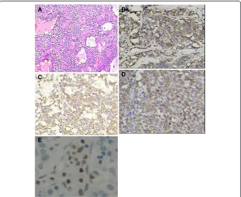
Immunohistochemical studies were performed in all 17 cases. Results were typically positive for vimentin, α1-antitrypsin, and neuron-specific enolase. Progesterone receptors, but usually not estrogen receptors, were variably present. Synaptophysin, cytokeratin, and chro-mogranin A were expressed only focally in a few tumors. Ki-67 was detected in three patients with pan-creatic parenchyma invasion. Figure 3 shows the histo-pathologic image results.
Follow-up
Follow-up included clinical examination, routine la-boratory tests, abdominal US, and CT or MRI every 3 months. The patients were followed up for a mean dur-ation of 48.2 months, (range 2 to 90 months) and all 17 patients were alive with no evidence of disease recur-rence or metastasis.
Predictive factors of malignancy
On univariate analysis, none of the features, including age, tumor size, tumor location, increased tumor mar-kers, and tumor characteristics, was predictive of malig-nant SPTs (Table 2).
Discussion
Solid pseudopapillary tumor of the pancreas is a rare neoplasm with a low malignant potential, usually affect-ing young women in the second or third decade of life.
The pathogenesis of the tumor is unknown, although its tendency to affect young women has suggested that sex hormones may be involved in the origin of SPT. How-ever, no differences in immunohistochemical stains for sex hormone-receptor proteins or in clinicopathologic characteristics had been found attributable to sex alone [5]. Sun et al. [6] reported that 62.5% of SPT patients had been infected by Hepatitis B virus (HBV), which may be involved in the pathogenesis of SPTs. However, this association has not been confirmed by other researchers.
The clinical presentation of SPTs is usually unspecific and two or more symptoms usually coexist. Most of the patients presented with unclear clinical features, in-cluding abdominal pain, abdominal discomfort, poor appetite, and nausea, which are related to tumor com-pression of the adjacent organs. Because patients lack distinctive symptoms, the majority of these tumors are diagnosed during complementary imaging investiga-tions, such as CT or US of the abdomen. On US or CT, the lesion is usually seen to be large, and its internal struc-ture ranges from cystic thick-walled or with an inner irregular margin to a predominantly solid mass with some cystic component [7]. On dynamic contrast-enhanced CT, the tumor is enhanced less than the adjacent nor-mal pancreas [8]. Magnetic resonance imaging is better than CT in differentiating the cystic or solid component inside the tumor and providing information about resectability. The use of FNAC, either percutaneously or endoscopic ultrasound guided, can help distinguish SPTs from other pancreatic tumors. However, seeding of the needle tract by neoplastic cells and such compli-cations as bleeding, pancreatic fistula, and biliary fistula during the procedure have also been reported [9].
Figure 1Contrast-enhanced CT shows a solid and cystic mass with a calcification (arrowed) located in the tail of
the pancreas.
Despite widespread availability of high-quality imaging systems, preoperative diagnosis was difficult. Only six patients were diagnosed as or suspected of SPTs in our series, and the misdiagnosis rate in other groups was reported as ranging from 38.5% to more than 70% [10,11]. According to our experience, data from CT or MRI scans combined with age and sex should be suffi-cient for the decision to operate, and FNAC should be performed where the radiological diagnosis is not clear enough.
Currently, complete aggressive surgical resection is the treatment of choice for SPTs, even in the case of local invasion or metastasis [12]. The surgical approach depends on the location, size, and nature of the
neoplasms, as well as the time of surgery [10]. Intra-operative frozen section may be helpful to ascertain the adequate of the resection margins. Extensive lymphatic dissection is not warranted, as SPTs rarely have lymph node metastases. For the case of local invasion or me-tastases, there is also a consensus that surgical therapy should be performed [13]. Because of the excellent out-comes after complete resection [14], surgeons should always aim for completeen-blocresection including ad-jacent structures preferably with microscopically clear margins. In our study, the infiltrated splenic vein and adjacent tissues were resected en bloc and a long-term survival was observed in these patients. Resection of distant metastases should be performed at the time of
primary resection or even for recurrences. This aggres-sive approach is supported in some studies, which showed that most patients were alive at long-term fol-low-up after extended resection [12,15].
In our study, four patients with splenic vein infiltra-tion or pancreatic parenchyma invasion were diag-nosed as malignant SPTs. Some studies have shown a correlation between tumor size above 5 cm, tumor ne-crosis, the male sex, and SPTs with malignant poten-tial [16,17]. However, several univariate analyses indicated that clinical factors, including sex, age, tumor size, tumor location, increased tumor markers, and tumor characteristics were not intensively related to the malignant potential of SPTs [4,10,18]. These results were consistent with that in our study. More-over, we found that positive immunoreactivity for Ki-67 was detected in three patients with pancreatic paren-chyma invasion. Our findings are similar to the report from Yang [10] and indicate that the detection of Ki-67 may correlate with the malignancy and poor outcome of SPTs. However, these results are only limited to
a small sample of SPTs, and more cases should be detected for Ki-67 and other new biomarkers in further studies.
Solid pseudopapillary tumors are readily diagnosable, based on their pathological and immunohistochemical features. The tumors contain a mixture of solid, cystic, and pseudopapillary patterns in various proportions. The solid portions of the tumor are composed of uniform and polygonal epithelioid cells with well-vascularized stroma and a discohesive arrangement [14]. Immunohistochemically, SPTs are typically posi-tive for vimentin, α1-antitrypsin, α1-antichymotrypsin, and neuron-specific enolase [19], but the unique immu-nohistochemical features with expression of CD56 and CD10 were not consistent in recent studies. Cells from SPTs may also reveal focal immunoreactivity for cyto-keratin and synaptophysin, demonstrate abnormal nu-clear localization of β-catenin and the presence of progesterone receptors and may express galectin-3, all of which are useful in differentiating SPTs from endo-crine pancreatic tumors [20].
The prognosis of SPTs is good, even with local recur-rence, as well as metastases or invasions. More than 95% of patients with SPTs limited to the pancreas are cured by complete surgical excision [20]. Local recur-rence is reported to be less than 10%, and usually within 4 years of surgery [11]. Recurrence, local inva-sion, and limited metastases are not contraindications for resection, and long-term survival has also been observed in patients with malignant SPTs. The overall 5-year survival was estimated to be 95% in a review of 718 patients reported in the English literature [21]. Owing to the favorable prognosis and excellent long-term survival, even in the presence of local recurrence or stable metastases, predictive factors of survival are difficult to identify.
Conclusions
Solid pseudopapillary tumors are infrequent, typically affecting young women without notable symptoms. Their behavior is relatively indolent and largely be-nign, however, surgical resection is warranted even in the presence of local invasion or metastases as patients demonstrate excellent long-term survival. Further studies should aim at acquiring more under-standing of SPTs and establishing guidelines for SPT diagnosis and treatment.
Consent
Written informed consent was obtained from the pa-tient for publication of this report and any accompany-ing images.
Table 2 Predictive factors of malignant SPTs
Clinicopathologic
Mean age (years) 34.4(19-55) 25.3(11-53) 0.11
Symptoms
Solid and cystic 1 9 0.11
Abbreviations
CP: central pancreatectomy; CT: computed tomography; FNAC: fine needle aspiration cytology; H&E: hematoxylin and eosin; MRI: magnetic resonance imaging; SPT: solid pseudopapillary tumor; US: ultrasonography.
Competing interests
The author(s) declare that they have no competing interests.
Authors’contributions
Wang X-G, Ni Q-F, Fei J-G, Zhong Z-X, Yu P-F designed and conducted the study, analyzed the data, and helped to write the manuscript. Yu P-F is the principal investigator, revised and edited the manuscript. All authors approved the final manuscript.
Author details
1
Department of Surgery, the Second Affiliated Hospital of JiaXing Medical College, JiaXing 314000, China.2Department of Abdominal Surgery, Zhejiang Cancer Hospital, 38# Guangji Road, Zhejiang Province, Hangzhou 310022, China.
Received: 27 November 2012 Accepted: 11 January 2013 Published: 6 February 2013
References
1. Franz VK:Papillary tumors of the pancreas: benign or malignant?In Tumors of the Pancreas. Atlas of Tumor Pathology. Edited by Franz VK. Washington, DC: US Armed Forces Institute of Pathology; 1959:32–33. 2. Bhanot P, Nealon WH, Walser EM, Bhutani MS, Tang WW, Logroñ R:Clinical,
imaging, and cytopathological features of solid pseudopapillary tumor of the pancreas: a clinicopathologic study of three cases and review of the literature.Diagn Cytopathol2005,33:421–428.
3. Kasem A, Ali Z, Ellul J:Papillary cystic and solid tumour of the pancreas: report of a case and literature review.World J Surg Oncol2005,3:62. 4. Goh BK, Tan YM, Cheow PC, Chung AY, Chow PK, Wong WK, Ooi LL:Solid
pseudopapillary neoplasms of the pancreas: an updated experience.
J Surg Oncol2007,95:640–644.
5. Sunkara S, Williams TR, Myers DT, Kryvenko ON:Solid pseudopapillary tumours of the pancreas: spectrum of imaging findings with histopathological correlation.Br J Radiol2012,85:e1140–e1144. 6. Sun GQ, Chen CQ, Yao JY, Shi HP, He YL, Hang WH:Diagnosis and
treatment of solid pseudopapillary tumor of pancreas: a report of 8 cases with review of domestic literature [in Chinese].Chin J Gen Surg 2008,17:902–907.
7. Igbinosa O:Pseudopapillary tumor of the pancreas. An algorithmic approach.JOP2011,12:262–265.
8. Vargas-Serrano B, Domínguez-Ferreras E, Chinchón-Espino D:Four cases of solid pseudopapillary tumors of pancreas: imaging findings and pathological correlations.Eur J Radiol2006,58:132–139.
9. Pettinato G, Di Vizio D, Manivel JC, Pambuccian SE, Somma P, Insabato L:
Solid-pseudopapillary tumor of the pancreas: a neoplasm with distinct and highly characteristic cytological features.Diagn Cytopathol2002,27:325–334. 10. Yang F, Jin C, Long J, Yu XJ, Xu J, Li J, Fu de L, Ni QX:Solid
pseudopapillary tumor of the pancreas: a case series of 26 consecutive patients.Am J Surg2009,198:210–215.
11. Yang F, Fu DL, Jin C, Long J, Yu XJ, Xu J, Ni QX:Clinical experiences of solid pseudopapillary tumors of the pancreas in China.J Gastroenterol Hepatol2008,23:1847–1851.
12. Lee JS, Han HJ, Choi SB, Jung CW, Song TJ, Choi SY:Surgical outcomes of solid pseudopapillary neoplasm of the pancreas: a single institution's experience for the last ten years.Am Surg2012,78:216–219. 13. Rebhandl W, Felderbauer FX, Puig S, Paya K, Hochschorner S, Barlan M,
Horcher E:Solid-pseudopapillary tumor of the pancreas (Frantz tumor) in children: report of four cases and review of the literature.J Surg Oncol 2001,76:289–296.
14. Tang LH, Aydin H, Brennan MF, Klimstra DS:Clinically aggressive solid pseudopapillary tumors of the pancreas. A report of two cases with components of undifferentiated carcinoma and a comparative clinicopathologic analysis of 34 conventional cases.Am J Surg Pathol 2005,29:512–519.
15. Hassan I, Celik I, Nies C, Zielke A, Gerdes B, Moll R, Ramaswamy A, Wagner HJ, Bartsch DK:Successful treatment of solid-pseudopapillary tumor of the pancreas with multiple liver metastases.Pancreatology2005,5:289–294. 16. Kang CM, Kim KS, Choi JS, Kim H, Lee WJ, Kim BR:Solid pseudopapillary
tumor of the pancreas suggesting malignant potential.Pancreas2006,
32:276–280.
17. Machado MC, Machado MA, Bacchella T, Jukemura J, Almeida JL, Cunha JE:
Solid pseudopapillary neoplasm of the pancreas: distinct patterns of onset, diagnosis, and prognosis for male versus female patients.
Surgery2008,143:29–34.
18. Lee SE, Jang JY, Hwang DW, Park KW, Kim SW:Clinical features and outcome of solid pseudopapillary neoplasm: differences between adults and children.Arch Surg2008,143:1218–1221.
19. Kosmahl M, Seada LS, Jänig U, Harms D, Klöppel G:Solid pseudopapillary tumor of the pancreas: its origin revisited.Virchows Arch2000,436:473–480. 20. Geers C, Moulin P, Gigot JF, Weynand B, Deprez P, Rahier J, Sempoux C:
Solid and pseudopapillary tumor of the pancreas: review and new insights into pathogenesis.Am J Surg Pathol2006,30:1243–1249. 21. Papavramidis T, Papavramidis S:Solid pseudopapillary tumors of the
pancreas: review of 718 patients reported in English literature.J Am Coll Surg2005,200:965–972.
doi:10.1186/1477-7819-11-38
Cite this article as:Wanget al.:Clinicopathologic features and surgical outcome of solid pseudopapillary tumor of the pancreas: analysis of 17 cases.World Journal of Surgical Oncology201311:38.
Submit your next manuscript to BioMed Central and take full advantage of:
• Convenient online submission
• Thorough peer review
• No space constraints or color figure charges
• Immediate publication on acceptance
• Inclusion in PubMed, CAS, Scopus and Google Scholar
• Research which is freely available for redistribution