R E S E A R C H
Open Access
Combination of electromagnetic navigation
bronchoscopy-guided biopsy with a novel
staining for peripheral pulmonary lesions
Kai Qian
1†, Yi Deng
2†, Cheng Shen
1†, Yong-Geng Feng
1, Bo Deng
1*and Qun-You Tan
1*Abstract
Background:The diagnosis of peripheral pulmonary lesions (PPLs) is a challenging task for pulmonologists, especially for small PPLs. Conventional localization of these small PPLs, which are > 1 cm away from the visceral pleura in operation, is quite difficult. Currently used methods inevitably damage the visceral pleura and may cause a series of complications, such as pneumothorax and hemothorax. Hence, the present study aimed to find out an intraoperative localization method with no damage to the visceral pleura.
Methods:We retrospectively reviewed 21 patients with PLLs who underwent electromagnetic navigation bronchoscopy (ENB)-guided biopsy plus a new methylene blue staining with the help of massage (Massage Staining) in our department between August 2017 and December 2018.
Results:The median age of these 21 patients was 51.3 ± 2.1 years. The diameter of the PPLs was 8.2 ± 2.3 mm. The rateof successful biopsy was 76.2%, and the rate of excellent or satisfactory of Massage Staining was 81.0%, while all lesions of these 21 cases were included in the range of staining, and the median distance from the edge of the stained site to the edge of the lesion was 29 ± 18 mm. The duration of ENB-guided biopsy plus Massage Staining was 26.7 ± 5.3 min, and the intraoperative blood loss was 3.3 ± 1.5 ml. No pneumothorax, hemorrhage, and tracheal injury occurred intraoperatively.
Conclusions:The ENB-guided biopsy combined with Massage Staining is an innovative one-stop strategy designed to enhance the precision of thoracic surgery. The Massage Staining avoids damage to the visceral pleura, causes the low incidence of complications, but yields precise localization of PPLs.
Keywords:Electromagnetic navigation bronchoscopy, Biopsy, Peripheral pulmonary lesions, Staining
Introduction
With the widespread application of computed tomog-raphy (CT), more cases with peripheral pulmonary lesions (PPLs), e.g., ground-glass nodules (GGNs), have been detected. Superficially localized solid nodules with pleural indentation can be visualized or palpable during surgery. However, the detection of pure GGNs and sub-solid nodules (SSNs) accompany with challenges. During operation, the approximate location of the pulmonary nodules can be determined by preoperative CT scan
images. However, there is often a deviation in terms of location of lesions before and after the collapse of the lungs, causing that lesions cannot be accurately located, leading to tremendous medical risk. Other conventional positioning methods include CT-guided implantation of hookwire or coils and CT-guided methylene blue stain-ing. Nevertheless, those methods damage the visceral pleura and induce pneumothorax, hemothorax, detach-ment, or movement from the localized object s[1].
Electromagnetic navigation bronchoscopy (ENB) is a promising technology that increases the diagnostic accuracy of peripheral lung and mediastinal lesions and is taken as a supplement to traditional
bronchos-copy, endotracheal ultrasound, and endotracheal
biopsy techniques into consideration [2]. Preoperative
© The Author(s). 2019Open AccessThis article is distributed under the terms of the Creative Commons Attribution 4.0 International License (http://creativecommons.org/licenses/by/4.0/), which permits unrestricted use, distribution, and reproduction in any medium, provided you give appropriate credit to the original author(s) and the source, provide a link to the Creative Commons license, and indicate if changes were made. The Creative Commons Public Domain Dedication waiver (http://creativecommons.org/publicdomain/zero/1.0/) applies to the data made available in this article, unless otherwise stated.
* Correspondence:superdb@163.com;tanqy001@163.com
†Kai Qian, Yi Deng and Cheng Shen contributed equally to this paper. 1Department of Thoracic Surgery, Institute of Surgery Research, Daping Hospital, Army Medical University, Chongqing, China
Full list of author information is available at the end of the article
Qianet al. World Journal of Surgical Oncology (2019) 17:158
pathological biopsy by using synchronized ENB con-tains significant clinical values for surgical treatment of pulmonary nodules.
In this study, we initially and innovatively conducted a novel Massage Staining without damage to the visceral pleura, combining with ENB-guided biopsy for diagnosis of PPLs.
Methods
Patients’selection
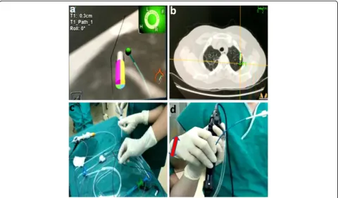
Patients were screened between August 2017 and October 2018 to undergo ENB-guided biopsy and Mas-sage Staining. The inclusion criteria were as follows: (1) patients who did not undergo pathological diagnosis and anti-tumor treatments before operation; (2) being Fig. 1Preoperative preparation.aPreoperative CT scan was carried out to determine the location of the lesion; the red arrow shows the lesion. bPreoperatively establish the navigation path by ENB, in which the green point is a lesion
resistant to ENB-guided biopsy; (3) the distance from the edge of the lesion to the visceral pleura ≤15 mm; (4) pa-tients who did not have any contraindications for pulmon-ary surgery, i.e., distant metastasis, bleeding tendency, blood clotting disorders, cardiopulmonary insufficiency, severe arrhythmia or hypertension, pulmonary hyperten-sion, and acute respiratory infection. This study was ap-proved by the Ethics Committee of the Daping Hospital, Army Medical University (Chongqing, China). Besides, all the patients signed a written informed consent form.
Instruments
The Super-D electromagnetic navigation system was purchased from Covidien (AAS000161-02; USA), in-cluding a 1.9 × 1070-mm edge positioning guide wire, a 2.8 × 1050-mm extended working channel for the locat-able guide, 1.8 × 1050-mm biopsy forceps, and a 1.8 × 1000-mm hollow cannula. Video-assisted thoracoscopic surgery (VATS) was performed using the STORZ thor-acoscopic system (KARL STORZ, Culver, CA, USA), and the robotic surgery was conducted by using the da Vinci Surgical System (Intuitive Surgical Inc., Sunny-vale, CA, USA).
Surgical procedure
The patients received general anesthesia and single-lumen intubation. After exploration with a broncho-scope, the locatable guide wire was inserted via an extended working channel to dilate the working chan-nel, where the virtual and actual bronchoscopy images were matched by Super-D electromagnetic navigation system. A navigation map to the target area was then automatically generated (Fig. 1a, b). Using the naviga-tion system, the posinaviga-tion of the locatable guide wire was corrected, and the locatable guide wire was
ad-vanced to the lesion site (Fig. 2a). Subsequently, the
locatable guide wire was retracted, the biopsy tool
was inserted through the guide wire’s expansion
chan-nel, and the target tissue was clamped out for frozen section diagnosis. Then, the locatable guide wire was again placed to the pleural adjacent to the lesion with
the guidance of the navigation system (Fig. 2b), a
cannula (with diameter of 1.8 mm) equal to the length of the locatable guide wire was inserted through the extended working channel, and methylene blue was
injected through the cannula (Fig. 2c) with a dose of
0.8 ml/cm per diameter of lesion. After that, the lo-catable guide wire was re-inserted and was confirmed to reach the presupposed location of the visceral pleura, and then, the locatable guide wire was repeat-edly used to massage the visceral pleura to complete
the staining process (Fig. 2d). Schematic diagram of a
novel staining method using Massage Staining is
shown in Fig. 3. The next surgical procedure was
performed as follows (Fig. 4): (1) if the benign lesions were diagnosed by biopsy, wedge resection was undertaken; (2) lobectomy was carried out for malig-nant lesions; and (3) if the lesions were extremely small to underwent biopsy, performing lobectomy depended on results of frozen section diagnosis.
Statistical analysis
Continuous variables were presented as median values. The chi-square test was applied to compare categorical variables among the groups, andPvalue < 0.05 was considered statis-tically significant. Statistical analyses were carried out by using SPSS 23.0 software (IBM, Armonk, NY, USA).
Fig. 3a–cSchematic diagram of a novel Massage Staining
Results
A total of 21 patients with PPLs [16 males (76.2%) and 5 females (23.8%); the patients’ mean age, 51.3 ± 2.1 years] underwent ENB-guided biopsy combined with the Mas-sage Staining. There were 8 (38.1%) cases with solid nodules, 8 (38.1%) cases with mixed ground-glass nodule (mGGN), 3 (14.3%) cases with pure ground-glass nodule (pGGN), and 2 (9.5%) cases with cavitary lesions. The diameter of the PPLs was 8.2 ± 2.3 mm. There were 6 (28.6%) cases in the right upper lobe, 1 (4.8%) case in the right middle lobe, 4 (19.0%) cases in the right lower
lobe, 5 (23.8%) cases in the left upper lobe, and 5 (23.8%) cases in the left lower lobe (Table1).
The intraoperative biopsy results of 16 patients (11 malignant tumors and 5 benign lesions) were consistent with postoperative pathology, which accompanied the successful biopsy rate of 76.2%.
In order to assess the effects of the Massage Staining, we classified outcomes into excellent, satisfactory, and unsatisfactory, respectively (Fig. 5). The results of Mas-sage Staining were excellent or satisfactory in 17 cases (Fig. 6a), although in the other 4 cases the result was Fig. 4The proposed strategy for surgical treatment by a combination of ENB-guided biopsy with Massage Staining. The red arrow represents the lesion is malignant according to the results of frozen section diagnosis
Table 1Patients’clinical characteristics
Variables Number of cases
Gender (male to female) 16:5
Age (years) 51.3 ± 2.1
The distance from the edge of the lesion to the visceral pleura (mm) 21 ± 8
The diameter of lesions (mm) 8.2 ± 2.3
ENB-guided biopsy 21
Mode of operation
Thoracoscopic wedge resection 7
VAST lobectomy 12
unsatisfactory, the manipulating still guided the approxi-mate extent of resection which is conducive to the de-tection of small lesions for frozen section diagnosis (Fig. 6b). The distance from the edge of the stained site to the edge of the lesions was 29 ± 18 mm. Additionally, no significant differences were noted in the results of the proposed Massage Staining method in terms of different sizes and imaging features of PPLs (P> 0.05) (Table 2). According to the results of frozen section diagnosis, 5 cases were diagnosed with the granulomatous or inflam-matory lesions and underwent thoracoscopic wedge re-section (TWR). One patient diagnosed with tuberculosis
was converted to thoracotomy, and wedge resection was performed. Besides, one of them was not precisely diag-nosed by ENB-guided biopsy, and according to the his-tory of medical imaging, VATS wedge resection was carried out. Moreover, 12 patients were diagnosed with non-small cell lung cancer (NSCLC). Therefore, VATS lobectomy and lymphadenectomy were undertaken, and 2 patients received lobectomy by the da Vinci Surgical System. No pneumothorax, hemorrhage, and tracheal in-jury occurred during ENB-guided biopsy combined with Massage Staining. The average duration of surgery for the ENB-guided biopsy plus Massage Staining was 26.7 ± 5.3 min, and the average blood loss during the surgery was 3.3 ± 1.5 ml.
Discussion
It is noteworthy that the correct diagnosis of pulmonary lesions is vital for the selection of an appropriate surgical Fig. 5Criteria for evaluation of the dyeing effect. Excellent: the
distance from the edge of the lesion to the edge of staining was less than 20 mm. Satisfactory: the distance from the edge of the lesion to the edge of staining was 20–40 mm. Unsatisfactory: the distance from the edge of the lesion to the edge of staining was more than 40 mm
Fig. 6Intraoperative observation of the proposed Massage Staining.aThe effects of staining where the catheter reached the visceral pleura.b The effects of staining where the distance from the catheter to the visceral pleura was about 0.8 cm
Table 2Characteristics and effects of ENB-guided biopsy combined with Massage Staining for peripheral pulmonary lesions
Characteristics Total Dye (+) Dye (−) Pvalue
Size of PPLs(mm) 0.092
≤9 8 6 2
9–12 7 7 0
12–15 5 4 1
≥15 1 0 1
Imaging features of PPLs 0.658
Solid nodule 8 7 1
mGGN 8 6 2
pGGN 3 2 1
Cavitary nodule 2 2 0
Dye (+)Excellent or satisfactory staining effects and wedge resection could be
performed for the lesion;Dye (-)Not satisfactory staining effect with diffused dye and wedge resection was not appropriate
approach. In the preoperatively diagnostic process, sensi-tivity and specificity of positron emission tomography-computed tomography (PET/CT) were 88% and 77%, respectively [3]. Meanwhile, the accuracy of percutan-eous CT-guided pulmonary biopsy is not satisfactory, and the puncture may lead to pneumothorax and metas-tasis [4]. The detection rate of bronchoscopy with
endo-bronchial ultrasound-guided transbronchial needle
aspiration (EBUS-TBNA) is closely associated with the size and location of the tumor, in which the detection rate is about 63% for lesion > 2 cm, and that is reduced to 34% for lesion < 2 cm [5].
Different from the above methods, ENB employs a three-dimensional reconstruction of CT scan data and sensor lo-cation technology to guide a steerable endoscopic probe to PPLs [6]. Multicenter prospective studies have shown that the rate of successful ENB-guided biopsy can reach to
91.8% [2]. ENB-combined transbronchial lung biopsy is
feasible and safe, provides larger samples, and has a higher diagnostic yield than transbronchial lung biopsy only [7].
Small peripheral pulmonary lesions (PPLs), which are 1 cm away from the visceral pleura, are usually invisible and untouchable during surgery. Conventional positioning and staining methods, e.g., CT-guided implantation of hook-wire or coils, inevitably damage the pleura, potentially causing a series of complications such as pneumothorax and hemothorax. Kleedehn et al. compared methylene blue staining and hookwire localization and found that the incidence rates of complications reached 54% and 46%, re-spectively [8]. Our intraoperative staining localization method is very important without damaging the pleura preoperatively and associating complications. Although in 19% of cases (4/21), the locatable guide wire cannot reach
the visceral pleura due to inflammation or tumor [9],
resulting in dissatisfaction staining; the manipulating still guided the approximate extent of resection which is con-ducive to detection of small lesions for rapid frozen biopsy. The average duration of manipulation time for ENB-guided biopsy plus Massage Staining was 26.7 ± 5.3 min. However, we believe that it is necessary to take the time to obtain the frozen biopsy results and staining for the precise resection range. Furthermore, Massage Stain-ing can save the time of detection of small lesions for rapid frozen biopsy.
In order to find out an appropriate dye marking, methy-lene blue, indigo carmine, and fibrin glue might be helpful. Indigo carmine dye has an advantage in that it can be vis-ible at least 3 days after marking, while methylene blue is dissipated after several hours of staining. Therefore, indigo carmine should be the first choice for patients who cannot promptly undergo surgery after staining [10]. The mixture of methylene blue and fibrin glue has been previously re-ported [11]. The fibrin glue can decrease the diffusion rate of methylene blue in the airway and form a palpable
blue-stained area as well [11]. However, that is relatively cumber-some and limited by materials. When fibrin glue is condensed, it is difficult to find small lesions after the oper-ation. With regard to Massage Staining with the small dose of methylene blue, it did not produce a large diffusion area after rubbing without condensation, which enabled us to distinguish the lesions during and after surgery.
Furthermore, the details are supposed to be focused on the following: (1) The results of Massage Staining were based on the distance from staining spot to the visceral pleura, rather than size and imaging features of PPLs. (2) The dose of the dye should be as small as possible. The point-like rubbing between the cannula and the pleura can effectively mark the position of the lesion and reduce the spread of the dye.
Conclusion
The ENB-guided biopsy plus Massage Staining is charac-terized by no damage to the visceral pleura, small dam-age to lung tissue, low incidence of complications, and
high precision, as well as “one-stop” diagnosis and
localization. Besides, this proposed strategy improves the rate of successful resection of PPLs in minimally invasive surgery. However, due to the small size of samples, it is essential to perform further multiple-center study to im-prove and optimize the proposed surgical technique.
Abbreviations
CBCT:Cone-beam CT; EBUS-TBNA: Endobronchial ultrasound-guided transbronchial needle aspiration; ENB: Electromagnetic navigation bronchoscopy; GGNs: Ground-glass nodules; LND: Lymph node dissection; NSCLC: Non-small cell lung cancer; PPLs: Peripheral pulmonary lesions; TWR: Thoracoscopic wedge resection; VATS: Video-assisted thoracoscopic surgery
Acknowledgements
We would like to thank Zhi-Yong Cheng and Bao-Qing Hu (Medtronic®) for providing assistance during the preparation of this manuscript.
Authors’contributions
KQ contributed in the study design, analysis, and interpretation of the data, as well as drafting of the manuscript. YD participated in the study design, analysis of the data, and drafting of the manuscript. CS contributed to the analysis and interpretation of the data and drafting of the manuscript. BD conducted a critical review of the manuscript and resources. QYT and YGF participated in the drafting of the manuscript. All authors studied and approved the final version of the manuscript.
Funding None
Availability of data and materials
The data used and/or analyzed during the current study are available from the corresponding author on reasonable request.
Ethics approval and consent to participate
Consent for publication N/A
Competing interests
The authors declare that they have no conflicts of interest.
Author details
1
Department of Thoracic Surgery, Institute of Surgery Research, Daping Hospital, Army Medical University, Chongqing, China.2Department of Oncology, Institute of Surgery Research, Daping Hospital, Army Medical University, Chongqing, China.
Received: 21 June 2019 Accepted: 5 September 2019
References
1. Yu KL, Tsai TH, Ho CC, Liao WY, Lin CK, Hsu CL, Shih JY. The value of radial endobronchial ultrasound-guided bronchial brushing in peripheral non-squamous non-small cell lung cancer. Sci Rep. 2018;8:5837.
2. Khandhar SJ, Bowling MR, Flandes J, Gildea TR, Hood KL, Krimsky WS, Minnich DJ, Murgu SD, Pritchett M, Toloza EM, et al. Electromagnetic navigation bronchoscopy to access lung lesions in 1,000 subjects: first results of the prospective, multicenter NAVIGATE study. BMC Pulm Med. 2017;17:59.
3. Jeong YJ, Yi CA, Lee KS. Solitary pulmonary nodules: detection,
characterization, and guidance for further diagnostic workup and treatment. AJR Am J Roentgenol. 2007;188:57–68.
4. Tuna T, Ozkaya S, Dirican A, Findik S, Atici AG, Erkan L. Diagnostic efficacy of computed tomography-guided transthoracic needle aspiration and biopsy in patients with pulmonary disease. Onco Targets Ther. 2013;6:1553–7. 5. Agarwal R, Srinivasan A, Aggarwal AN, Gupta D. Efficacy and safety of
convex probe EBUS-TBNA in sarcoidosis: a systematic review and meta-analysis. Respir Med. 2012;106:883–92.
6. Gex G, Pralong JA, Combescure C, Seijo L, Rochat T, Soccal PM. Diagnostic yield and safety of electromagnetic navigation bronchoscopy for lung nodules: a systematic review and meta-analysis. Respiration. 2014;87:165–76. 7. Taton O, Bondue B, Gevenois PA, Remmelink M, Leduc D. Diagnostic yield
of combined pulmonary cryobiopsies and electromagnetic navigation in small pulmonary nodules. Pulm Med. 2018;2018:6032974.
8. Kleedehn M, Kim DH, Lee FT, Lubner MG, Robbins JB, Ziemlewicz TJ, Hinshaw JL. Preoperative pulmonary nodule localization: a comparison of methylene blue and hookwire techniques. AJR Am J Roentgenol. 2016;207:1334–9. 9. Haghighi B, Choi S, Choi J, Hoffman EA, Comellas AP, Newell JD Jr, Lee CH,
Barr RG, Bleecker E, Cooper CB, et al. Imaging-based clusters in former smokers of the COPD cohort associate with clinical characteristics: the SubPopulations and intermediate outcome measures in COPD study (SPIROMICS). Respir Res. 2019;20:153.
10. Krimsky WS, Minnich DJ, Cattaneo SM, Sarkar SA, Harley DP, Finley DJ, Browning RF, Parrish SC. Thoracoscopic detection of occult indeterminate pulmonary nodules using bronchoscopic pleural dye marking. J Community Hosp Intern Med Perspect. 2014;4.
11. Luo K, Lin Y, Lin X, Yu X, Wen J, Xi K, Lin P, Zhang L. Localization of peripheral pulmonary lesions to aid surgical resection: a novel approach for electromagnetic navigation bronchoscopic dye marking. Eur J Cardiothorac Surg. 2017;52:516–21.
Publisher’s Note
Springer Nature remains neutral with regard to jurisdictional claims in published maps and institutional affiliations.