O R I G I N A L R E S E A R C H
Butyrate production in patients with end-stage
renal disease
This article was published in the following Dove Press journal: International Journal of Nephrology and Renovascular Disease
Matty L Terpstra1–3 Marjan J Sinnige1,3
Floor Hugenholtz4
Hessel Peters-Sengers4
Ester BM Remmerswaal1,3
Suzanne E Geerlings2
Frederike J Bemelman1
1Department of Internal Medicine,
Division of Nephrology, Renal Transplant Unit, Amsterdam UMC, University of Amsterdam, Amsterdam, Netherlands;
2Department of Internal Medicine,
Division of Infectious Diseases, Amsterdam Infection & Immunity Institute (AI&II), Amsterdam UMC, University of Amsterdam, Amsterdam,
The Netherlands;3Department of
Experimental Immunology, Amsterdam Infection & Immunity Institute (AI&II), Amsterdam UMC, University of Amsterdam, Amsterdam, The
Netherlands;4Center for Experimental
and Molecular Medicine, Amsterdam UMC, University of Amsterdam, Amsterdam, Netherlands
Background: Chronic kidney disease (CKD) is associated with a decreased intestinal
barrier function, causing bacterial translocation over the intestinal wall and triggering a systemic inflammatory response. Butyrate, a short-chain fatty acid produced by certain bacterial strains, is considered instrumental to keep the intestinal barrier intact. There are indications that a decreased amount of these specific bacterial species is part of the cause of the decreased intestinal barrier function in CKD. The aim of this study is (i) to determine if Dutch patients with end-stage renal disease (ESRD) have a decreased amount of butyrate-producing species and butyrate-butyrate-producing capacity and (ii) whether this correlates with systemic inflammation.
Methods: We used qPCR to evaluate the most abundant butyrate-producing species
F. prauznitzii, E. rectale and Roseburia spp. and the BCoAT gene, which reflects the butyrogenic capacity of the intestinal microbiota. Fecal samples were collected from healthy kidney donors (n=15), preemptive renal transplant recipients (n=4) and dialysis patients (n=31). Markers of inflammation (CRP and IL-6) and intestinal permeability (D-lactate) were measured in plasma.
Results:Patients with ESRD did not have a significantly decreased amountF. prauznitzii,
E. rectaleandRoseburia spp. or the BCoAT gene. Neither was there a significant correlation with CRP, IL-6 or D-lactate. On the individual level, there were some patients with decreased BCoAT levels and increased levels of CRP, IL-6 and D-lactate.
Conclusions: Patients with ESRD do not have a decreased amount of the most abundant
butyrate-producing species nor a decreased butyrate-producing capacity.
Keywords: renal failure, intestinal barrier function, butyrate, intestinal microbiota,
inflammation
Introduction
Despite the improvements that have been made in treatment modalities for patients with chronic kidney disease (CKD), morbidity and mortality rates remain high. Cardiovascular disease is the leading cause of morbidity and mortality and accounts
for 50% of deaths in CKD patients regardless of biological age.1This
(dispropor-tional) rate of cardiovascular mortality is hypothesized to stem from the presence of
specific nontraditional risk factors that accompany the loss of renal function, such
as the alterations in the gut that are seen in CKD.2
Numerous studies have pointed out intestinal alterations in CKD, including
changes of the microbes, a decreased intestinal barrier function and inflammation
of the intestinal wall.3,4 This decreased intestinal barrier function may lead to
bacterial translocation and endotoxemia, which is known to potentiate systemic Correspondence: Matty L Terpstra
Department of Internal Medicine, Division of Nephrology, Renal Transplant Unit, Amsterdam UMC, University of Amsterdam, Meibergdreef 9, 1100 DD Amsterdam, The Netherlands Tel +31 20 566 680 Email m.terpstra@amc.nl
International Journal of Nephrology and Renovascular Disease
Dove
press
open access to scientific and medical research
Open Access Full Text Article
International Journal of Nephrology and Renovascular Disease downloaded from https://www.dovepress.com/ by 118.70.13.36 on 23-Aug-2020
inflammation and atherosclerosis.5Systemic inflammation is associated with progression of CKD and is a mediator of a variety of complications in CKD such as anemia, cogni-tive impairment, insulin resistance and cardiovascular
disease,4,6–8which points out the importance of a healthy
intestinal barrier. Microbes appear also to have essential functions for the maintenance of this intestinal barrier function. One of these functions is the production of butyrate.
Butyrate, a short-chain fatty acid, is an important energy source for the colonocytes. It is synthesized from butyryl-CoA by the enzymes butyrate kinase and butyryl-CA: acet-ate CoA-transferase (BCoAT). Since the majority of human colonic butyrate-producing species use BCoAT for the last step of butyrate formation, this is considered as the
domi-nant system for butyrate production.9Butyrate is known to
promote cell differentiation, suppresses colonic infl
amma-tion and facilitate the transcripamma-tion of Claudine-1, which
enhances the tight-junction assembly.3,10 Therefore,
buty-rate might attenuate bacterial translocation and enhance the gut barrier function. It is found in little amounts in foods such as parmesan cheese and butter; nonetheless, intestinal levels are mainly dependent on bacterial fermentation of
dietary fiber by specific bacterial species.11 Butyrate can
also be administrated, which appeared to be beneficial in
a variety of gastrointestinal conditions such as diverticulitis
and ulcerative colitis.12,13
A decreased amount of butyrate could be part of the cause of the decreased intestinal barrier function and
thereby the systemic inflammation and high rate of
cardio-vascular disease in CKD. Studies in the Chinese population
have pointed out a decreased amount of specific
butyrate-producing species in different stages of CKD (non-dialysis),
which also appeared to be correlated with inflammatory
markers and was even associated with the progression of
CKD.14,15Thus, butyrate could be an interesting potential
therapeutic target to decrease the systemic inflammatory
response. However, literature on butyrate levels in CKD is scarce; the current available evidence is only based on the previously mentioned Chinese studies and there are no data on the western CKD population, which hinders the transla-tion to actual interventransla-tion studies. In additransla-tion to this, the actual ability from the intestinal microbiota to synthesize butyrate has never been evaluated in CKD.
With our study, we aim to:
(I) Evaluate the presence of the most abundant buty-rate-producing species in the Dutch population
with end-stage renal disease (ESRD), which is the most severe form of CKD, and healthy controls.
(II) Evaluate whether the abundance of these
butyrate-producing species correlates with inflammatory
markers and the intestinal barrier function. (III) Measure the actual capacity of the intestinal
microbiota to produce butyrate in these patients.
Materials and methods
Patients and samples
We enrolled healthy kidney donors, preemptive renal trans-plant recipients (transtrans-plant before the need to start dialysis) and patients undergoing dialysis treatment. Participants had to be able to collect stool samples to be included in this study. Participants who had received antibiotic treatment within the past 6 months were excluded; otherwise, there were no exclusion criteria. This study was conducted according to the Declaration of Helsinki and all participants gave written informed consent prior to enrolment.
From the kidney donors and the preemptive renal transplant recipients, samples were obtained one day before the scheduled renal transplantation. From each par-ticipant, blood and fecal samples were obtained and stored in the Biobank Renal Diseases in the Amsterdam UMC. This Biobank has been approved by the Biobank Ethical Committee of the Amsterdam UMC. Participants were asked to collect the fecal samples at home or in the Amsterdam UMC. Samples were kept at 7°C and were transported to the hospital in a cooler bag, after which they
were immediately stored at −80°C. In addition to this,
participants were asked questions about the use of anti-biotics, prebiotics and dietary habits.
After the initial analysis of our patient samples, we performed an additional analysis of fecal samples from 7 young healthy volunteers at two time points (0 and at 3 months) to analyze the stability of the measured bacterial species and the butyrate-producing capacity of the intest-inal microbiota.
DNA extraction
DNA was extracted from approximately 200 mg feces by using the QIAamp DNA Stool Mini Kit (Qiagen, Hilden, Germany). Samples were kept frozen until DNA extraction by manufacturer manual for extraction of bacterial DNA was started. After extraction, DNA concentration was determined by using the NanoDrop photospectrometer
International Journal of Nephrology and Renovascular Disease downloaded from https://www.dovepress.com/ by 118.70.13.36 on 23-Aug-2020
(model ND2000, Thermo Fisher, Waltham, MA USA), after which the DNA was stored at 4°C until further processing.
qPCR
We used qPCR to quantify the total amount of bacterial DNA (16S), the three most abundant butyrate-producing
species: F. prausnitzii, E. rectale, Roseburia spp. and
the BCoAT gene (reflecting the ability of the intestinal
microbiota to produce butyrate) in the fecal samples of each of the participants. For each of these outcome measures, a qPCR was set up by using the standard
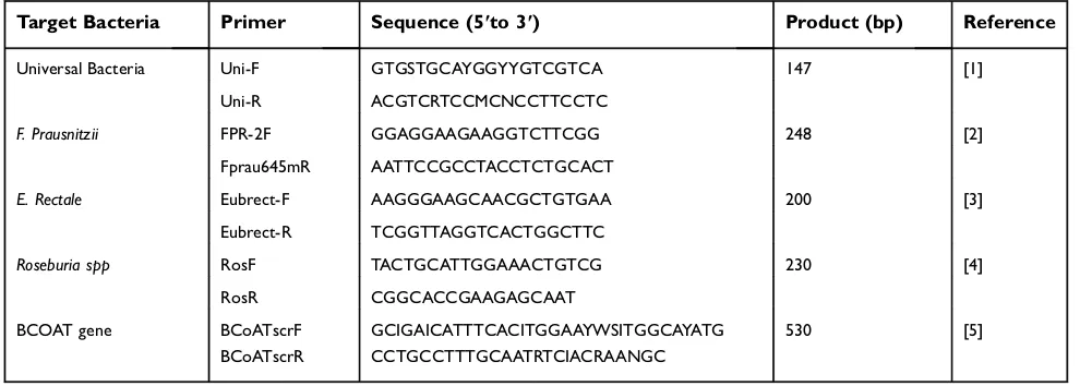
curve method for absolute quantification. Primers, listed
in supplemental Table S1, were synthesized by Sigma
Aldrich. Amplification of the product for standard curve
design was done at the BioRad 48 well PCR machine (S1000 thermal cycler, Bio-Rad, Hercules, CA, USA) qPCR assays were performed in a 96-well optical plate on the Step One Plus Cycler (StepOnePlus, Applied
Biosystems, Foster City, CA, USA). All assays were carried out in triplicate. The reaction mixture consisted of 2 ng DNA, 5 µM of each primer, SensiFast SYBR No ROX Kit (BioLine, London, UK) SybrGreen and PCR Grade water (Roche, Basel, Switzerland). The copy number of target DNA was determined by serially diluting standards running on the same plate. Data was
analyzed using StepOne Software 2.1 (Applied
Biosystems, Foster City, CA, USA).
Biochemical analysis
CRP, IL-6 (inflammatory markers) and D-lactate (reflecting the
intestinal barrier function) were measured in plasma. An auto-matic and validated immunoturbidimetric assay was per-formed to determine CRP levels by using a Roche Cobas C8000 with a c702 module (Roche Diagnostics, Risch-Rotkreuz, Zwitserland). IL-6 was measured by using an ELISA assay. Plasma samples were measured in duplicates by using the Sanquin PeliKine human IL6 ELISA with
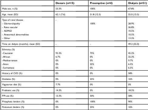
Table 1Baseline characteristics
Donors (n=15) Preemptive (n=4) Dialysis (n=31)
Male sex, n (%) 53.3% 100% 67.8%
Age, mean (SD) 62.1 (7.6) 51.8 (12.3) 55.0 (15.5)
Type of renal disease - Glomerulopathy - Reno vascular - ADPKD
- Anatomical abnormalities - Other
-100% 28.9%
36.8% 10.5% 10.5% 13.2%
Time on dialysis (months), mean (SD) - - 49.3 (55.0)
Ethnicity (%) -Caucasian -African -Mediterranean -Asian -Surinamese
93.3% 6.7% 0% 0% 0%
75% 0% 0% 25% 0%
45.2% 32.3% 9.7% 6.5% 6.5%
History of CVD (%) 0% 0% 58%
Diabetes (%) 0% 25% 16%
Vegetarian diet (%) 7.7% 0% 0%
Probiotic use (%) 14.3% 0% 18.5%
PPI use (%) 13.3% 50% 58%
Phosphate binders (%) 0% 100% 90%
Potassium binders (%) 0% 25% 16%
Abbreviations:SD, standard deviation; ADPKD, autosomal dominant polycystic kidney disease; CVD, cardiovascular disease; PPI, proton pump inhibitor.
Dovepress Terpstra et al
International Journal of Nephrology and Renovascular Disease downloaded from https://www.dovepress.com/ by 118.70.13.36 on 23-Aug-2020
additional reagents from the PeliKine Toolset (Sanquin, Amsterdam, the Netherlands). Absorption was determined by
ELISA plate reader (BioRad, model 680). Quantification of the
D form of lactic acid in the plasma samples was performed by using the highly sensitive method of ultra-performance liquid chromatography-electrospray ionization-tandem mass
spectro-metry, as described before.16The detection limit of this assay
was 0.06 mmol/L, and D-lactate values below 0.06 are dis-played as 0. In healthy subjects, D-lactate levels are below this detection limit.
Statistical analysis
All data analysis was conducted using IBM SPSS
Statistics 23; graphs and figures were created using
Graphpad Prism 7. The assumption of normality of each of the variable was assessed both visually and trough normality tests, of which the skewness, kurtosis and
Shapiro–Wilk test were performed. For positive
right-skewed variables, a log transformation was checked for improvement. We used parametric tests in case of normal distributed variables or nonparametric tests in case of
a persisting non-normal distribution or categorical
variables.
(I) ESRD vs healthy donors
A one-tailed independent sample T-test or the Mann–
Whitney U test was used to compare copy numbers of each of the bacterial species, the BCoAT gene, CRP, IL-6 and D-lactate levels between the dialysis group and the healthy kidney donors. Due to the small num-ber of preemptive renal transplant recipients, no
statis-tical comparison could be made between the
preemptive renal transplant recipients and the other study groups.
(II) Correlation with inflammatory markers
Depending on a normal or non-normal distribution, the
Pearson (r) or the Spearman (ρ) coefficient was calculated
for each of the butyrate-producing species, the BCoAT gene and CRP, IL-6 and D-lactate levels to evaluate a possible correlation. In this analysis, all study participants were included (n=50). Sensitivity analysis was performed for the dialysis group. Since in this analysis multiple correlations
were tested,p-values <0.007 were considered as statistically
significant after Bonferroni correction.17
In an additional analysis, both the correlation (Pearson or Spearman) between each of the butyrate-producing species and the BCoAT gene were also evaluated, as well as the correlation between CRP, IL-6 and D-lactate.
16S
F. Praus
Eubact. R.
Roseburia spp
BCoAT
107
106
105
104
103
102
101
100
10-1
Healthy donor
Preemptive NTX
Dialysis
C
o
pies/ng inp
ut gD
N
A
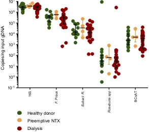
Figure 1Butyrate-producing species and the butyrate-producing capacity in healthy donors, preemptive renal transplant recipients and dialysis patients. Copy numbers/
nanogram input gDNA of the total amount of bacterial DNA (16S), the three most abundant butyrate-producing species and the BCoAT gene (median + IQR).
Abbreviation:NTX = kidney transplant recipient.
International Journal of Nephrology and Renovascular Disease downloaded from https://www.dovepress.com/ by 118.70.13.36 on 23-Aug-2020
(III) Stability over time
The Wilcoxon signed rank test was used to compare the copy numbers of each of the butyrate-producing species and the BCoAT gene between the two time points.
Ethics approval and informed
consent
All participants gave informed consent for participation in the Biobank Renal Diseases. This Biobank has been approved by the Biobank Ethical Committee of the Amsterdam UMC.
Results
We included 15 healthy kidney donors, 4 preemptive renal transplant recipients and 31 dialysis patients. Demographic characteristics are displayed in Table 1. Copy numbers of the total amount of bacterial DNA (16S), each of the butyrate-producing species and the BCoAT gene are expressed per ng input DNA and are displayed in Figure 1. Median copy num-bers/ng input DNA for each group and for each of the bacterial species and the BCoAT gene are displayed in Table 2. There
were no significant differences between the groups for either of
the producing species or considering the butyrate-producing capacity.
As expected, CRP, IL-6 and D-lactate were elevated in the dialysis group compared to the healthy kidney donors
(p<0.05;p<0.001;p<0.05, respectively). There was no
sta-tistically significant correlation between either of the
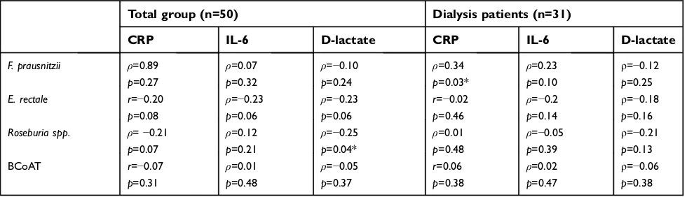
buty-rate-producing species or the BCoAT gene and CRP, IL-6 or D-lactate (Table 3; Figure 2). The weak negative correlation
between Roseburia spp. and D-lactate (ρ=−0.25, p=0.04)
was not significant after Bonferroni correction.
Sensitivity analysis for only the dialysis group did reveal
a positive correlation betweenF. prausnitzii, and CRP (ρ=0.34,
p=0.03); however, this was not significant after Bonferroni
correction (Table 3). There were 7 dialysis patients with values below the 25th percentile of the BCoAT gene. These patients
did not share any specific patient characteristics (Table 4). The
patients with elevated levels of D-lactate did not share specific
patients characteristics either (Table S2).
As expected there was a significant positive correlation
between F. prausnitzii, E. rectale, Roseburia spp. and the
BCoAT gene (ρ=0.48 p=0.001, r=0.56 p=<0.0001, ρ=0,38
p=0.003, respectively) and between CRP and IL-6 (ρ=0.64,
p=<0.0001), as displayed in supplemental Figure 1. There was
a weak positive correlation between D-lactate and IL-6
T able 2 Butyrate-pr oducing species, in fl ammator y mark ers and the intestinal barrier function 16S copies/ng input gDNA F. pr ausnitzii copies/ ng input gDNA E rectale copies/ng input gDNA Roseburia spp . copies/ ng input gDNA BCoA T copies/ng input gDNA CRP mg/ l IL-6 pg/ml D-lactate mmol/l Health y con-tr ols (n=15) 2,828,150 (1,904,217 – 3,465,626) 356,986 (127,028 – 478,816) 14,625 (8845 − 75 283) 30 (13 – 102) 2493 (949 – 9609) 0.8 (0.5 – 2.2) 1.4 (1.3 – 1.6) <0.1 (0 – 0) Pre emptiv e (n=4) 3,680,962 (3,166,966 – 6,10,362) 600,164 (331,419 – 1,003,237) 35,026 (7532 – 196,945) 63 (15 – 656) 4862 (1761 – 23,368) 0.9 (0.1 – 35.8) 2.5 (1.5 – 11.5) <0.1 (0 – 0.1) Dialysis gr oup (n=31) 3,027,599 (1 576,133 – 3,544,898) 297,402 (82,416 – 524,638) 12,896 (4928 – 34,306) 29 (7 – 66) 3656 (410 – 7684) 2.2 (0.9 – 4.2)* 2.7 (1.9 – 3.8)*
<0.1 (0–
0.13)* Notes: Median (Inter quartile range) cop y numbers 16S, the measur ed butyrate-pr oducing species and the BCoA T gene and le vels of in fl ammator y mark ers and D-lactate. Fo r comparison betwee n the health y contr ols and dialysis gr oup , either the one-tailed independent sample T -test or the Mann – Whitne y U test was used: * p <0.05.
Dovepress Terpstra et al
International Journal of Nephrology and Renovascular Disease downloaded from https://www.dovepress.com/ by 118.70.13.36 on 23-Aug-2020
(ρ=0.36, p=0.005), but not between D-lactate and CRP
(ρ=0.16,p=0.13) (Figure S1).
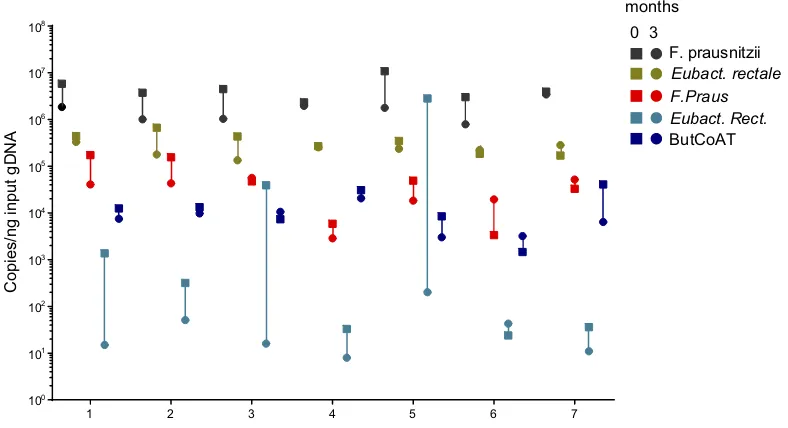
Additional longitudinal analysis in healthy
volunteers
We evaluated 7 young and healthy individuals at two time points: 0 and 3 months. Table 5 shows the demographics of these healthy volunteers. Figure 3 shows the copy numbers of each of the bacterial species and the BCoAT gene at the two different time points.
Copy numbers of the BCoAT gene,F. prausnitziiand
E. rectale were relatively stable, as there was no signifi -cant difference between the two time points, whilst copy
numbers ofRoseburia spp. didfluctuate (p=0.03).
Discussion
In our study, we found no difference in the amount of the measured butyrate-producing species between healthy kid-ney donors, preemptive renal transplant recipients and dialysis patients, neither was there a decreased capacity to produce butyrate in either of the patient groups. There was no correlation between the butyrate-producing species
or the butyrate-producing capacity and inflammatory
mar-kers or D-lactate on a population level. As expected,
dialysis patients did have increased levels of the infl
am-matory markers and D-lactate.
Our results of the Dutch ESRD (CKD stage V) patients are in contrast with the decreased amount of butyrate-producing species that has been reported in Chinese patients
with different stages of CKD (stage I–V).14,15 There are
several explanations for these conflicting results. Instead of
per gram of isolated DNA, the results from the Chinese studies were expressed per gram of stool, an outcome
mea-sure that is easily influenced by factors such as the
effectiveness of the DNA-isolation step, but also by the consistency of the stool sample, which especially in CKD patients can differ because of the different recommendations
about restrictions influid intake. Besides stool consistency,
the composition of the intestinal microbiota is known to be
influenced by many factors such as dietary habits, ethnicity,
smoking and medication (among others antibiotics and
pro-ton pump inhibitors).18–21In this prospect, it is difficult to
compare the Chinese population to the Dutch population as there are many differences considering environmental,
beha-vioral and genetic factors, which may have influenced test
results. Our study is, to the best of our knowledge, thefirst
study evaluating butyrate-producing species in the western CKD population. It is unclear whether demographic charac-teristics explain the difference in test results between the Chinese and Dutch patient population.
The strength of our study is that this is thefirst time that the
ability of the intestinal microbiota to produce butyrate has
been evaluated in CKD patients. Quantification of the
BCoAT gene has been pointed out as a valid tool to evaluate the butyrate-producing capacity of the intestinal microbiota
and thus all butyrate-producing species.22The additional
ana-lysis we performed in healthy volunteers also points out that the BCoAT gene is a robust outcome measure, as it appears to be relatively stable over time. Our results are reported in copy numbers per nanogram input gDNA, which is less easily
influenced by technical issues than the expression per gram
of feces. We used D-lactate to evaluate the intestinal barrier function. D-lactate is the metabolic product of bacteria, and in normal conditions, its serum level is very low, below our
detection limit of 0.06 mmol/L.23,24 When the intestinal
mucosa is damaged, the intestinal permeability increases and serum D-lactate levels will increase. Therefore, D-lactate is considered as a sensitive marker assessing the intestinal barrier
Table 3Correlation between butyrate-producing species, the butyrate-producing capacity, inflammatory markers and the intestinal
barrier function
Total group (n=50) Dialysis patients (n=31)
CRP IL-6 D-lactate CRP IL-6 D-lactate
F. prausnitzii ρ=0.89
p=0.27
ρ=0.07
p=0.32
ρ=−0.10
p=0.24
ρ=0.34
p=0.03*
ρ=0.23
p=0.10
ρ=−0.12
p=0.25
E. rectale r=−0.20
p=0.08
ρ=−0.23
p=0.06
ρ=−0.23
p=0.06
r=−0.02
p=0.46
ρ=−0.2
p=0.14
ρ=−0.18
p=0.16
Roseburia spp. ρ=−0.21
p=0.07
ρ=0.12
p=0.21
ρ=−0.25
p=0.04*
ρ=0.01
p=0.48
ρ=−0.05
p=0.39
ρ=−0.21
p=0.13
BCoAT r=−0.07
p=0.31
ρ=0.01
p=0.48
ρ=−0.05
p=0.37
r=0.06
p=0.38
ρ=0.02
p=0.47
ρ=−0.06
p=0.38
Abbreviations:r, Pearson coefficient;ρ, Spearman coefficient. *p<0.05. None of the parameters were statistically significantly different after Bonferroni correction.
International Journal of Nephrology and Renovascular Disease downloaded from https://www.dovepress.com/ by 118.70.13.36 on 23-Aug-2020
0 . 0 0 . 2 0 . 4 0 . 6 0 . 8 1 . 0 0 5 1 0 1 5 2 0
0 1 2 3
0 5 1 0 1 5 2 0
0 1 2 3 4 5
0 5 1 0 1 5 2 0
0 . 0 0 . 5 1 . 0 1 . 5
0 5 1 0 1 5
0 1 2 3
0 5 1 0 1 5
0 1 2 3 4 5
0 5 1 0 1 5
0 . 0 0 . 2 0 . 4 0 . 6 0 . 8 1 . 0 0
5 1 0 1 5 2 0
0 1 2 3
0 5 1 0 1 5 2 0
0 1 2 3 4 5
0 5 1 0 1 5 2 0
0 . 0 0 . 5 1 . 0 1 . 5
0 5 1 0 1 5
0 1 2 3
0 5 1 0 1 5
0 1 2 3 4 5
0 5 1 0 1 5 Log(c opi e s/ng i n put gDNA) Log(c opies/ng i n put gDNA) Log(c o pi es/ng input gDNA) Log(c opi es/ng input gDNA) Log(CRP) F.Prausnitzii ρ=0.89 P=0.27 Log(IL-6) Log(c opi es /ng input gDNA ) ρ=0.07 P=0.32 F.Prausnitzii F.Prausnitzii ρ=-0.10 P=0.24 Log(c opies /ng input gDNA ) Log(CRP) r=-0.2 P=0.08
Eubact. Rect. Eubact. Rect.
Log(IL-6) Log(D-lactate) Log(c opi es /ng input gDNA ) ρ=-0.23 P=0.06 Log( c opi es /ng input gDNA
) Eubact. Rect.
Log(D-lactate) ρ=-0.23 P=0.06 Log(CRP) Roseburia spp. ρ=-0.21 P=0.07 Log(IL-6) Roseburia spp. Log(c opies /ng input gDNA ) ρ=0.12 P=0.21 Roseburia spp. ρ=-0.25 P=0.04 Log(c opies /ng input gDNA ) Log(D-lactate) Log(CRP) BCoAT Log(IL-6) Log(c o pies /n g input gDNA ) Log( copi es/ ng input gDNA ) ρ=0.01 P=0.48 r=-0.07 P=0.31 BCoAT Log(D-lactate) ρ=-0.05 P=0.37 BCoAT Healthy donor Preemptive NTX Dialysis
Figure 2Correlation between butyrate-producing species, the BCoAT gene, inflammatory markers and D-lactate.
Abbreviations:r, Pearson coefficient;ρ, Spearman coefficient; log, natural logarithm +1; NTX, kidney transplant recipient.
Dovepress Terpstra et al
International Journal of Nephrology and Renovascular Disease downloaded from https://www.dovepress.com/ by 118.70.13.36 on 23-Aug-2020
T able 4 Baseline characteristics of patients with a low butyr ogenic capacity Ag e ye a rs M/F Ethn Renal disease CVD DM Time on dia-lysis months Ve g diet PPI PO4 binder K binder F. pra usnitzii copies/ng input gDNA E. rectale copies/ ng input gDNA Roseburia spp .
copies/ng input gDNA
BCoA T copies/ ng input gDNA CRP mg/L IL-6 pg/mL D-lactate mmol/L 38 M Black Reno- vascular Y es No 12 No Y es Y es Y es 99,178 1646 7 89 2.3 1.7 0.2 63 M Black Glomer -ulopath y Y es No 36 No Y es Y es No 297,402 5540 9 243 0.5 1.8 <0.1 62 M Asian FSGS No No 10 No Y es Y es No 349,171 2883 0 353 19 8.0 0.4 73 M Black Reno- vascular Y es No 24 No No Y es No 145,251 17,381 61 360 3.1 3.4 <0.1 66 F Cauc Hyper -o xaluria No No 112 No No Y es No 1450 0 0 409 2.3 10.1 1.2 40 M Cauc
Dense deposit disease
No No 7 No Y es Y es No 692 4928 0 338 <0.3 1.22 <0.1 28 M Asian Glomerulo- path y No No 96 No No No No 1,417,213 12,896 118 217 4.2 2.7 <0.1 Note: Characteristics of patients with low (<25th per centile) values of the BCoA T gene. Abbre viations : M, male; F, female, Ethn, ethnicity; FSGS, focal segmental glomerulosclerosis ; veg diet, vegetarian diet; CVD , cardiovascular disease; DM, diabetes mellitus; PPI, pr oton pump inhibitor ; P04, phosphate; K, potassium; Cauc, Caucasian.
International Journal of Nephrology and Renovascular Disease downloaded from https://www.dovepress.com/ by 118.70.13.36 on 23-Aug-2020
function.25,26To the best of our knowledge, D-lactate is cur-rently the best available method to evaluate the intestinal barrier function in patients with CKD. There are no validated methods in CKD, and most other markers evaluating the
intestinal barrier function are influenced by renal function.27
It is assumed that serum D-lactate levels are not influenced by
renal function since studies have shown that D-lactate is metabolized in peritoneal dialysis patients without urinary
production.28,29
Our study also has some limitations. Since we specifically
focused on the intestinal butyrate production, we quantified
the most abundant butyrate-producing species and the butyro-genic capacity of the intestinal microbiota. Quantitative PCR is a valid tool to measure actual copy numbers and to answer
our research question. However, it is impossible to speculate about the composition of the intestinal microbiome in ESRD
and the influence or competition with other bacterial species.
In addition to this, all samples were collected at one time point, which we do consider as a limitation considering the
interpretation of results fromRoseburia spp. Furthermore, the
included dialysis population was heterogeneous, from differ-ent ethnical backgrounds, with a wide variety of comorbid-ities and medication such as proton pump inhibitors. In the recent years, several studies have pointed out that a variety of medication, including these proton pump inhibitors,
pro-foundly alter the intestinal microbiota.30In addition to this,
the most common comorbidities among dialysis patients, including diabetes and cardiovascular disease, are also
asso-ciated with alterations in the intestinal microbiome.31,32Other
factors that might influence the intestinal microbiota are the
dietary restrictions imposed to these patients and also the
dialysis procedure itself. Intradialytic hypotension,
a common complication in hemodialysis patients, is known to result in regional hypo-perfusion that can also affect the gut
mucosa.33,34It is unknown whether this disturbance of the
homeostatic environment also affects the composition of the intestinal microbiome and butyrate-producing species. Accordingly, even though our patient population does truly
reflect the general dialysis population, many factors may
influence intestinal health in dialysis patients, and in this
study, it is impossible to speculate about the influence of all
the separate factors.
Table 5Demographics healthy volunteers included in
longitudi-nal alongitudi-nalysis
Healthy volunteers (n=7)
Male sex, n (%) 42%
Age, mean (SD) 30 (3.42)
Ethnicity (%)
-Caucasian 100%
History of CVD (%) 0%
Diabetes (%) 0%
Vegetarian diet (%) 14.3%
Use of PPI (%) 0%
Antibiotics (%) <6 months 0%
Abbreviations:SD, standard deviation; CVD, cardiovascular disease; PPI, proton
pump inhibitor.
1 2 3 4 5 6 7
100
101
102
103
104
105
106
107
108
0 3
F. prausnitzii
Eubact. rectale F.Praus Eubact. Rect.
ButCoAT months
Copies/ng input
gDNA
Figure 3Longitudinal analysis of the most abundant butyrate-producing species and the BCoAT gene in healthy volunteers. Longitudinal analysis of 7 healthy volunteers,
numbered 1–7. Copy numbers/nanogram input gDNA of the total amount of bacterial DNA (16S), the three most abundant butyrate-producing species and the BCoAT gene. Squares represent thefirst measured time point for each of the individuals, circles the second time point three months later.
Dovepress Terpstra et al
International Journal of Nephrology and Renovascular Disease downloaded from https://www.dovepress.com/ by 118.70.13.36 on 23-Aug-2020
We included only 4 preemptive renal transplant recipients (ESRD not on dialysis). These patients did not start with renal replacement therapy yet, and thus had a less severe form of
ESRD. As we did notfind a significant difference between the
dialysis patients and the healthy controls and results from these 4 patients were comparable to the dialysis patients, we do not expect that including more preemptive renal transplant recipients will change our results and conclusions.
Butyrate production
F. prausnitzii, E. rectaleandRoseburia spp. are the
buty-rate-producing species with the highest relative
abundance.35 Nevertheless, it is unknown what the exact
contribution of each of these butyrate-producing species is to the total butyrate production. Our data suggest that
whilstF. prausnitziiis by far the most abundant
butyrate-producing species, E. rectale may deliver a greater
con-tribution to the total butyrate production, as the correlation
between the BCoAT gene and E. rectalewas higher than
the correlation between the BCoAT gene and
F. prausnitzii. Thus, calculating the exact production of butyrate may be far more complex than the sum of the most abundant butyrate producing species.
Individual results
On a group level, no significant differences between healthy
controls and ESRD patients in either of the outcome
mea-sures reflecting the intestinal butyrate production and no
correlation between these outcome measures and the markers
reflecting intestinal permeability or the systemic infl
amma-tory response were found. On an individual level, there were patients with a decreased amount of the butyrate-producing species, a decreased capacity to produce butyrate and ele-vated levels of CRP, IL-6 and D-lactate. These patients did
not share specific patient characteristics. Since in our patient
group only a few patients had increased inflammatory
mar-kers and/or increased D-lactate levels, it is possible that our sample size was too small to detect a correlation between
butyrate production and inflammatory markers and the
intest-inal barrier function.
Thus, our results point out that a decreased amount of butyrate may be a realistic concern in some but not all
ESRD patients, but a clear subgroup cannot be defined.
Additionally, not all dialysis patients appear to have a decreased intestinal barrier function and not all patients
have increased inflammatory markers. Further research is
necessary to determine which factors influence the
intest-inal butyrate production on the individual level but also
whether butyrate administration may be a possible
thera-peutic target to prevent and treat the systemic infl
amma-tory response specifically in these patients.
Conclusion
In conclusion, we did not find a significant difference in
the amount of the three most abundant butyrate-producing species or in the butyrate-producing capacity between patients with ESRD and healthy kidney donors, neither was there a correlation with CRP, IL-6 or D-lactate. Among the dialysis patients, the variability was high and we did identify patients with low amounts of the butyrate-producing species, a low butyrogenic capacity and high
inflammatory markers or D-lactate. Further research is
warranted to determine which factors influence the
intest-inal butyrate production on individual level, especially in hemodialysis patients.
Abbreviation list
CKD, chronic kidney disease; ESRD, end-stage renal dis-ease; SD, standard deviation; ADPKD, autosomal domi-nant polycystic kidney disease; CVD, cardiovascular
disease; PPI, proton pump inhibitor; r, Pearson coefficient;
ρ, Spearman coefficient; M, male; F, female, Ethn,
ethni-city; FSGS, focal segmental glomerulosclerosis; veg diet, vegetarian diet; CVD, cardiovascular disease; DM, dia-betes mellitus; PPI, proton pump inhibitor; P04, phos-phate; K, potassium; Cauc, Caucasian.
Acknowledgments
We would like to thank Nelly van der Bom for her help with processing of the samples and Marit van Sandwijk for her help with the inclusion of patients. Samples in this study have been stored in the Biobank Renal Diseases, this Biobank is funded by Astellas Pharma.
Author contributions
All authors contributed to data analysis, drafting and
revis-ing the article, gave final approval of the version to be
published, and agree to be accountable for all aspects of the work.
Disclosure
Frederike J Bemelman reports grants from Astellas, during the conduct of the study. The authors report no other
conflicts of interest in this work.
International Journal of Nephrology and Renovascular Disease downloaded from https://www.dovepress.com/ by 118.70.13.36 on 23-Aug-2020
References
1. Shastri S, Sarnak MJ. Cardiovascular disease and CKD: core curri-culum 2010.Am J Kidney Dis. 2010;56(2):399–417. doi:10.1053/j. ajkd.2010.03.019
2. Jovanovich A, Isakova T, Stubbs J. Microbiome and cardiovascular disease in CKD. Clin J Am Soc Nephrol. 2018. doi:10.2215/ CJN.12691117
3. Vaziri ND. Effect of synbiotic therapy on gut-derived uremic toxins and the intestinal microbiome in patients with CKD.Clin J Am Soc Nephrol. 2016;11(2):199–201. doi:10.2215/CJN.13631215
4. Vaziri ND, Goshtasbi N, Yuan J, et al. Uremic plasma impairs barrier function and depletes the tight junction protein constituents of intestinal epithelium.Am J Nephrol. 2012;36(5):438–443. doi:10.1159/000343886 5. Stoll LL, Denning GM, Weintraub NL. Potential role of endotoxin as a proinflammatory mediator of atherosclerosis. Arterioscler Thromb Vasc Biol. 2004;24(12):2227–2236. doi:10.1161/01. ATV.0000147534.69062.dc
6. Stenvinkel P. Inflammation in end-stage renal disease: the hidden enemy. Nephrology (Carlton). 2006;11(1):36–41. doi:10.1111/ j.1440-1797.2006.00541.x
7. Amdur RL, Feldman HI, Gupta J, et al. Inflammation and progression of CKD: the CRIC Study. Clin J Am Soc Nephrol. 2016;11 (9):1546–1556. doi:10.2215/CJN.13121215
8. Nowak KL, Chonchol M. Does inflammation affect outcomes in dialysis patients?Semin Dial. 2018;31(4):388–397. doi:10.1111/sdi.12686 9. Louis P, Duncan SH, McCrae SI, Millar J, Jackson MS, Flint HJ.
Restricted distribution of the butyrate kinase pathway among butyrate-producing bacteria from the human colon. J Bacteriol. 2004;186(7):2099–2106.
10. Morrison DJ, Preston T. Formation of short chain fatty acids by the gut microbiota and their impact on human metabolism.Gut Microbes. 2016;7(3):189–200. doi:10.1080/19490976.2015.1134082
11. Stilling RM, van de Wouw M, Clarke G, Stanton C, Dinan TG, Cryan JF. The neuropharmacology of butyrate: the bread and butter of the microbiota-gut-brain axis?Neurochem Int. 2016;99:110–132. doi:10.1016/j.neuint.2016.06.011
12. Hallert C, Bjorck I, Nyman M, Pousette A, Granno C, Svensson H. Increasing fecal butyrate in ulcerative colitis patients by diet: con-trolled pilot study.Inflamm Bowel Dis. 2003;9(2):116–121. 13. Krokowicz L, Stojcev Z, Kaczmarek BF, et al. Microencapsulated sodium
butyrate administered to patients with diverticulosis decreases incidence of diverticulitis–a prospective randomized study.Int J Colorectal Dis. 2014;29(3):387–393. doi:10.1007/s00384-013-1807-5
14. Jiang S, Xie S, Lv D, et al. Alteration of the gut microbiota in Chinese population with chronic kidney disease. Sci Rep. 2017;7 (1):2870. doi:10.1038/s41598-017-02989-2
15. Jiang S, Xie S, Lv D, et al. A reduction in the butyrate producing species Roseburia spp. and Faecalibacterium prausnitzii is associated with chronic kidney disease progression.Antonie Van Leeuwenhoek. 2016;109(10):1389–1396. doi:10.1007/s10482-016-0737-y
16. Mason S, Reinecke CJ, Kulik W, van Cruchten A, Solomons R, van Furth AM. Cerebrospinal fluid in tuberculous meningitis exhibits only the L-enantiomer of lactic acid.BMC Infect Dis. 2016;16:251. doi:10.1186/s12879-016-1987-z
17. Gordi T, Khamis H. Simple solution to a common statistical problem: interpreting multiple tests.Clin Ther. 2004;26(5):780–786. 18. Minalyan A, Gabrielyan L, Scott D, Jacobs J, Pisegna JR, Gastric T.
Intestinal microbiome: role of proton pump inhibitors. Curr Gastroenterol Rep. 2017;19(8):42. doi:10.1007/s11894-017-0577-6
19. Donaldson GP, Lee SM, Mazmanian SK. Gut biogeography of the bacterial microbiota. Nat Rev Microbiol. 2016;14(1):20–32. doi:10.1038/nrmicro3552
20. Vandeputte D, Falony G, Vieira-Silva S, Tito RY, Joossens M, Raes J. Stool consistency is strongly associated with gut microbiota richness and composition, enterotypes and bacterial growth rates. Gut. 2016;65(1):57–62. doi:10.1136/gutjnl-2015-309618
21. Hughes V. Microbiome: cultural differences. Nature. 2012;492 (7427):S14–S15. doi:10.1038/492S14a
22. Louis P, Flint HJ. Development of a semiquantitative degenerate real-time pcr-based assay for estimation of numbers of butyryl-coenzyme A (CoA) CoA transferase genes in complex bac-terial samples. Appl Environ Microbiol. 2007;73(6):2009–2012. doi:10.1128/AEM.02561-06
23. Ewaschuk JB, Naylor JM, Zello GA. D-lactate in human and rumi-nant metabolism. J Nutr. 2005;135(7):1619–1625. doi:10.1093/jn/ 135.7.1619
24. Brandt RB, Siegel SA, Waters MG, Bloch MH. Spectrophotometric assay for D-(-)-lactate in plasma. Anal Biochem. 1980;102(1):39–46.
25. Tan SJ, Yu C, Yu Z, et al. High-fat enteral nutrition reduces intestinal mucosal barrier damage after peritoneal air exposure. J Surg Res. 2016;202(1):77–86. doi:10.1016/j.jss.2015.12.010
26. Wang H, Gong J, Wang W, et al. Are there any different effects of Bifidobacterium, Lactobacillus and Streptococcus on intestinal sensa-tion, barrier function and intestinal immunity in PI-IBS mouse model? PLoS One. 2014;9(3):e90153. doi:10.1371/journal. pone.0090153
27. Terpstra ML, Singh R, Geerlings SE, Bemelman FJ. Measurement of the intestinal permeability in chronic kidney disease. World J Nephrol. 2016;5(4):378–388. doi:10.5527/wjn.v5.i4.378
28. Yasuda T, Ozawa S, Shiba C, et al. D-lactate metabolism in patients with chronic renal failure undergoing CAPD. Nephron. 1993;63 (4):416–422. doi:10.1159/000187245
29. Adeva-Andany M, Lopez-Ojen M, Funcasta-Calderon R, et al. Comprehensive review on lactate metabolism in human health. Mitochondrion. 2014;17:76–100. doi:10.1016/j.mito.2014.05.007 30. Imhann F, Vich Vila A, Bonder MJ, et al. The influence of proton
pump inhibitors and other commonly used medication on the gut microbiota. Gut Microbes. 2017;8(4):351–358. doi:10.1080/ 19490976.2017.1284732
31. Tilg H, Moschen AR. Microbiota and diabetes: an evolving relationship. Gut. 2014;63(9):1513–1521. doi:10.1136/gutjnl-2014-306928
32. Ahmadmehrabi S, Tang WHW. Gut microbiome and its role in cardiovascular diseases. Curr Opin Cardiol. 2017;32(6):761–766. doi:10.1097/HCO.0000000000000445
33. Chou JA, Kalantar-Zadeh K, Mathew AT. A brief review of intradia-lytic hypotension with a focus on survival. Semin Dial. 2017;30 (6):473–480. doi:10.1111/sdi.12627
34. van der Sande FM, Dekker MJ, Leunissen KML, Kooman JP. Novel insights into the pathogenesis and prevention of intradialytic hypotension. Blood Purif. 2018;45(1–3):230–235. doi:10.1159/ 000485160
35. Dillon SM, Kibbie J, Lee EJ, et al. Low abundance of colonic butyrate-producing bacteria in HIV infection is associated with microbial translocation and immune activation. AIDS. 2017;31 (4):511–521. doi:10.1097/QAD.0000000000001366
Dovepress Terpstra et al
International Journal of Nephrology and Renovascular Disease downloaded from https://www.dovepress.com/ by 118.70.13.36 on 23-Aug-2020
Supplementary materials
0 . 0 0 . 2 0 . 4 0 . 6 0 . 8 1 . 0
0 1 2 3 4 5
0 5 1 0 1 5
0 5 1 0 1 5 2 0
0 1 2 3 4 5
0 1 2 3
Log(I
L-6)
Log(CRP)
ρ=0.64
P
<0.0001
Healthy donor
Preemptive NTX
Dialysis
Log(I
L
-6)
Log(D-lactate)
ρ=0.36
P
=0.005
Log(D-lactate)
Log(CRP
)
ρ=0.16
P
=0.13
Log(copies BCoAT)
Log(I
c
opies
F.
P
raus
nit
zii
)
ρ=0.48
P
=0.001
Log(copies BCoAT)
Log(c
opies
E
ubac
t.
Rec
t
.)
r=0.56
P
<0.001
Log(copies BCoAT)
Log(c
opies
Ros
eburi
a s
pp.
)
ρ=0.38
P
=0.003
0 . 0 0 . 2 0 . 4 0 . 6 0 . 8 1 . 0
0 1 2 3
0 5 1 0 1 5
0 5 1 0 1 5 2 0
0 5 1 0 1 5
0 5 1 0 1 5
Figure S1Correlation between outcomes measures. The correlation between CRP, IL-6 and D-lactate and the correlation between each of the butyrate producing species
and the BCoAT gene.
Abbreviations:r, Pearson coefficient;ρ, Spearman coefficient; log, natural logarithm +1; NTX, kidney transplant recipient.
International Journal of Nephrology and Renovascular Disease downloaded from https://www.dovepress.com/ by 118.70.13.36 on 23-Aug-2020
Table S1Primers used in qPCR
Target Bacteria Primer Sequence (5ʹto 3ʹ) Product (bp) Reference
Universal Bacteria Uni-F GTGSTGCAYGGYYGTCGTCA 147 [1]
Uni-R ACGTCRTCCMCNCCTTCCTC
F. Prausnitzii FPR-2F GGAGGAAGAAGGTCTTCGG 248 [2]
Fprau645mR AATTCCGCCTACCTCTGCACT
E. Rectale Eubrect-F AAGGGAAGCAACGCTGTGAA 200 [3]
Eubrect-R TCGGTTAGGTCACTGGCTTC
Roseburia spp RosF TACTGCATTGGAAACTGTCG 230 [4]
RosR CGGCACCGAAGAGCAAT
BCOAT gene BCoATscrF
BCoATscrR
GCIGAICATTTCACITGGAAYWSITGGCAYATG CCTGCCTTTGCAATRTCIACRAANGC
530 [5]
Dovepress Terpstra et al
International Journal of Nephrology and Renovascular Disease downloaded from https://www.dovepress.com/ by 118.70.13.36 on 23-Aug-2020
T able S2 Baseline characteristics of patients with a decr eased intestinal barrier function Ag e ye a rs M/F Ethn Renal disease CVD DM Time on dia-lysis months Ve g diet PPI PO4- binder K – binder F. pr aus copies/ ng input gDNA Eubact. Rect .
copies/ng input gDNA
Roseburia spp . copies/ ng input gDNA BCoA T copies/ ng input gDNA CRP mg/l IL-6 pg/ml D-lactate mmol/l 69 M Cauc Glomerulo- path y No No N.A. No No Y es No 547,389 4471 2 4862 47.3 14.3 0.14 62 M Asian FSGS No No 10 No Y es Y es No 349,171 2883 0 353 19 8.0 0.38 40 F Cauc Glomerulo- phaty No No 24 No Y es Y es No 258,156 4434 9 1778 2.2 3.0 0.23 39 M Black Reno- vascular Y es No 72 No No No No 82,416 168,575 56 40,568 0.8 1.6 0.15 62 M Med ADPKD No No 48 No Y es Y es No 709,905 38,521 34 5530 2.4 4.6 0.13 77 F Caus Reno- vascular Y es No 96 No Y es Y es No 578,255 7150 298 5873 1.1 3.8 0.24 66 M Cauc Hyper -o xaluria No No 112 No No Y es No 1450 0 0 409 2.3 10.1 1.2 61 F Cauc ADPKD No No 24 No No Y es Y es 103,497 64,107 34 10,558 0.5 1.9 0.20 38 M Black Reno- vascular Y es No 12 No Y es Y es Y es 99,178 1646 7 89 2.3 1.7 0.1 Note: Characteristics of patients with ele vated D-lactate le vels. Abbre viations : M, male; F, female, Ethn, ethnicity; Cauc, Caucasian; FSGS, focal segmental glomeruloscler osis; ADPKD , autosomal dominant polycystic kidne y dis ease; veg diet, vegetarian diet; CVD , cardiovascular disease; DM, diabetes mellitus; PPI, pr oton pump inhibitor ; P04, phosphate; K, potassium; Med, Mediterranean; N.A., not applicable.
International Journal of Nephrology and Renovascular Disease downloaded from https://www.dovepress.com/ by 118.70.13.36 on 23-Aug-2020
References
1. Maeda H, Fujimoto C, Haruki Y, et al. Quantitative real-time PCR using TaqMan and SYBR Green for Actinobacillus actinomycetemco-mitans, Porphyromonas gingivalis, Prevotella intermedia, tetQ gene and total bacteria.FEMS Immunol Med Microbiol. 2003;39(1):81–86. 2. Ramirez-Farias C, Slezak K, Fuller Z, et al. Effect of inulin on the human gut microbiota: stimulation of Bifidobacterium adoles-centis and Faecalibacterium prausnitzii. Br J Nutr. 2009;101 (4):541–550.
3. Balamurugan R, Rajendiran E, George S, et al. Real-time polymerase chain reaction quantification of specific butyrate-producing bacteria, Desulfovibrio and Enterococcus faecalis in the feces of patients with colorectal cancer.J Gastroenterol Hepatol. 2008;23(8 Pt 1):1298–1303. 4. Larsen N, Vogensen FK, van den Berg FW, et al. Gut microbiota in human adults with type 2 diabetes differs from non-diabetic adults. PLoS One. 2010;5(2):e9085.
5. Louis P, Flint HJ. Development of a semiquantitative degenerate real-time pcr-based assay for estimation of numbers of butyryl-coenzyme A (CoA) CoA transferase genes in complex bac-terial samples.Appl Environ Microbiol. 2007;73(6):2009–2012.
International Journal of Nephrology and Renovascular Disease
Dove
press
Publish your work in this journal
The International Journal of Nephrology and Renovascular Disease is an international, peer-reviewed open-access journal focusing on the patho-physiology of the kidney and vascular supply. Epidemiology, screening, diagnosis, and treatment interventions are covered as well as basic
science, biochemical and immunological studies. The manuscript man-agement system is completely online and includes a very quick and fair peer-review system, which is all easy to use. Visit http://www.dove-press.com/testimonials.php to read real quotes from published authors.
Submit your manuscript here:https://www.dovepress.com/international-journal-of-nephrology-and-renovascular-disease-journal
Dovepress Terpstra et al
International Journal of Nephrology and Renovascular Disease downloaded from https://www.dovepress.com/ by 118.70.13.36 on 23-Aug-2020