Copyright 0 1986 by the Genetics Society of America
THE EVOLUTIONARY HISTORY
OF DROSOPHILA
BUZZATZI. XII. THE GENETIC BASIS
OF STERILITY I N
HYBRIDS BETWEEN D. BUZZATIZ AND ITS SIBLING D.
SERIDO
FROM ARGENTINA
HORACIO NAVEIRA AND ANTONIO FONTDEVILA
Departamento de Genitica y Microbiologh, Universidad Auto’noma de Barcelona, Bellaterra (Barcelona), Spain
Manuscript received December 4, 1985 Revised copy accepted July 17, 1986
ABSTRACT
The genetic basis of hybrid sterility has been investigated in backcross seg- mental hybrids between two sibling species, Drosophila burratii and D. serido.
Asynapsis of homologous bands in hybrid polytene chromosomes has been used to identify the D. serido chromosome segments introgressed into the D. buzzatti
genome. All the investigated chromosomes contain male sterility factors. For autosomes, sterility is produced when an introgressed D. serido chromosome
segment, or combination of segments, reaches a minimum size. On the other hand, any introgressed X chromosome segment from D. serido, irrespective of its size, produces either male hybrid sterility or inviability.
YBRID sterility is an important and widespread mechanism of genetic
H
isolation in Drosophila. In general, speciation is made possible by mech- anisms of reproductive isolation, which are based on those genetic changes that differentiate incipient species up to the point where gene exchange is pre- vented. TEMPLETON (1 98 1) postulates three basic architectures for the genetics of reproductive isolation: many segregating units, each one of small effect (typeI); one or a few major segregating units, commonly with many epistatic mod- ifiers (type 11); and complementary or duplicate loci (type 111). T h e evidence reported so far points to a relative abundance of type I architectures in Dro- sophila (DOBZHANSKY 1936; PONTECORVO 1943a,b; SPENCER 1944; SPIETH
1949; WEISBROT 1963, KILIAS and ALAHIOTIS 1982; COYNE 1984), but there may be some other cases of type I1 architectures as well, although the evidence is not conclusive (PRAKASH 1972; VAL 1977). However, the nature, actual number and mode of interaction of those “segregating units” is a matter of high speculation. This is so because genetic dissection of sterility factors re- quires appropriate markers all over the genomes of closely related species which, additionally, must be able to produce hybrids. COYNE (1984) suggested that the scarcity of these studies was due to the difficulty of finding appropriate pairs of recently diverged species. But, even in those cases where backcross hybrids between two sufficiently related species are subjected to study, the
842 H . NAVEIRA AND A. FONTDEVILA
conclusions are vitiated by a lack of sufficient markers; that usually makes it
impossible to distinguish between one and several genetic factors per chro- mosome arm. Moreover, those studies cannot provide precise information either o n the architecture of factors in the same or different chromosomes or
on their nature.
D. buzzatii and
D.
serido are two closely related species of theD.
repletagroup for which phylogenetic relationships have been recently established (RUIZ, FONTDEVILA and WASSERMAN 1982). Hybrids are not found in nature, but laboratory crosses between D. serido females and
D.
buzzatii males produce sterile hybrid males and fertile hybrid females. Reciprocal crosses never pro- duce offspring. Using a planned scheme of successive backcrosses between hybrid females and D. buzzatii males, it is possible to select recombinant strains ofD.
buzzatti, such that each one is introgressed with a different chromosomal segment of D. serido in heterozygous condition (segmental hybrids). Identifi- cation and selection of segmental hybrids is feasible due to the availability of appropriate marking techniques, mainly interspecific chromosome asynapsis (NAVEIRA, PLA and FONTDEVILA 1986). In Drosophila the degree of asynapsis between homologues can be increased by various environmental and genetic methods (PERJE 1955; BEERMAN 1962; BERENDES 1963), but it is always very much enhanced in interspecific hybrids, even when their banding sequences may be identical (KERKIS 1936; DOBZHANSKY 1957; KAMBYSELLIS 1970; EV- GENEV 1971). Recently, RIEDE and RENZ (1983) have shown that pairing of homologous chromosome bundles in hybrids is a property that resides in each specific polytene band, and that the frequency of asynapsis between homolo- gous bands is strongly negatively correlated with the nucleotide sequence ho- mology between them. In hybrids betweenD.
buzzatii and D. serido, asynapsis is strong and present in all the chromosomes. With very few exceptions, any hybrid region will exhibit asynapsis in more than 60% of the cells, whereas asynapsis never occurs in more than 5% of the regions in the controls ofD.
buzzatii and D. serido (NAVEIRA, PLA and FONTDEVILA 1986). This property allows an easy identification of the introgressed chromosome segments, and, consequently, the fine-scale dissection of the genetic architecture of interspe- cific sterility. Our aim has been to find out what is the minimum number of genetic differences necessary to produce a sterile hybrid male. Briefly, our results show that every piece of theD.
serido X chromosome tested yields either sterile or inviable males when introgressed into aD.
buzzatii genetic back- ground. This is not true for autosomes, where small segments from D. seridoyield fertile males when introgressed, and sterility is only produced when these introgressed segments reach a minimum critical size.
MATERIALS AND METHODS
Biological material: D. buzzatii and D. serido are two sibling cactophilic species which coexist in many of the arid and semiarid zones of Andean Bolivia and of Northwest Argentina. In South America, D. serido has a range of distribution larger than that of D. buzzatii, extending into Paraguay and up to the northeastern region of Brazil (Caa- tinga). D. buzzatii is also found in the Old World and Australia.
HYBRID STERILITY IN DROSOPHILA 843
December 1979 by the senior author (A.F.) and A. RUIZ. The strain of D. buzzatii was derived from a single wild inseminated female (isoline) collected at the Sierra de San Luis, Argentina. That of D. serido was derived from an inseminated female taken from a population cage founded by the combination of F1 progenies of 59 wild-inseminated females (isolines) also collected at Sierra de San Luis. Both strains were kept by mass- culturing thereafter. They share the same arrangement in chromosomes X, 3 and 4. Chromosome 2 differs by a series of inversions (j9Z9m9) in D. serido that cover about two-thirds of its length, whereas D. buzzatii exhibits the st and j arrangements. Chro- mosome 5 differs by a single inversion (g) in D. buzzatii including about one-third of its length (RUIZ, FONTDEVILA and WASSERMAN 1982).
All stocks and experimental cultures were kept at 25'.
Genetic markers: Detection of segmental hybrids has been possible using two kinds of independent chromosomal markers: first, diagnostic electromorphs for each species, marking specific chromosome regions; and second, the presence of asynapsis of homol- ogous polytene chromosomes in the backcross hybrids (Figures 1 and 2). This kind of asynapsis is frequently characterized by the presence of chromatin fibers connecting both homologues (incomplete pairing), a feature never observed in intraspecific spon- taneous asynapsis (Figure 1, arrows). Asynapsis appears in every hybrid cell, although the frequency of pairing varies from band to band (EVGENEV and POLIANSKA 1976). Each band of the polytene chromosomes in the D. serido X D. buzzatii hybrids has a certain probability (usually very low) of being paired (NAVEIRA, PLA and FONTDEVILA
1986). Six to eight karyotypes were examined per larva. The absence of asynapsis in all the cells was taken to mean that the studied segment was homozygous. Using this criterion, the probability of a hybrid segment not being detected was always less than 0.01.
Cytological methods: D. buzzatii and D. serido have the standard D. repleta group polytene karyotype, consisting of five rod-like chromosomes and a tiny dot-like chro- mosome. Number 1 corresponds to the X, numbers 2-5 to the long acrocentric chro- mosomes, and number 6 to the dot. Cytological maps of both species were constructed by RUIZ, FONTDEVILA and W A S ~ L I ~ M A N (1982) from the D. repleta maps of WHARTON (1 942). Each chromosome is subdivided into cytological intervals, identified by capital letters and numbers. Each interval contains a series of bands, which are identified by lowercase letters, in alphabetical order from telomere to centromere.
Polytene chromosomes from the salivary glands of third instar larvae (Figure 1) and from the Malpighian tubes of adult males (Figure 2) were examined according to the following procedures:
Salivary glands: Third instar larvae were dissected in acetic alcohol (3:1), and their salivary glands were extracted and placed on a slide in a small drop of lactic-acetic orcein (1:l) for half an hour; the preparation was then covered with a coverslip and squashed. Slides were kept at 4 O
.
Malphighian tubes: Well-fed, adult males were etherized and dissected in 50% acetic acid, and their Malpighian tubes were extracted and placed in acetic alcohol (3:l) for 3 min, then transferred to acetic orcein for 2 min and, finally, to lactic-acetic orcein (1:l) for 1 min. Afterwards, the preparation was covered with a coverslip, squashed and kept at 4 O
.
Malpighian chromosomes of adults are harder to identify than salivary chromosomes of larvae (Figures 1 and 2), and it is difficult to determine the precise extremes of the introgressed segments by observing asynapsis in Malpighian chromosomes. However, it is always possible to identify the asynapsed chromosomes and, more important, to locate the introgressed segment in them with a precision of one or two cytological intervals (10-20 bands) at both ends. This accuracy is enough to corroborate the indirect evi- dence that sterile males are always hybrids for an introgressed segment longer than a critical minimum size.
#-
ca
&-
- I
- ‘ > .
--
>- I -
*%
LCL
c
1
FIGURE 1 .-Polytene chromosomes of salivary glands from segmental hybrid larvae. Long ar- rows signal the asynapsed segments. and short arrows point at some of the bands connected by chromatin fibers (incomplete pairing). a, Segmental hybrid female for the interval XA-C4b from D. scrido. b, Segmental hybrid for 4 C 3 b E 3 a . c, Segmental hybrid for 3C4c-E2f. d, Segmental hybrid for 2F4b-H. e, Segmental hybrid for 5A-CId. Bar represents I O pm.
HYBRID STERILITY IN DROSOPHILA 845
FIGURE 2.-Polytene chroniosonies of Malpighian tubes from hybrid sterile adult males. Arrows point at both ends of asynapsed segments. a, Segmental hybrid for the interval 3A-A5e. b. Hybrid for the whole chromosome 5, showing the inversion loop 5st/g. c, Segmental hybrid for 3A4f--E3d. Bar represents 10 pm.
loci were analyzed: Octanol dehydrogenase (Odh), Alcohol dehydrogenase (Adh) and Phosphoglucomutase (Pgm), with slight modifications of the method of POULIK ( 1 957). Odh is on chromosome 2; Adh on chromosome 3, in the cytological interval 3F4c-F4f; and Pgm, on chromosome 4, in the interval 4Ala-Alg (NAVEIRA, PLA and FONTDEV-
ILA 1986).
846 H. NAVEIRA AND A . FONTDEVILA
20ppD.~.(MS) x 20.ddD.b.(Mb)
r_____i
P
l L
I
"1[Zdd D. b. x 1 q (Mb/Mb)] [2qpD.b. x I d(Mb/Mb)],,
INTRASPECIFIC
STERILITY TESTS
(CONTROL)
t -
[I p (M b/Ms) x 2 ddD. b.]
[19(Mb/Ms) x 2 ddD.b-1, (ecn+,)
[I p(Mb/Mb) x 2 ddD.b.1, (c)
I L
I L m
"I
[Id(Mb/Ms) X 2 Q Q D.b.1
[ld(Mb/Mb) x 2qpD.b.l I L
I L
HYBRID BACKCROSS (BC,+,)
STERILITY T E S T S WITH CONTROL (C)
[ 1 ; MC ; EM ]
FIGURE 3.-Arrow diagram showing the experimental method to obtain segmental hybrids and
to test them for male sterility. Tests are performed in the unspecified backcross generation (BC.) in which each particular D. serido segment (M') becomes introgressed. M b stands for the homolo- gous D. buzzatii interval. Selection of an introgression line in hybrid females is performed analyzing polytene chromosomes in larvae (L) from each backcross (BC.) trio offspring (10 X 266). The number of analyzed females (m,
+
m2) in trios ranges from 20 to 30 each generation. Hybrid sterility tests are performed analyzing the offsprings of 30 males in trios (ml+
m2 = 30) for polytene chromosomes in fertile male larva offspring and for Malpighian chromosomes (MC) plus electrophoretic markers (EM) in sterile males. Offspring analysis of sister hybrid females (BC,+,)is used as a test for introgressed segments not found in fertile males.
Several groups (usually five) of 20 D. serido virgin females a n d 20 D. buzzatii males, each 2-4 days old, were placed in food vials a n d transferred t o fresh food every week. Hybrid females (F, hybrids) were backcrossed t o D. buzzatii males in t h e same way (20
X 20). From each first backcross, 20-30 females (BCI offspring) were randomly chosen a n d individually backcrossed t o two D. buzzatii males (10 X 288). T h e offspring of each
of these backcrossed females (BC2) is called a n introgression subline. The polytene chromosomes of eight larvae from each subline were analyzed in order t o find o u t t h e chromosomal constitution o f t h e backcrossed parent female. The probability is lower than 0.01 that a maternal D. serido chromosome element would not be detected. T h u s , backcrossed females were divided into two groups: those that were homozygous D . buzzatii f o r all t h e chromosomes ( M h / M b ) a n d those that were heterozygous (hybrid) for chromosomes or chromosome segments (M'/Mb). Sublines f r o m homozygous females were always discarded. Sublines from hybrid females whose segmental karyotypes seemed interesting were selected. Twenty t o 30 females were then chosen in each selected subline (BC2 hybrids) a n d were individually backcrossed once m o r e t o two D.
HYBRID STERILITY IN DROSOPHILA 847
state in an otherwise unaltered D. buzzatii genome (segmental hybrids). Thus, the length of the introgressed segments in these sublines ranges from one or a few polytene bands to a whole chromosome, and the sterility effects of a wide array of individual segments can be checked.
Observations performed in the introgression sublines: The sex-ratio (number of sons us. daughters) and the frequency of sterile males were estimated in the offspring of each selected hybrid female, using the first 50 progeny to emerge and the first 30 males, respectively. The karyotypes of various fertile and sterile individuals were then determined.
Sterility tests: Thirty offspring males, 6-7 days old, from each hybrid female were placed individually with two D. buzzatii females in small food vials (Figure 3). T h e cultures were examined 8 days later. Five to ten of the males from cultures without larvae were dissected, and their seminal vesicles were checked for the presence of sperm in order to verify their sterility. In addition, those males from sublines introgressed with autosomal segments were used either to analyze the polytene chromosomes of their Malpighian tubes or to detect the biochemical markers by electrophoresis. Males from sublines introgressed with X segments could not be detected cytologically because they were hemizygous for the X chromosome, for which we have no allozyme marker avail- able.
Females of these same five to ten cultures with no larvae on the eighth day, together with all the other trios without progeny on that same date, were transferred to fresh food vials and kept until their death. Each one of the 30 males was finally identified as fertile or sterile, according to the presence or absence of progeny in the vials. Whenever a male produced progeny, the polytene chromosomes of eight larvae were analyzed, and the male karyotype was inferred from these data.
Controls of these sterility tests were performed by analyzing the progeny of nonhy- brid sisters of the selected hybrid females (Figure 3). At least two replicates were run of each subline, thus making a total of 60 males examined for the effect of a given chromosome segment.
Karyotypic analysis: Karyotypes of fertile individuals were inferred by direct chro- mosomal analysis of third instar larvae in their progeny. On the other hand, karyotypes of sterile individuals could be inferred indirectly, by comparing the karyotype of their mother with those of their fertile sibs. The karyotype of the mother shows the maxi- mum size of segment that would have been transferred to her sons in the absence of crossing over. Since the karyotypes of the fertile sons show a set of recombinant seg- ments that do not prevent fertility, comparing the two would reveal whether there exists any maternal segment that is never present in the fertile sons. This segment would be a good candidate for causing hybrid male sterility. This indirect method was checked by analyzing either the electrophoretic pattern of the sterile males or the polytene chromosomes of their Malpighian tubes.
RESULTS
A summary of t h e results is presented here, a n d a m o r e detailed account of the data is available upon request. Table 1 gives a set of overlapping
D.
serido chromosomal segments found in fertile hybrid males f r o m offspring sublines. Segments included in a larger segment already found in another fertile male are not shown in this table. Recombination between homologous segments is very frequent in F,'s a n d first backcrosses, b u t it is r a t h e r infrequent in t h e following backcrosses when t h e introgressed segments become very small. T h e s e small introgressed segments a r e very rarely broken u p by crossing over a n d segregate as single units.848 H. NAVEIRA AND A. FONTDEVILA
TABLE 1
Chromosomal structure of fertile segmental hybrid males
Cytological intervals of single intro- gressed segments
2F4b-H
2B3e-Bld (21’)
3A-C3a 3D4c-G 1 e
3F 1 f-H 3D5a-FI h 3C 1 b-D4e ?C39-D3~
4Flb-H 4D3d-Fle 4C la-Elg 4D2c-Fl b 4A4c-DI d 4B3a-D3c 4Bla-Dla 4A-B3d 4E3a-F4d 4A5a-C3b 4C2a-D3a 5A-C2a 5A 2e-C3c 5Gla-H 5A4d-C4f
6 (whole)
Percentage size relative to chromosome 2 18.2 17.4 34.5 30.6 27.5 20.5 19.0 14.7 23.2 22.9 21.3 21.3 20.5 18.6 17.0 16.7 16.7 16.3 10.1 27.9 27.5 26.3 13.2 3.9 Cytological intervals
of two simultaneous introgressed segments
Same chromosome 3A-A4f; Flg-H 3A-A5d; Dlc-E3d 3A-A4f; D5c-Fla ?A-A4f; C4c-El f
4Elb-Fla; G2e-H 4Elb-Fle; Gla-G3d
Different chromosomes 3A-Cla; 4F4e-C4a 4C3b-E3a; 5A-A5d 3A5-Clc; 4G3a-H 4C3b-E3a; 6 (whole) 4C3b-E3a; 6 (whole) 4Bla-Dla; 6 (whole) 3C4c-E2b; 6 (whole) 3C4c-D4b; 6 (whole) 5A-A5d; 6 (whole) 4C2a-D3a; 6 (whole) 4G3b-H; 5A-A3c
Percentage size relative to chromosome 2 37.2 31.0 28.3 26.3 20.8 20.3 37.2 31.4 24.8 24.0 24.0 20.9 16.7 15.9 15.1 13.9 11.6
cur anywhere along the chromosome. A set of overlapping segments covering the whole of chromosomes 3 and 4 has been found in fertile hybrid males. T h e chromosomal intervals covered by these segments are given in Table 1 and are depicted graphically in Figure 4. Therefore, major sterility factors cannot be mapped in these chromosomes using the overlapping method de- scribed in NAVEIRA, PLA and FONTDEVILA (1986), and we may conclude that, in chromosomes 3 and 4 , no single gene exists that is able to produce inter- specific sterility by itself. T h e same seems to be true for chromosome 6, the dot, since male hybrids for this chromosome are always fertile.
HYBRID STERILITY IN DROSOPHILA 849
U
-I -
FIGURE 4.--Graphical distribution of some D. serido chromosomal segments introgressed into D. burzatii genomic background. Bars (1-1) represent length and position of introgressed seg- ment relative to each D. buzzatii chromosome. Segments producing male hybrid sterility are drawn above each D. buzzatii chromosome, while those not producing sterility are drawn below. Each row with a single segment corresponds to an individually introgressed segment in a subline. Two segments united by dots in a row means that they are introgressed simultaneously in the same subline. D. buzzatii chromosomes are drawn following RIJIZ, FONTDEVILA and WASSERMAN (1 982),
and inverted parts due to cytological evolution are shown upside down. In chromosomes 2 and 5 , the extent of inverted segments due to species-specific inversions for D. serido (2j9, Zm9, 21') and for D. burzatii (5g) is shown.
In sharp contrast, no recombinant D. serido segments of chromosome X have ever been found in fertile hybrid males, suggesting that sterility factors of
850 H . NAVEIRA AND A . FONTDEVILA
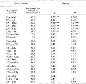
TABLE 2
Frequency of sterile males (J) and sex-ratio (SR) in offspring sublines from hybrid females introgessed with chromosomal segments that were never
recorded in their fertile sons
Hybrid females Offspring
Percentage size Cytological relative to
interval chromosome 2 b SR
X (whole) XC 1 e-H XA-D 1 d XA-D3c XA-C 1 a XF5c-H XE3b-E4h XA-A2a 2 (whole)
?A-G 1 e 2Clg-FJd
3B2f-H 3C4c-H 3C 1 b-GZa 3A-D3c
3D5a-GZa
4A-D3c 4D4e-G3a 4Bla-E I g
5 (whole)
5A-C4f 5C3b-FZh 86.8 64.3 39.0 34.1 20.9 18.2 5.0 3.9 100.0 56.6 79.1 76.0 56.6 52.7 46.1 34.1 34.1 32.1 29.8 88.7 39.5 34.1 0.73*** 0.40 0.05*** 0.10***
o.oo***
0.23*** 0.45 0.56 0.52 0.49 0.50 0.58 0.50 0.44 0.52 0.60 0.40 0.57 0.52 0.52 0.44 0.57 0.58* 0.78 0.27*** 0.36*** 0.45*** 0.74 0.41*** 0.93 1.08 1.12 0.87 0.85 1.15 1.17 0.82 0.85 1.00 0.92I .06
1.27 0.89
x 2 probability values (P) are as follows:
*
P < 0.05;**
P < 0.01;***
P <0.001. The remaining figures for& and SR are statistically nonsignificantly differ- ent from the expected values of 0.50 and 1.00, respectively.
been found in approximately one-half of the females of each offspring subline, as expected if the parental female is a segmental hybrid. A similar segregation in males should give one-half of the males with the segment. None of these males has been found among the fertiles, and consequently, they must be in the class of steriles. This indirect evidence of a relationship between these large autosomal segments and male hybrid sterility is also reinforced by several independent observations. First, frequencies of sterile males are not signifi- cantly different from 0.50 (Table 2), as expected in the backcross offspring of
a hybrid female for a dominant male-sterility factor. Second, the analysis of Malpighian polytene chromosomes (5-1 0 per subline) revealed that, within the limitations of this technique (see MATERIALS AND METHODS), the expected seg-
HYBRID STERILITY I N DROSOPHILA
TABLE 3
Critical sizes (%) relative to chromosome 2, showing the differential effect of the chromosomes in the determination of hybrid male
sterility
85 1
Size
Chromosome 3 Chromosome 4 Chromosome 5
Maximum for fertility 34.5 23.2 27.9
Minimum for sterility 34.1 29.8 34.1
gotes. As an example, in each of the sublines, 2Clg-F3d, 3D5a-G2a; 3Clb-G2a; 3C4c-H and 4A-D3c, more than 95% of the sterile males have always been found heterozygous for the electrophoretic markers (see
MATERIALS AND METHODS). These evidences, when combined, are sufficient to
substantiate the hypothesis that segments in Table 2 produce sterility when introgressed.
One feature that distinguishes autosomal segments producing sterile males from those found in fertiles is size. Tables 1 and 2 show also the sizes of the introgressed segments relative to the total length of chromosome 2 for com- parison among autosomes. Generally,
D.
serido segments producing male ste-rility are longer than those carried by fertile males. In Table 3 we have shown the longest segment that maintains fertility and the shortest segment that pro- duces sterility in chromosomes 3, 4 and 5. T h e size effect is chromosome- dependent and may suggest a nonuniform distribution of sterility factors among chromosomes. Chromosome 4 is the most effective in producing steril- ity because it shows the lowest minimum size (29.8) for sterility. This minimum is 34.1 for chromosomes 3 and 5 . In chromosome 3 there is an overlap be- tween the maximum size for fertility (34.5) and the minimum size for sterility (34.1). This may simply reflect that sterility factors are not evenly distributed in this chromosome. Thus, the longest tested segment (3A-C3a) producing fertility is situated in a different chromosomal region from the shortest tested segment (3D5a-GZa) producing sterility (Figure 4).
852 H. NAVEIRA AND A. FONTDEVILA
TABLE 4
Crosses between sublines introgressed with segments that do not produce sterility in hybrid males
Cross
Frequency of sterile
(1) x (2) sons
3D4c-Gle X 2B3e-BId 3A-Cla X 3F2a-H 3D4c-Gle X 5A-CId 3D4c-Gle X 3A-Cla 3D4c-Gle X 4F1b-H 3A-Cla X 3D5a-Flh 3D5a-FIh X 2B3e-Bld 4Bla-Dla X 4Flb-H 3D5a-Flh X 4Flb-H 3D5a-FI h X 3F2a-H 4Flb-H X 2B3e-Bld 4D2c-Flb X 4F3a-H SA-Cld X 2B3e-Bld 5A-A5d X 2B3e-BId 5A-A5d X 4Flb-H 5A-A5d X 3F2a-H 5A-A5d X 3D4c-Gle 4F4e-G4a X 4Bla-Dla 3CIb-D4e X 4Bla-Dla 3CIb-D4e X 3F2a-H 3F2a-H X 2B3e-Bld 3A-Cla X 4F4e-G4a 3A5c-CIc X 4C3a-H 4C3b-E3a X 5A-A5d
0.23 0.30 0.23 0.23 0.37 0.27 0.23 0.23 0.20 0.23 0.30 0.23 0.07 0.00 0.03 0.00 0.00 0.03 0.00 0.07 0.07 0.03 0.00 0.00
Frequency of fertile sons No. of (1/2) karyotypes
over Added size examined of the two sterile
(1) (2) (1/2) (0) segments males
0.20 0.30 0.20 0.30 0.30 0.20 0.23 0.27 0.30 0.16 0.33 0.20 0.37 0.17 0.33 0.20 0.27 0.20 0.13 0.40 0.30 0.20 0.30 0.20 0.20 0.30 0.20 0.33 0.27 0.23 0.20 0.30 0.27 0.23 0.23 0.27 0.17 0.30 0.23 0.23 0.27 0.23 0.30 0.17 0.17 0.37 0.30 0.20
0.00 0.27 0.00 0.27 0.00 0.27 0.00 0.27 0.00 0.17 0.00 0.20 0.00 0.23 0.00 0.24 0.00 0.33 0.00 0.24 0.00 0.20 0.00 0.27 0.20 0.23 0.20 0.27 0.23 0.24 0.30 0.20 0.20 0.30 0.13 0.34 0.17 0.36 0.20 0.34 0.17 0.33 0.23 0.27 0.20 0.26 0.27 0.23
0.480 0.539 0.566 0.593 0.538 0.492 0.379 0.402 0.437 0.457 0.406 0.395 0.434 0.286 0.344 0.364 0.418 0.255 0.360 0.442 0.426 0.372 0.248 0.314 5/5 5/5 5/5 5/5 415 5/5 515 5/5 515 5/5 5/5 5/5 212 111 212 2/2
01 1 ( 1 ) Segment contributed by the male; (2) segment contributed by the female; (112) both seg- ments simultaneously; (0) D. burratti. Sizes are relative to chromosome 2
70% of fertile males) to sterility (0%) is very sharp with a minimum increase in segment length.
HYBRID STERILITY IN DROSOPHILA 853 gressed sizes than d o crosses with segments of other autosomes. Some com- bined segments (i.e., SA-Cld X 2B3e-Bld; 3Clb-D4e X 3F2a-H; 3F2a-H X 2B3e-Bld) may be classified as semisteriles they produce some sterile hybrid males (0.07). However, the transition from fertility to sterility is not gradual, and it shows in general a large discontinuity in the frequency of fertile sons, ranging from the expected numbers (0.25) to zero.
These results not only confirm the size-dependent threshold effect of the D.
serido introgressed segments but also prove that two segments can cooperate in an additive way, no matter whether they are introgressed in the same (cis- action) or in different autosomes (truns-action), to produce sterility once their cumulative size exceeds a critical value.
Sex chromosome effects deserve an independent and close examination. Ta- ble 2 and Figure 4 show that any introgressed segment produced sterility, irrespective of its size and localization in the chromosome. Besides, introgres- sion of X chromosome segments is frequently associated with small sex ratios. This suggests that X segments may also be involved in determining the viability of males. Sublines XA-D 1 d, XA-D3c and XA-C 1 a are specially illuminat- ing in this respect (Table
2).
Their sex ratios are significantly smaller than one, which probably indicates a high mortality in males. Interestingly, most or all of the viable males are fertile (fs = 0.05, 0.10 and 0.00, respectively), but none of them carries the introgressed X segment or any of its recombinant products. This segment is still segregating, as witnessed by chromosomal anal- ysis of fertile hybrid females. T h e most plausible explanation for these data is that hybrid males for any of these X chromosome segments are inviable. In other cases, X chromosome segments are responsible for male sterility and male viability, as well, but the relationship between size and these male effects is not straightforward.DISCUSSION
Asynapsis is not only a characteristic of mitotic chromosomes but also of meiotic pairing (EVGENEV and POLIANSKA 1976), which apparently reduces the probability of two nonconspecific homologous chromosome segments being involved in crossover events. This property is beneficial for our purposes. Many different chromosome segments from D. serido in the D. buzzutii background were initially obtained, due to the high recombination frequency in F1 and first backcross hybrid females. Later, they were isolated and their effect on fertility was checked, taking advantage of the reduction of recombination fre- quency in segmental hybrids.
854 H . NAVEIRA AND A. FONTDEVILA
other authors has provided comparable information, or, at least, not so explic- itly. Nearly all of them have been performed at the chromosomal level, like ours, but their results are in terms of a few marked chromosome segments. Their power of resolution depends on the number of genetic markers avail- able, always very small. In our case, we are only limited by the frequency of recombination that allows us to obtain a large array of chromosomal segments marked by their somatic asynapsis. However, we are still unable to explain some important differences with other authors’ results.
Our results might be an example of the type I architecture described by
TEMPLETON (1 98 1). However, their relationship to earlier evidence provided by other authors on sterility in Drosophila interspecific hybrids (for a review, see EHRMAN 1962), is not straightforward. Several different kinds of interspe- cific F1 sterility have been found so far. T h e F1 may consist of (1) both sterile males and sterile females, as in the case of the hybrids between
D.
melanogasterand
D.
simulans, where sterility is produced by a small number of “polygenic sets” (MULLER and PONTECORVO 1940, 194 1 ; PONTECORVO I943b);(2)
sterile males and fertile females, as in our study; (3) both fertile males and fertile females, but where sterile males appear among the F2 and/or backcrosses dueto some special combinations of chromosomes of both species. There is still another kind of hybrid sterility (4) exemplified by
D.
paulistorum, where the sterility of backcross hybrid males between races, or incipient species, is due to the presence of microorganisms (EHRMAN 1960; SOMERSON et al. 1984).T h e second (2) and third (3) classes have been traditionally considered as equivalent, insofar as they deal with male-specific sterility factors. But they result most probably from different genetic determinants, or, at least, differ- entially organized. Thus, amongst the third class one frequently finds Y-auto- soma1 interactions as the main genetic determinants of hybrid sterility (STONE
1947; ALEXANDER, LEA and STONE 1952; HENNIC 1977; SCHAEFER 1978;
ZOUROS 1981), although in other cases the results are much more confusing and difficult to interpret (DOBZHANSKY 1974, 1975), perhaps corresponding to a more advanced stage of evolutionary divergence. In contrast, sterility of the class (2) seems to be brought about by a relatively high number of genes carried by all the chromosomes. This is so, for example, in the hybrids between
D.
pseudoobscura andD.
persimilis, where sterility is due to a minimum of eight loci (DOBZHANSKY 1936), and the effectiveness of a chromosome in producing sterility is generally proportional to its length (WU and BECKENBACH 1983), a result that agrees quite well with o u r findings (NAVEIRA, HAUSCHTECK-JUNGENand FONTDEVILA 1984). The same is t r u e for hybrids between
D.
mauritianaand
D.
simulans, where sterility is produced by at least five loci (one on each chromosome), although probably there are many more (COYNE 1984).HYBRID STERILITY IN DROSOPHILA 855
inant sterility only when accumulated in critical amounts; that is, when the introgressed segments exceed roughly a size equivalent to 25-3596 of the chromosome 2 length. Differences between chromosomes, or within a given chromosome, in this respect, may or may not reflect underlying differences in the distribution of these nonspecific factors. For example, it cannot be con- cluded that the number of these factors is greatest in chromosome 4 because the length of a chromosome is not closely related to the length of its DNA, and packing ratios of chromatin (euchromatin vs. heterochromatin) may also vary within the chromosome.
T h e results reported in this paper are concerned with a single type of in- trogression into D. buzzatii. However, reciprocal introgression experiments into
D . serido using different stocks, and introgression experiments designed to test the role of specific
D.
serido inversions in the determination of hybrid sterility, seem to produce essentially similar results (H. NAVEIRA and A. FONTDEVILA, unpublished results).The final version of this paper was written while A.F. was spending a sabbatical leave at the University of Georgia (UGA) in Athens sponsored by a personal grant from the US-Spain Joint Committee for Scientific and Technological Cooperation. We greatly acknowledge the helpful comments on this paper suggested by WYATT ANDERSON and JOHN MCDONALD from the Depart- ment of Genetics (UGA) and also by BRIAN CHARLESWORTH and another anonymous reviewer. This work has been funded by research grant 0910/81 awarded to A.F. by the Comisibn Asesora de Investigacibn Cientifica y Ticnica, Spain, and also by a research scholarship to H.N. from the Programa de Formacibn de Personal Investigador, Ministerio de Educacibn y Ciencia, Spain.
LITERATURE CITED
ALEXANDER, M. L., R. B. LEA, and W. S. STONE, 1952 Interspecific gene variability in the virilis group. Univ. Tex. Publ. 5204: 106-113.
BEERMAN, N., 1962
BERENDES, H. D., 1963
Riesenchromosomen. Protoplasmatologia, Vol. 6D. Springer-Verlag, W e n . Asynapsis in the salivary gland chromosomes of D . hydei. Genetica 33:
Genetic basis of male sterility in hybrids between two closely related species
Studies on hybrid sterility. 11. Localization of sterility factors in Droso-
The X-chromosome in the larval salivary glands of hybrids Drosophila
Genetic analysis of hybrid sterility within the species Drosophila pseu-
Analysis of reproductive isolation within a species of Drosophila. Proc.
288-300.
COYNE, J. A., 1984
DOBZHANSKY, TH., 1936
of Drosophila. Proc. Natl. Acad. Sci. USA 81: 4444-4447.
phila pseudoobscura hybrids. Genetics 21: 113-135.
insularis X Drosophila tropicalis. Chromosoma 8: 691-698.
doobscura. Hereditas 77: 81-88.
DOBZHANSKY, TH., 1957
DOBZHANSKY, TH., 1974
DOBZHANSKY, TH., 1975
Natl. Acad. Sci. USA 72: 3638-3641.
EHRMAN, L., 1960 The genetics of hybrid sterility in Drosophila paulistorum. Evolution 14: 212-
Hybrid sterility as an isolating mechanism in the genus Drosophila. Q. Rev.
The pattern of polytene chromosome conjugation and crossing-over in
223.
EHRMAN, L., 1962
Bioi. 37: 279-302.
EVGENEV, M. B., 1971
856
EVGENEV, M. B. AND G. G. POLIANSKA, 1976 The pattern of polytene chromosome synapsis in
Gene interactions in germ cell differentiation of Drosophila. Adv. Enzyme
H. NAVEIRA AND A. FONTDEVILA
Drosophila hybrids. Chromosoma 57: 285-295.
Regul. 15: 363-371.
demic to Hawaii. J. Exp. Zool. 175: 169-180. HENNIG, W., 1977
KAMBYSELLIS, M. P., 1970 Compatibility in insect tissue transplantations. 1. Ovarian species en-
KERKIS, J., 1936
KILIAS, G. and S. N. ALAHIOTIS, 1982
Chromosome conjugation in hybrids between D . melanogaster and D. simulans.
Am. Nat. 7 0 81-86.
Genetic studies on sexual isolation and hybrid sterility in
MULLER, H. J. and G. PONTECORVO, 1940 Recombinants between Drosophila species the F1 hy- brids of which are sterile. Nature 146: 199-200.
MULLER, H. J. and G. PONTECORVO, 1941 Recessive genes causing interspecific sterility and other disharmonies between D. melanogaster and D. simulans. Genetics 27: 157.
NAVEIRA, H., E. HAUSCHTECK-JUNGEN, and A. FONTDEVILA, 1984 The evolutionary history of D. buzzatii. VII. Spermiogenesis of inversion heterozygotes in backcross hybrids between Droso- phila buzzatii and Drosophila serido. Genetica 65: 205-214.
The evolutionary history of D. buzzatii. XI. A new method for cytogenetic localization based on asynapsis of polytene chromosomes in in- terspecific hybrids of Drosophila. Genetica. In press.
Analysis of asynapsis in salivary chromosomes of larvae from different strains of Drosophila funebris. Acta Zoologica 36: 101-1 12.
Viability interactions between chromosomes of Drosophila melanogaster
and Drosophila simulans. Genetics 45: 51-66.
Hybrid sterility in artificially produced recombinants between Drosophila
Starch gel electrophoresis in a discontinuous system of buffers. Nature 180:
Origin of reproductive isolation in the absence of apparent genic differentia-
Study on the somatic pairing of polytene chromosomes. Chromo-
The evolutionary biology of D. buzzatii.
111. Cytogenetic relationships between two sibling species of the buzzatii cluster. Genetics 101:
long-term cage populations of D. melanogaster. Evolution 36: 121-1 31.
NAVEIRA, H., C. PLA, and A. FONTDEVILA, 1986
PERJE, A. M., 1955
PONTECORVO, G., 1943a
PONTECORVO, G., 1943b
melanogaster and Drosophila simulans. Proc. R. Soc. Edinb. Sect. B. 61: 385-397. POULIK, M. D., 1957
1477-1479.
tion in a geographic isolate of Drosophila pseudoobscura. Genetics 72: 143-155. RIEDE, I. and M. RENZ, 1983
soma 88: 116-123. PRAKASH, S., 1972
RUIZ, A., A. FONTDEVILA, and M. WASSERMAN, 1982
503-5 18. SCHAEFER, U., 1978
SOMERSON, N. L., L. EHRMAN, J. P. KOCKA, and F. J. GOTTLIEB, 1984
Sterility in Drosophila hydei X D . neohydei hybrids. Genetica 4 9 205-214. Streptococcal L-forms isolated from Drosophila paulistorum semispecies cause sterility in male progeny. Proc. Natl. Acad. Sci. USA 81: 282-285.
The genetic basis of differences between two species of Drosophila. Am.
SPIETH, H. T., 1949 Sexual behavior and isolation in Drosophila. 11. The interspecific mating
STONE, W. S., 1947 TEMPLETON, A. R., 1981
Ecol. Syst. 12: 23-48. SPENCER, W. P., 1944
Nat. 78: 183-188.
behavior of species of the willistoni group. Evolution 3: 67-81.
HYBRID STERILITY I N DROSOPHILA 857
VAL, R. C., 1977
WEISBROT, D. R., 1963
WHARTON, L. T., 1942
Wu, C. 1. and A. T. BECKENBACH, 1983
Genetic analysis of the morphological differences between two infertile species
Studies on differences in the genetic architecture of related species of
Analysis of the repleta group of Drosophila. Univ. Tex. Publ. 4228 23-
Evidence for extensive genetic differentiation between the sex-ratio and the standard arrangement of Drosophila pseudoobscura and D . persimilis and identification of hybrid sterility factors. Genetics 105 7 1-86.
An autosome-Y chromosome combination that causes sterility in D. mojauensis
X D . arizonensis hybrids. Drosophila Inform. Serv. 5 6 167-168.
Communicating editor: J. R. POWELL
of Hawaiian Drosophila. Evolution 31: 61 1-629.
Drosophila. Genetics 4 8 1121-1 139.
52.