Dissertation zur Erlangung des Doktorgrades
der Fakultät für Chemie und Pharmazie
der Ludwig-Maximilians-Universität München
Optimization of shielding and targeting domains within
sequence-defined, cationic carriers for pDNA delivery
Stephan Wolfgang Morys
aus Forchheim, Deutschland
Erklärung
Diese Dissertation wurde im Sinne von § 7 der Promotionsordnung vom 28. November 2011 von Herrn Prof. Dr. Ernst Wagner betreut.
Eidesstattliche Versicherung
Diese Dissertation wurde eigenständig und ohne unerlaubte Hilfe erarbeitet.
München, 09.11.2017
...………
Stephan Morys
Dissertation eingereicht am: 09.11.2017
1. Gutachter: Prof. Dr. Ernst Wagner
2. Gutachter: Prof. Dr. Wolfgang Frieß
„Science, my lad, is made up of mistakes, but they are mistakes which it is useful to make, because they lead little by little to the truth.”
Table of contents
1
Introduction ... 11
1.1 Non-viral gene therapy ... 11
1.1.1 The delivery pathway of non-viral nucleic acid carriers ... 13
1.1.2 Cationic carriers: From polydisperse polymers towards sequence-defined oligomers ... 18
1.1.2.1 SPS as a method to develop sequence-defined cationic vectors for structure activity relationships ... 19
1.1.2.2 Tailoring topologies and functionalizing oligomers to improve nucleic acid delivery ... 21
1.1.2.2.1 Polyplex shielding ... 25
1.1.2.2.2 Approaches of pre-PEGylation, post-PEGylation ... 26
1.1.2.3 Receptor targeting ... 27
1.1.2.3.1 HGFR targeting ... 28
1.1.2.3.2 EGFR targeting ... 28
1.2 Aim of the thesis ... 30
2
Materials and Methods ... 32
2.1 Materials ... 32
2.1.1 Equipment for solid-phase synthesis ... 34
2.1.2 Plasmid DNA ... 34
2.1.3 Cell culture ... 35
2.2 Methods ... 36
2.2.1.1 General procedure for solid phase synthesis (SPS) ... 36
2.2.1.1.1 Loading of a 2-chlorotrityl chloride resin with an Fmoc protected amino acid ... 36
2.2.1.1.2 Procedure of a manually conducted solid phase synthesis ... 36
2.2.1.1.3 Kaiser test ... 38
2.2.1.1.4 Procedure of an automated solid phase synthesis ... 38
2.2.1.2 Cleavage of oligomers and reagents ... 39
2.2.1.2.1 General cleavage of oligomers ... 39
2.2.1.2.2 Cleavage of oligomers containing oleic acid ... 40
2.2.1.2.3 Cleavage of NPys containing PEGylation reagents ... 40
2.2.1.3 Synthesis of oligomers ... 40
2.2.1.3.1 Synthesis of untargeted, PEGylated 2-arm oligomers... 40
2.2.1.3.2 Synthesis of untargeted 2-arm oligomers containing Pro-Ala-Ser repeats ... 41
2.2.1.3.3 Synthesis of untargeted 3-arm oligomer ... 42
2.2.1.3.4 Synthesis of cmb containing two and 3-arm oligomers ... 42
2.2.1.3.5 Synthesis of PEGylated 2-arm oligomers with GE11 ligand and alanine ... 43
2.2.1.3.6 Synthesis of the T-shaped lipo-oligomer for post-modification ... 44
2.2.1.3.7 Synthesis of improved T-shaped lipo-oligomers ... 44
2.2.1.4 Synthesis of PEGylation reagents for polyplex post-modification ... 45
2.2.1.4.1 Synthesis of monovalent PEGylation reagents. ... 46
2.2.1.4.2 Synthesis of bivalent PEGylation reagents ... 46
2.2.2.1 Post-modification with PEGylation reagents ... 47
2.2.3 pDNA binding assays ... 47
2.2.4 Particle size and zeta potential ... 47
2.2.5 Transmission electron microscopy (TEM) of polyplexes ... 48
2.2.6 Ethidium bromide compaction assay and polyanionic stress test ... 48
2.2.7 Stability of polyplexes in serum and media ... 49
2.2.8 Polyplex stability in the presence of salt ... 49
2.2.9 Polyplex adhesion to erythrocytes or serum ... 49
2.2.10 UV spectrometrical investigation of polyplex modification ... 50
2.2.11 Ellman’s assay of oligomers ... 50
2.2.12 Ellman´s assay of polyplexes ... 50
2.2.13 Release of 3-nitro-2-thiopyridone ... 51
2.2.14 EGF and HGF receptor measurement ... 51
2.2.15 In vitro pCMVLuc gene transfer and metabolic activity of transfected cells (MTT assay) ... 52
2.2.16 In vitro pCMVLuc gene transfer and metabolic activity of transfected cells (MTT assay) with addition of endosomolytic chloroquine or LPEI ... 53
2.2.17 Cellular association of pDNA polyplexes ... 53
2.2.18 Cellular internalization of pDNA polyplexes ... 54
2.2.19 In vivo gene transfer ... 54
2.2.20 Iodide uptake activity after hNIS gene delivery ... 55
2.2.21 MALDI-TOF mass spectrometry ... 56
2.2.22 Proton NMR spectroscopy... 56
2.2.24 ESI mass spectrometry ... 57
2.2.25 Statistical analysis ... 57
3
Results ... 58
3.1 Influence of defined hydrophilic blocks within oligoaminoamide copolymers: compaction versus shielding of pDNA nanoparticles ... 58
3.1.1 Peptide and oligomer synthesis ... 59
3.1.2 Physicochemical polyplex characterization ... 60
3.1.3 Steric shielding ... 63
3.1.4 DNA compaction ... 65
3.1.5 Serum stability ... 67
3.1.6 Tumor cell interactions in vitro ... 69
3.1.7 Tumor cell interactions in vitro without and with targeting ... 70
3.1.8 Tumor cell interactions in vivo without and with targeting ... 73
3.2 EGFR targeting and shielding of pDNA lipopolyplexes via bivalent attachment of a sequence-defined PEG agent ... 75
3.2.1 pDNA nanoparticle design, peptide and oligomer syntheses ... 76
3.2.2 Physicochemical polyplex characterization ... 81
3.2.3 Luciferase gene transfections... 90
3.2.4 Cellular binding and internalization of bivalent post-PEGylated polyplexes ... 96
3.2.5 Iodide uptake activity after hNIS gene delivery ... 100
3.3 Lipo-oligomers optimized towards enhanced lipopolyplex stability ... 102
3.3.1 Library design and oligomer synthesis ... 102
3.3.2.1 Size and zeta potential of unmodified as well as post-modified
lipopolyplexes ... 105
3.3.2.2 pDNA compaction in buffer and after polyanionic stress ... 109
3.3.2.3 Steric stability of unmodified as well as PEGylated lipopolyplexes under physiological salt conditions ... 111
3.3.2.4 Serum stability of unmodified as well as post-modified lipopolyplexes ... 113
3.3.3 Luciferase gene transfections... 115
3.3.4 Ellman’s assay to determine free thiols for polyplex post-modification 117 3.3.5 Cellular polyplex uptake ... 119
3.3.6 Gene transfer after enhanced endosomal escape ... 121
4
Discussion ... 124
4.1 Influence of defined hydrophilic blocks within oligoaminoamide copolymers: compaction versus shielding of pDNA nanoparticles ... 124
4.2 EGFR targeting and shielding of pDNA lipopolyplexes via bivalent attachment of a sequence-defined PEG agent ... 127
4.3 Lipo-oligomers optimized towards enhanced lipopolyplex stability ... 130
5
Summary ... 136
6
Appendix ... 138
6.1 Abbreviations ... 138
6.2 Serum stability of optimized T-shapes determined by DLS ... 141
6.3 Summary of SPS derived oligomers ... 144
6.4 Summary of SPS derived shielding reagents ... 144
6.5 Analytical data ... 145
6.5.2 MALDI-TOF MS of the targeting peptides cmb and GE11 ... 146
6.5.3 1H NMR spectra of oligomers ... 147
6.5.4 RP-HPLC of oligomers ... 161
6.5.5 Mass spectra of oligomers... 168
6.5.5.1 Full mass spectra of oligomers ... 169
6.5.6 Mass spectra of shielding reagents ... 176
6.5.6.1 Full mass spectra of shielding reagents ... 176
6.5.7 RP-HPLC of shielding reagents ... 178
7
References ... 181
8
Publications ... 196
1
Introduction
This chapter should give a brief introduction into the research field of bioreducible
polycationic carriers for nucleic acid delivery.
1.1 Non-viral gene therapy
This chapter gives a brief introduction into chemically designed, artificial vectors for
nucleic acid delivery. It does not aim at giving a full review of the advances in polymer-based gene therapy since its invention in the 1960’s. This, as well as an appropriate
review of advances in viral gene therapy and a comparison of both would exceed the
intended introduction of this Ph.D. thesis.
However, Lächelt and Wagner [1] as well as Herzog and colleagues [2] reviewed
recent advances in detail.
So far, genetic disorders like mucoviscidosis [3], severe combined immunodeficiency (SCID) [4], haemophilia [5], β-thalassemia [6], as well as adrenoleukodystrophy (ALD) [7], metachromatic leukodystrophy [8], aromatic L-amino acid decarboxylase (AADC) deficiency [2] among others [9-11] have been tackled by classical, viral gene therapy. Thereby, classical gene therapy addresses these diseases by inserting functional DNA into the human genome in order to replace defect gene sections.
However, it took until 2012 until the first therapeutic product, Glybera, was approved by the European Medicines Agency (EMA) for the treatment of lipoprotein lipase deficiency (LPLD). Facing high therapy costs, the company will not extend the admission of Glybera® after its ended in October 2017. However, in the meantime Strimvelis®, for the treatment of adenosine deaminase (ADA)‐deficient severe combined immunodeficiency (SCID), was approved on the european market [12].
1.1.1 The delivery pathway of non-viral nucleic acid carriers
Non-viral nucleic acid carriers face several obstacles prior to an efficient intracellular delivery. These barriers are schematically illustrated (cf. Scheme 1) to exhibit the very complex delivery pathway of polymer-based systems. The following chapter addresses the critical steps of nucleic acid complexation (1), cellular binding and uptake (2), endosomal escape (3), cargo release and intracellular trafficking (4) towards the compartment of further processing.
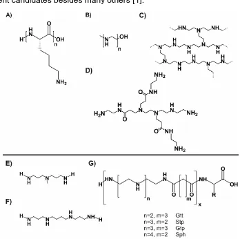
A large size, anionic charge, as well as sensitivity towards degradation by nucleases are rather inefficient properties for the delivery of naked nucleic acids to their target location [15, 16], however, it rarely is possible [17, 18]. To overcome these issues, chemically engineered cationic polymers along with cationic lipids [19-21] were developed. By complexing nucleic acid, they were intended to shade the cargo in the extracellular environment and prevent them from degradation, but also facilitate the nucleic acid to be transported into the cytoplasm [22]. Within cationic polymers, polylysine (pLL) [23, 24], which was first evaluated clinically as a delivery vehicle for pDNA [25], the branched [26-29] and linear [30-33] versions of polyethyleneimine (PEI) as well as dendritic polyamidoamine (PAMAM) [34-36] (cf. Figure 1A-D) represent the most prominent candidates besides many others [1].
These cationic carriers, all comprise basic amines, are partially protonated at neutral pH and are thereby able to bind and compact negatively charged nucleic acids via electrostatic interaction. This complexation is entropy driven and leads to the formation of nano-sized complexes, so-called “polyplexes” [37, 38].
These polyplexes need to exhibit a certain size as well as stability for the successful delivery. Nanoparticles with a size of 5.5 nm or below suffer from rapid clearance by the kidney [39], while particles with a size between 20 and 400 nm can penetrate into highly vascularized solid tumors as a result of the enhanced permeability and retention (EPR) effect due to passage through leaky vessels of the tumor tissue [40-42]. However, the extent of passive tumor accumulation via EPR effect is strongly dependent on the size-threshold of the porous tumor vasculature which varies within different types of cancer [43-45].
As already mentioned before, also stability within the biological environment represents a crucial property for polyplex delivery. Here the positive surface charge of unshielded polyplexes can mediate interaction with proteins and electrolytes, causing polyplex dissociation or severe aggregation due to counterion exchange. This stability issue can be addressed by increased cationic charge or by the introduction of crosslinking domains (e.g. terminal cysteines) via formation of bio-reducible disulfide bonds [46-50], or the introduction of hydrophobic elements [51-53].
[55, 57-67]. As polyplex targeting represents a crucial part of this Ph.D. work, the two key targets (EGFR and HGFR) are described in more detail later on (cf.1.1.2.3).
Polyplex shielding, often impaired by the introduction of a targeting domain, represents another issue to be addressed as polyplexes with a positive surface charge can undergo aggregation or dissociation with electrolytes or proteins within the bloodstream. In addition, a positive surface charge can lead to the activation of the immune system. However, as polyplex shielding presents a key topic of this thesis, it is elucidated in more detail later on (cf. 1.1.2.2.1).
After having reached the cytoplasm, the delivered polyplexes now have to release the cargo. For this reason, particle stability needs to be well balanced between sufficient extracellular stability and fast nucleic acid release at the target site. Here, previously mentioned disulfide bonds can be reduced by cytosolic GSH, releasing the cargo and the single polymers, thereby reducing cytotoxicity due to an increased biodegradability of smaller units.
1.1.2 Cationic carriers: From polydisperse polymers towards
sequence-defined oligomers
Artificial vectors as potent nucleic acid vehicles need to comprise different functionalities to be bioresponsive.
Previously, cationic vectors like pLL, PEI, PAMAM were generated by different kinds of polymerization techniques and resulted in polydisperse polymers. With improved chemistries, such as controlled radical polymer synthesis or specific ligation strategies, products with decreased polydispersity and more highly controlled architecture of carries were obtained [89-94].
Further development of cationic delivery systems requires clear-cut structure-activity relationships to be drawn. Therefore, a technique to obtain polymers with a precisely defined sequence is needed. A series of researchers have applied the well-established method of solid-phase assisted synthesis (SPS) to develop linear [84, 85, 95-103] and branched [75, 104-109] peptide-based as well as lipid-based [110-113] nucleic acid carriers. Recently, also artificial amino acids have been assembled to sequence-defined oligomers as shuttles for pDNA and siRNA (cf. Figure 1E-G) [49, 51, 64, 113-122].
1.1.2.1 SPS as a method to develop sequence-defined cationic vectors for structure activity relationships
This chapter is partly based on:
Krhac Levacic A., Morys, S., Wagner E. Solid-phase Supported Design of Carriers for Therapeutic Nucleic Acid Delivery. Bioscience Reports 2017, 37 (5).
achieved by introduction of novel protecting groups [129].
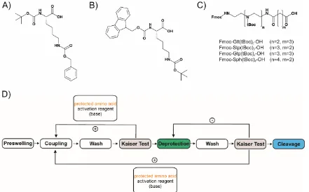
A schematical cycle of SPS is given in Figure 2D pointing out the different, repetitive steps to obtain a fully deprotected peptide at the end of the synthesis.
Besides the previously mentioned, mostly lysine-based, oligomers [95, 96, 98, 100, 101, 104] the Fmoc peptide SPS strategy has been adopted for the synthesis of sequence-defined oligo(ethylenamino)amides (Figure 2C). Instead of natural amino acids, artificial oligoamino acids such as Stp (succinyl tetraethylene pentamine), Gtp (glutaroyl-tetraethylene pentamine) or Sph (succinyl pentaethylene hexamine) in Fmoc, tBoc-protected forms [49, 131, 132] can be used for manual as well as automated SPS, the latter requiring a peptide synthesizer [133]. These building blocks introduced the diaminoethane motif of LPEI, a well-established nucleic acid binding and endosomal buffering domain, for solid phase synthesis.
1.1.2.2 Tailoring topologies and functionalizing oligomers to improve nucleic acid delivery
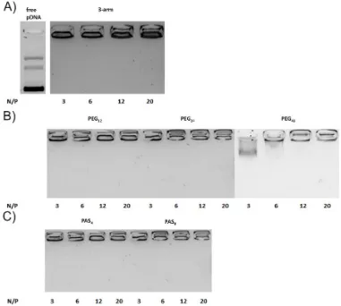
With these artificial amino acids, a library of more than 1100 oligomers has been established and the oligomers have been tested for different nucleic acids (pDNA, siRNA, miRNA, mRNA) to evaluate the best suitable carriers. The choice of the artificial amino acids mentioned above, significantly influenced the nucleic acid binding and endosomal buffer ability of the first oligomers generated. Different topologies, including linear [134] as well as branched [49] structures, incorporating different artificial cationic building blocks were generated and evaluated [50, 132]. However, Stp (Fmoc-Stp(boc)3-OH) was introduced into most of the later mentioned oligomers, as it can be obtained by a highly reproducible synthesis with good yield, nevertheless providing the required key features for a successful gene delivery. To achieve a more sophisticated multifunctionality, different topologies with the diamino acid lysine as a branching point were developed (cf. Figure 3).
Structures consisting of three Stp enriched cationic arms (3-arm) [49, 64, 133], as well as oligomers with PEG of a defined length instead of a third cationic arm (PEGylated 2-arm) were investigated (cf Figure 3A,B). The latter topology facilitated the introduction of a targeting domain like folic acid [135-137], as well as peptidic ligands like cmb, targeting the HGFR [64] or the GE11 peptide [138], targeting EGFR as well as other peptidic ligands [139, 140].
histidines and cationic building blocks thereby led to a significantly improved buffer capacity at endosomal pH. Increased buffering enhanced cellular electrolyte influx, finally leading to endosomal burst due to osmotic swelling. In the 1990’s Behr et al. already pointed out that this effect, also known as the proton sponge effect, importantly contributes to LPEI’s intracellular performance [71]. Similar findings were made with highly branched HK rich peptides by Mixson et al. [105, 106], demonstrating that the proton sponge effect is also transferrable to sequence-defined vehicles, finally resulting in improved transduction efficacy in vitro as well as in vivo.
Figure 3 Common topologies of oligomers generated by SPS. A) histidine-rich 3-arm, B) histidine-rich shielded 2-arm, C) histidine-rich 4-arm. D)-F) represent fatty acid containing i-shape, T-shape and U-shapes, respectively. C represents cysteine, H histidine, K lysine and Y tyrosine. HD represents hydrophobic domains like aliphatic fatty acids or cholanic acid. TD represents a targeting domain such as peptides or small molecules targeting receptors overexpressed on tumor cell surfaces. SD represents shielding domains like PEG or (Pro-Ala-Ser) repeats. BD represents the cationic binding domain, in sequence-defined oligomers; Gtt glutaryl-triethylene tetramine, Stp succinyl tetraethylene pentamine, Gtp glutaryl-tetraethylene pentamine and Sph succinyl pentaethylene hexamine could be introduced, however, Stp was introduced mostly.
These findings then were transferred to the previously mentioned PEGylated 2-arm topology, resulting in significantly improved in vitro performance, not requiring the addition of the endosmolytic reagent chloroquine for successful gene delivery in vitro
3-arm oligomer these polyplexes even mediated sufficient stability for HGFR directed pDNA delivery in vivo [64, 65].
Within time, the existing histidine-rich 4-arm topology was further improved by the introduction of additional lysines between the cationic building blocks and the histidines, contributing to enhanced nucleic acid binding with its free - amine. This resulted in increased pDNA compaction and improved gene delivery in vitro as well as
in vivo [142].
Besides the artificial amino acids and lysine all the above-mentioned topologies contained terminal cysteines. These were proven, within the evaluation of the first oligomers generated by SPS, to mediate significantly improved polyplex stability, resulting in enhanced nucleic acid delivery. An explanation for this is given due to the crosslinking abilities of cysteines by forming bioreducible disulfide bonds. Thereby, larger cationic chains exhibiting LPEI-like properties were generated within the polyplex [134]. The importance of disulfide formation and its characteristics is summarized in a review by Klein and Wagner [46], pointing out, that the general toxicity of LPEI [143] could be overcome by the assembly of shorter cationic oligomers to potent nucleic acid shuttles via disulfide crosslinking. Previous investigations on SPS derived oligolysine previously came to similar conclusions [95].
oligomer. In a series of experiments, it turned out, that also here, the combination of centrally placed fatty acids and terminal cysteines worked best in combination with peripheral tyrosine trimers. Nucleic acid delivery and polyplex stability could be further improved by increased hydrophobicity as well as - stacking between tyrosines of neighbored oligomers, resulting in extended polyplex circulation time and siRNA delivery in vivo [52]. The latter could be justified by an additional effect on endosomal buffering by the newly introduced tyrosines [52]. Consequently, tyrosine trimers were also incorporated into PEGylated 2-arm oligomers [137, 141], suffering from reduced polyplex stability mediated by PEGylation [133].
Recently, the introduction of cholanic acid into T-shaped oligomers mediated notable gene knockdown after siRNA delivery in vitro as well as in vivo, while not exhibiting lytic activity [111]. These oligomers also comprised a bioreducible disulfide building block to destabilize the polyplex after its uptake via GSH mediated endosomal cleavage of the central hydrophobic domain. This again resulted in enhanced siRNA release from the endosome and increased gene knockdown.
1.1.2.2.1 Polyplex shielding
During nucleic acid delivery, an excess of the positively charged carrier is usually required for nucleic acid complexation. By mixing nucleic acid and cationic carrier, nanoparticles with positive surface potential are generated.
This positive charge offers advantages for the formed polyplexes, as it facilitates binding to negatively charged cell surfaces [145, 146] and contributes to endosomal escape after cellular uptake [147, 148].
Putting these positively charged nanoparticles into living organisms, these cationic carriers may mediate undesired interactions in the extracellular space. Positively charged polyplexes might lead to activation of the complement system, blood cells or other blood components [143, 149-151].
Polyplex surface shielding by introduction of a hydrophilic shielding domain into these artificial vectors has shown to reduce these interactions. Polyethylene glycol (PEG), with its hydrophilic properties, resulting in good solubility, is the most prominent and well-established shielding agent [152]. It has been successfully used for shielding of polyplexes in numerous instances, including solid-phase derived oligomers [59, 149, 153-158].
Due to reduced extracellular interaction of the polyplex, circulation time within the blood and biodistribution to the target tissue may greatly improve [152, 153]. However, its non-biodegradability, as well as recently reported formation of anti-PEG antibodies, gave reasons for the investigation of alternatives. Therefore, besides PEG, also poly(N-(2-hydroxypropyl)methacrylamide) (pHPMA) [142, 159, 160], hydroxyethyl starch (HES) [161] and polysarcosine [162] have been investigated as alternative hydrophilic shielding agents for polyplex shielding. Within this Ph.D. work, also a peptidic sequence composed of Pro-Ala-Ser (PAS) repeats has been examined [133] (cf. Figure 4).
Figure 4 Chemical structures of selected reagents used for shielding. A) poly(N-(2-hydroxypropyl)methacrylamide) (HPMA), B) polyethylene glycol (PEG), C) polysarcosine, D) repetitive PAS blocks (Pro-Ala-Ser)
To overcome the disadvantages of PEGylation, several approaches of implementing PEG directly during synthesis of the cationic oligomers (referred to as pre-PEGylation) as well as after formation of PEG- free polyplexes (referred to as post-PEGylation), were investigated.
1.1.2.2.2 Approaches of pre-PEGylation, post-PEGylation
shielding domain on the solid support, requires careful consideration of the right chain length of PEG.
In another approach, to avoid difficulties with nucleic acid compaction, non-PEGylated polyplexes were first formed and then PEG was introduced by receptor targeted PEGylation reagents via different chemical strategies. These approaches can be divided into thiol-dependent bioreducible and acid labile routes.
Post-modification of cationic vehicles with PEG via thiol chemistry was first investigated by Blessing et al. on LPEI [168]. The herein used thiol-maleimide chemistry was transferred to T-shaped oligomers containing terminal cysteines. By addition of folate [169], transferrin [170], or EGF [171] receptor targeted PEGylation reagents, polyplexes were successfully shielded and mediated tumor-specific siRNA delivery in vitro as well as in vivo.
Introducing acid labile hydrazone linked PEG onto cationic LPEI polyplexes via NHS chemistry was previously investigated by Fella et al. [156], also other pre-PEGylation of OEI polymers with acid labile PEG derivates depicted the improved endosomal release of polyplexes PEGylated with acid labile reagents [172-174].
The acid labile post-modification approach has recently been transferred onto pDNA polyplexes, composed of sequence-defined oligomers, via an acid labile AzMMan linker [142]. Here, post-modification with PEG and HPMA significantly improved polyplex stability with a fully recovered transfection efficacy in vitro as well as in vivo
after cleavage in the acidic tumor environment.
1.1.2.3 Receptor targeting
previously mentioned PEG-Dilemma [183, 184]. In the following subchapters the two growth factor (EGF and HGF) receptors, targeted within different projects of this thesis are depicted in more detail.
1.1.2.3.1 HGFR targeting
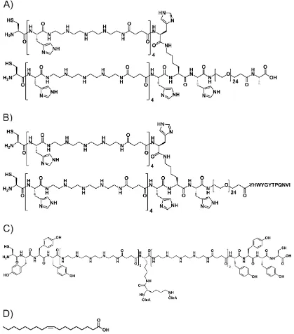
The receptor tyrosine kinase HGFR/cMet is over-expressed in epithelial-derived tumors as well as in stromal and interstitial cell-derived tumors such as sarcomas [185]. When the natural ligand hepatocyte growth factor (HGF) binds to its receptor, cMet, it thereby stimulates cell motility and migration, triggers mitogenesis and morphogenesis and thus promotes oncogenesis and tumor progression. Therefore, cMet signaling has been addressed by different cancer treatment approaches: (1) Antagonists preventing binding of HGF to its receptor, (2) cytosolic active tyrosine kinase inhibitors (TKI) as well as (3) inhibitors of the downstream cascade after receptor activation have been investigated [186]. Therapeutic drug-antibody conjugates have been applied to target cMet over-expressing cancer tissues [187]. Also, in vivo imaging by application of HGFR specific antibodies [188-190] or two phage display-derived peptides is mentioned [181, 191]. Recently, in our laboratory, one of these peptides (KSLSRHDHIHHH) was introduced into PEGylated 2- and 4-arm oligomers, mediating HGFR specific cellular polyplex uptake as well as in vivo gene delivery of pCMV-Luc [64] and pCpG-hCMV-NIS [65]. The latter coding for the human NIS (Natrium Iodide Symporter) facilitated application of therapeutic 131I, leading to significantly reduced tumor growth and longed survival rate in Huh7 tumor-bearing mice.
1.1.2.3.2 EGFR targeting
The overexpression of epidermal growth factor receptor (EGFR) offers another well-established targeting strategy for the specific delivery of polyplexes [61-63, 68, 138, 192].
its tyrosine kinase activity, as well as various monoclonal antibodies [180] (e.g. cetuximab and panitumumab) inhibiting ligand binding leading to enhanced receptor internalization and thereby promoted cytotoxicity [195]. EGFR represents an interesting target for the directed delivery of polyplexes. For polyplex targeting, murine EGF was successfully applied for LPEI polyplexes [55, 168, 196, 197]. Regarding the ratio of cation to targeting ligand, a short peptide offers advantages when used for oligomer targeting. Li et al. reported a phage display-derived peptide (GE11) exhibiting efficient and specific binding to the EGFR [198]. The hydrophobic GE11 peptide with a sequence of YHWYGYTPQNVI revealed less mitogenic activity compared to EGF, the original substrate of EGFR. GE11 as a targeting ligand for PEGylated LPEI conjugates has been widely explored [61, 62, 68, 192] without inducing receptor activation [62]. Mickler et al. investigated uptake mechanisms for EGF-PEG-PEI/pDNA and GE11-PEG-PEI/pDNA polyplexes, concluding that GE11 mediated uptake happens via more time consuming clathrin-mediated endocytosis, while EGF-PEG-PEI polyplexes are promptly taken up by EGFR activated endocytosis. However, the delayed, GE11 dependent uptake turned out to be as efficient as the faster EGFR activating pathway [68].
1.2 Aim of the thesis
The recent development of a solid-phase synthesis platform for the assembly of sequence-defined oligo(ethanamino)amides enables quick and easy synthesis of cationic oligomers complexing and delivering nucleic acids [131]. By introducing artificial amino acids [131], based on the diaminoethane motif of PEI which is well known for its nucleic acid binding and endosomal buffering abilities [26], differently shaped oligomers were synthesized. Into these oligomers, different functionalities, for shielding, polyplex stabilization and targeting can be introduced via SPS.
As the first aim of this thesis, the effect of different PEG lengths within 2-arm oligomers on biophysical properties and their biological performance in vitro as well as in vivo
were to be investigated, incorporating pDNA as cargo. Also, an alternative to PEG, a more hydrophilic shielding motif sequentially comprising the natural amino acids proline-alanine-serine (PAS) was to be examined. 2-arm oligomers containing four and eight repetitions as shielding domain were to be compared in parallel to the PEGylated 2-arm oligomers equipped with 12, 24 or 48 EO units. The oligomers generated by SPS for this study should contain histidines for improved endosomal buffering [50], and cysteines for bioreducible crosslinking via intermolecular disulfide formation. Also, a peptide (cmb), mediating HGF dependent polyplex uptake, was to be introduced in a set of oligomers.
2
Materials and Methods
2.1 Materials
The solvents, reagents and buffers used for the experiments are summarized in Table 1, Table 2 and Table 3.
Table 1 Solvents used for experimental procedures
Solvent CAS-No. Supplier
Acetonitrile1,11 75-05-8 VWR Int. (Darmstadt, Germany)
Chloroform2,11 67-66-3 VWR Int. (Darmstadt, Germany)
Chloroform-d3,11 865-49-6 Euriso-Top (Saint-Aubin Cedex, France)
Deuterium oxide3 7789-20-0 Euriso-Top (Saint-Aubin Cedex, France)
Dichloromethane4 75-09-2 Bernd Kraft (Duisburg, Germany)
N,N-Dimethylformamide5 68-12-2 Iris Biotech (Marktredewitz, Germany)
Ethanol absolute4,11 64-17-5 VWR Int. (Darmstadt, Germany)
Ethyl acetate7,11 141-78-6 Staub & Co. (Nürnberg, Germany)
n-Heptane8,11 142-82-5 Grüssing (Filsum, Germany)
n-Hexane8 110-54-3 Brenntag (Mülheim/Ruhr, Germany)
Methanol4 67-56-1 Fisher Scientific (Schwerte, Germany)
Methyl-tert-butyl ether9 1634-04-4 Brenntag (Mülheim/Ruhr, Germany)
N-Methyl-2-pyrrolidone5 872-50-4 Iris Biotech (Marktredewitz, Germany)
Tetrahydrofuran4 109-99-9 Fisher Scientific (Schwerte, Germany)
Water10 7732-18-5 In-house purification
1 HPLC grade; 2 DAB grade, distilled before use; 3 NMR grade (> 99.9 %); 4 analytical grade; 5 peptide grade; 6 BioReagent grade (> 99.9 %); 7 purum, distilled before use; 8 purissimum; 9 synthesis grade; 10 purified, deionized; 11only used within synthesis of Fmoc-Stp(boc)
3-OH
Table 2 Reagents used for experimental procedures
Reagent CAS-No. Supplier
1-Hydroxybenzotriazole hydrate 123333-53-9 Sigma-Aldrich (Munich, Germany) 2-Chlorotritylchloride resin 42074-68-0 Iris Biotech (Marktredewitz, Germany) 5,5′-Dithiobis(2-nitrobenzoic acid) 69-78-3 Sigma-Aldrich (Munich, Germany) 5β-Cholanic acid 546-18-9 Sigma-Aldrich (Munich, Germany) Agarose NEEO Ultra 9012-36-6 Carl Roth (Karlsruhe, Germany) Boc-L-Cys(NPys)-OH 76880-29-0 Bachem (Bubendorf, Switzerland) Boc-L-Cys(Trt)-OH 21947-98-8 Iris Biotech (Marktredewitz, Germany) Bromophenol blue 115-39-9 Sigma-Aldrich (Munich, Germany) Chloroquine diphosphate 50-63-5 Sigma-Aldrich (Munich, Germany)
4′,6-Diamidin-2-phenylindol (DAPI) 28718-90-3 Sigma-Aldrich (Munich, Germany)
Reagent CAS-No. Supplier
Ethidium bromide 1239-45-8 Sigma-Aldrich (Munich, Germany) Fmoc-L-Asn(Trt)-OH 132388-59-1 Iris Biotech (Marktredewitz, Germany) Fmoc-L-Arg(Pbf)-OH 154445-77-9 Iris Biotech (Marktredewitz, Germany) Fmoc-L-Cys(Trt)-OH 103213-32-7 Iris Biotech (Marktredewitz, Germany) Fmoc-L-Gln(Trt)-OH 132327-80-1 Iris Biotech (Marktredewitz, Germany) Fmoc-L-Gly-OH 29022-11-5 Iris Biotech (Marktredewitz, Germany) Fmoc-L-His(Trt)-OH 109425-51-6 Iris Biotech (Marktredewitz, Germany) Fmoc-L-Ile-OH 71989-23-6 Iris Biotech (Marktredewitz, Germany) Fmoc-L-Leu-OH 35661-60-0 Iris Biotech (Marktredewitz, Germany) Fmoc-L-Lys(Boc)-OH 71989-26-9 Iris Biotech (Marktredewitz, Germany) Fmoc-L-Lys(Fmoc)-OH 78081-87-5 Iris Biotech (Marktredewitz, Germany) Fmoc-L-Lys(Dde)-OH 204777-78-6 Iris Biotech (Marktredewitz, Germany) Fmoc-L-Pro-OH 71989-31-6 Iris Biotech (Marktredewitz, Germany) Fmoc-L-Ser(tBu)-OH 71989-33-8 Iris Biotech (Marktredewitz, Germany) Fmoc-L-Thr-OH 73731-37-0 Iris Biotech (Marktredewitz, Germany) Fmoc-L-Trp(Boc)-OH 43824-78-6 Iris Biotech (Marktredewitz, Germany) Fmoc-L-Tyr(tBu)-OH 71989-38-3 Iris Biotech (Marktredewitz, Germany) Fmoc-L-Val-OH 68858-20-8 Iris Biotech (Marktredewitz, Germany) Fmoc-N-amido-dPEG12-acid 756526-01-9 Quanta Biodesign (Powell, Ohio, USA)
Fmoc-N-amido-dPEG24-acid 756526-01-9 Quanta Biodesign (Powell, Ohio, USA)
Fmoc-OSu 82911-69-1 Iris Biotech (Marktredewitz, Germany) Fmoc-STODTA-OH 172089-14-4 Sigma-Aldrich (Munich, Germany) Fmoc-Stp(Boc3)-OH - In-house synthesis [131, 199]
GelRed - Biotium Inc. (Hayward, CA, USA) HBTU 94790-37-1 Multisyntech (Witten, Germany) Heparin sodium 5000 I.E/mL 9041-08-1 ratiopharm GmbH (Ulm,.Germany) HEPES 7365-45-9 Biomol (Hamburg, Germany) Hydrazine monohydrate 7803-57-8 Merck Millipore (Darmstadt, Germany) Hydrochloric acid solution (1 M) 7647-01-0 Sigma-Aldrich (Munich, Germany) LPEI 9002-98-6 In-house synthesis [197] MTT 298-93-1 Sigma-Aldrich (Munich, Germany)
Table 3 Buffers used for experimental procedures
Buffer Composition
10 mM HCl SEC solvent 693 mL water, 300 mL acetonitrile, 7 mL 1M HCl solution
Electrophoresis loading buffer 6 mL glycerine, 1.2 mL 0.5 M EDTA solution (pH 8.0), 2.8 mL H2O, 20 mg bromophenol blue
Ellman buffer 0.1 M sodium phosphate buffer (pH 8.0), 1 mM EDTA HBG 20 mM HEPES, 5 % glucose, pH 7.4
TBE buffer 89 mM Trizma® base, 89 mM boric acid, 2 mM
EDTA-Na2
Citrate-buffered erythrocytes for erythrocyte adhesion assays were kindly provided by Klinikum der Universität München, Großhadern (Munich, Germany).
2.1.1 Equipment for solid-phase synthesis
Automated parallel synthesis or synthesis supported with microwave irradiation was carried out using a Biotage Syro Wave (Biotage, Uppsala, Sweden) peptide synthesizer. Disposable polypropylene (PP) syringe microreactors with the volume sizes 2 mL, 5 mL, and 10 mL were purchased from Multisyntech (Witten, Germany). It was conducted with polytetrafluoroethylene (PTFE) filters. The recommended size of the reactors was chosen according to the amount of resin. For manual solid-phase synthesis microreactors with polyethylene filters (Multisyntech, Witten, Germany) were used. Reactions were carried out under steady shaking with an overhead shaker.
2.1.2 Plasmid DNA
The plasmid pCMVLuc (encoding for firefly luciferase under control of the CMV promoter) was purchased from Plasmid Factory (Bielefeld, Germany). The concentration of nucleic acid solutions was determined photometrically using an Eppendorf BioPhotometer (Eppendorf, Hamburg, Germany). Cy5-labeled nucleic acids were produced with a Cy5-labelling kit obtained from Mirus Bio (Madison, WI, USA).
2.1.3 Cell culture
Cell culture work was carried out by Sarah Urnauer (Klinikum der Universität München, AG Spitzweg) and Ana Krhac Levacic, (Pharmaceutical Biotechnology, LMU). Cell culture media, antibiotics and fetal bovine serum (FBS) were purchased from Invitrogen (Karlsruhe, Germany) or Sigma Aldrich (Munich, Germany). The individual media used for the different cell cultures are summarized in Table 4. All media were supplemented with 10 % FBS, 4 mM stable glutamine, 100 U/mL penicillin and 100 μg/mL streptomycin. Cell lines were cultured at 37 °C and 5 % CO2 in an incubator with a relative humidity of 95 %.
Exponentially growing cells were detached from the culture flasks using Millipore water, supplemented with 0.05 % trypsin-EDTA (Invitrogen, Karlsruhe, Germany), followed by resuspension in the required culture media. Cell suspensions were seeded at the desired density for each experiment. Luciferase cell culture lysis buffer and D-luciferin sodium salt were purchased from Promega (Mannheim, Germany).
Table 4 Overview of the used cell lines and corresponding culture media
Cell line Description Medium
Neuro2A Mouse neuroblastoma cells DMEM, low glucose DU145 Human prostate cancer cells RPMI-1640
2.2 Methods
2.2.1 Synthesis of oligomers and PEGylation reagents via solid phase
synthesis (SPS)
2.2.1.1 General procedure for solid phase synthesis (SPS)
2.2.1.1.1 Loading of a 2-chlorotrityl chloride resin with an Fmoc protected amino
acid
After swelling 750 mg of a 2-chlorotrityl chloride resin (1.2 mmol chloride) in dry DCM for 10 min, the first Fmoc protected amino acid (T-shape: 0.3 eq. Fmoc-L-Cys(Trt)-OH; 3-arm: 0.3 eq. Fmoc-L-Cys(Trt)-OH or Fmoc-L-Lys(Dde)-OH; untargeted PEG/PAS shielded 2-arm: 0.4 eq. Dde-L-Lys(fmoc)-OH; targeted 2-arm: 0.2 eq. Fmoc-L-Lys(Dde)-OH (cmb) or 0.3 eq. Fmoc-L-Ile-OH (GE11); GE11 targeted PEGylation reagents: 0.3 eq. Fmoc-L-Ile-OH and a threefold molar excess of DIPEA over Fmoc protected amino acid were added to the resin for 1 h. The reaction solvent was drained and a mixture of DCM/MeOH/DIPEA (80/15/5) was added for at least 30 min. After the removal of the reaction mixture, the resin was washed with DMF and DCM 5 times each.
About 30 mg of the resin were removed and dried to determine the loading of the resin. Therefore, an exact amount of resin was treated with 1 mL deprotection solution (20 % piperidine in DMF) for 1 h. Afterwards, the solution was diluted and absorption was measured at 301 nm. The loading was then calculated according to the equation: resin load [mmol/g] = (A*1000)/(m [mg]*7800*df) with df as dilution factor.
The resin was treated twice with 20 % piperidine in DMF and twice with 20 % piperidine and 2 % DBU in DMF to remove the Fmoc protection group. Reaction progress was monitored by Kaiser test (cf. 2.2.1.1.3). Afterwards, the resin was washed with DMF, DCM and n-hexane and dried in vacuo.
2.2.1.1.2 Procedure of a manually conducted solid phase synthesis
excess (based on the quantity of free amines) whilst an identical excess of HOBt and PyBOP was used for preactivation. DIPEA was added with an eightfold excess (also related to free amines). HOBt and PyBOP were dissolved in 5 mL of DMF/g of resin and the Fmoc protected amino acid was dissolved in 5 mL of DCM/g of resin. The corresponding amount of DIPEA was added, the solutions are mixed for preactivation and added to the resin. Routinely coupling time was chosen as 1 h, using an overhead shaker for steady shaking. After each coupling step (as well as after each step of deprotection), three washes with DMF and with DCM (10 mL/g of resin) were carried out. 20 % (v/v) piperidine/DMF was applied for Fmoc-removal four times per 10 min by default (10 mL/g resin). Coupling and deprotection were verified by testing for free amines qualitatively using Kaiser test (cf. 2.2.1.1.3). If the result was unsatisfying the previous coupling or deprotection step was repeated. After a completed cycle (coupling and deprotection, with washing steps in between), the prodecure was repeated until the desired oligomer is obtained. After the last coupling, the resin was dried and cleavage conducted (cf. 2.2.1.2). Synthesis conditions for manual synthesis are summarized in Table 5 and synthesis is displayed schematically in Scheme 2
Table 5 General steps of a manually conducted synthesis cycle
Step Description Solvent Volume Time
1 Coupling DCM/DMF 50/50 10 mL/g resin 60 min 2 Wash DMF and DCM 10 mL/g resin 3 x 1 min each
3 Kaiser test - - -
4 Fmoc deprotection 20 % piperidine/DMF 10 mL/g resin 4 x 10 min 5 Wash DMF and DCM 10 mL/g resin 3 x 1 min each
6 Kaiser test - - -
2.2.1.1.3 Kaiser test
Free amines of deprotected amino acids on the resin were determined qualitatively by the Kaiser test [130]. Therefore, a small sample of DCM washed resin was transferred into an Eppendorf reaction tube. One drop of each 80 % phenol in EtOH (w/v), 5 % ninhydrin in EtOH (w/v) and 20 μM potassium cyanide (KCN) in pyridine (mixture of 1 mL aqueous 0.001 M KCN solution and 49 mL pyridine) were added. The tube was incubated at 99 °C for 4 min under shaking. A deep blue color indicated the presence of free amines.
2.2.1.1.4 Procedure of an automated solid phase synthesis
Synthesis conditions for automated synthesis are summarized in Table 6 and are schematically displayed in Scheme 3.
Table 6 General steps of an automated synthesis cycle using the Biotage Syro Wave synthesizer
Step Description Solvent Volume Time
1
Coupling NMP/DMF 5 mL/g resin
60 min at RT or 12 min at 50 °C Wash DMF 8 mL/g resin 2 x 1 min Recoupling NMP/DMF 5 mL/g resin
60 min at RT or 12 min at 50 °C 2 Wash DMF 8 mL/g resin 5 x 1 min 3 Fmoc deprotection 40 % piperidine/DMF 7 mL/g resin 5 x 10 min 4 Wash DMF 8 mL/g resin 5 x 1 min
Scheme 3 Illustration of an automated synthesis using the Biotage Syro Wave synthesizer
2.2.1.2 Cleavage of oligomers and reagents
2.2.1.2.1 General cleavage of oligomers
solution–acetonitrile (7 : 3) as solvent. The relevant fractions were lyophilized, obtaining HCl salts of all oligomers.
2.2.1.2.2 Cleavage of oligomers containing oleic acid
The cleavage of t-shape oligomers containing oleic acid off the resin require an optimized protocol [200]. Therefore a mixture of TFA–EDT–H2O–TIS (94 : 2.5 : 2.5 : 1.0; 10 mL g−1 resin cooled to 4 °C prior to addition) was applied for 30 min, followed by immediate precipitation in 50 mL of pre-cooled MTBE–n-hexane (1 : 1). The oleic acid containing oligomers were then purified by SEC without further delay. A Äkta purifier system (GE Healthcare Bio-Sciences AB, Uppsala, Sweden), a Sephadex G-10 column and 10 mM hydrochloric acid solution–acetonitrile (7 : 3) as solvent were used. The relevant fractions were lyophilized, obtaining HCl salts of all oligomers.
2.2.1.2.3 Cleavage of NPys containing PEGylation reagents
The cleavage of the structures off the resin was performed by incubating the dried resin with TFA–TIS–H2O (95 : 2.5 : 2.5) for 90 min followed by immediate precipitation in 50 mL of pre-cooled MTBE–n-hexane (1 : 1). Purification was conducted with SEC, using a Äkta purifier system (GE Healthcare Bio-Sciences AB, Uppsala, Sweden), a Sephadex G-10 column and 10 mM hydrochloric acid solution–acetonitrile (7 : 3) as solvent. The relevant fractions were lyophilized, obtaining HCl salts of all PEGylation reagents.
2.2.1.3 Synthesis of oligomers
Unless mentioned otherwise, oligomers were synthesized using a 2-chlorotrityl resin preloaded with the first C-terminal amino acid of the respective topology (cf. 2.2.1.1.1) as solid support. All sequences and topologies of oligomers can be found in Table 21.
2.2.1.3.1 Synthesis of untargeted, PEGylated 2-arm oligomers
(Oligomers 1088,1091,1120)
scale on a 2-chlorotrityl chloride resin, preloaded with Dde-L-Lys(fmoc)-OH. Fmoc was removed, as described in 2.2.1.1.1, prior to synthesis. To reduce costs, coupling of Fmoc-N-amido-dPEG12/24-OH was carried out manually by adding the reagents dissolved in NMP/DMF and applying microwave irradiation for 50 °C for 12 min. After washing five times for one min, a Kaiser test has been performed. The peptides were placed separately into the automated peptide synthesizer, starting with a deprotection step. From now on synthesis was conducted with microwave irradiation as described in 2.2.1.1.4. L-His(Trt)-OH, L-Lys(Fmoc)-OH, L-His(Trt)-OH, Fmoc-L-Lys(Fmoc)-OH, Fmoc-Stp(Boc)3-OH [131, 199] and Boc-L-Cys(Trt)-OH were attached in the order as described in Table 21. Prior to cleavage, the Dde group protecting the - amine of the C-terminal lysine was removed by 15 cycles of 4 % hydrazine in DMF (v/v) for 3 min each. After a final washing step, the resins, now containing the completed oligomers, were dried and cleavage was conducted as described in 2.2.1.2.1.
2.2.1.3.2 Synthesis of untargeted 2-arm oligomers containing Pro-Ala-Ser repeats
(Oligomers 1094, 1097)
each. After a final washing step, the resins, now containing the completed oligomers, were dried and cleavage was conducted as described in 2.2.1.2.1.
2.2.1.3.3 Synthesis of untargeted 3-arm oligomer
(Oligomer 689)
In the case of non-shielded three-arm oligomer, a Cys(Trt)-OH preloaded 2-chlorotrityl resin was used and automated microwave-assisted synthesis (cf. 2.2.1.1.4) was carried out to obtain the sequence mentioned in Table 21.
2.2.1.3.4 Synthesis of cmb containing two and 3-arm oligomers
(Oligomers 442, 694, 901, 996, 1000, 1078)
For hepatocyte growth factor (HGF) receptor/cMet targeted delivery, oligomers containing a cMet binding peptide (cmb) were synthesized. Chlorotrityl chloride resin preloaded with Fmoc-Lys(Dde)-OH was used for the synthesis of 2 x 0.1 mmol. After Fmoc removal with 5 times 20% piperidine, and a Kaiser test for verification of the deprotection, the cmb ligand (KSLSRHDHIHHH) was synthesized with a Syro Wave (Biotage, Uppsala, Sweden), following the synthesis protocol as previously described in 2.2.1.1.4. The terminal lysine was bocylated with 10 eq. Di-tert-butyldicarbonate and 20 eq DIPEA in NMP for 60 min to terminate synthesis at this arm of the lysine. After this coupling, an analytical cleavage (cf. 2.2.1.2.1) of a small fraction for MALDI-TOF mass spectroscopy was done to verify the identity of the peptide (cf. 6.5.2).
microwave cavity of the Syro Wave automated synthesizer and the cationic backbone again was built with Fmoc-L-His(Trt)-OH, Fmoc-L-Lys(Fmoc)-OH, Fmoc-Stp(Boc)3-OH performing double couplings. The sequence then was terminated with a double coupling of Boc-L-Cys(Trt)-OH for 1 h per coupling at room temperature. Instead of a hydrophilic shielding block, cmb-3-arm was built of three cationic arms containing alternating Fmoc-L-His(Trt)-OH, Fmoc-Stp(Boc)3-OH, Fmoc-L-Lys(Fmoc)-OH, and was terminated by coupling Boc-L-Cys(Trt)-OH at room temperature. Synthesis followed the same coupling procedure as of the 3-arm (689), with the only difference that the C-terminal arm was not started with a Fmoc-L-Cys(Trt)-OH, but was attached to the H2N-cmb-K, previously synthesized on the solid support. The exact sequences can be found in Table 21 and deprotected oligomers were obtained after cleavage, following the standard protocol (cf. 2.2.1.2.1).
2.2.1.3.5 Synthesis of PEGylated 2-arm oligomers with GE11 ligand and alanine
(Oligomers 835 and 440)
Synthesis of GE11 targeting peptide (YHWYGYTPQNVI) was carried out on a resin preloaded with Fmoc-L-Ile-OH. The peptide sequence was assembled with an automated Syro Wave peptide synthesizer equipped with a microwave cavity (cf. 2.2.1.1.4) in 0.1 mmol scale size. After the last coupling cycle, an analytical cleavage (cf. 2.2.1.2.1) of a small fraction for MALDI-TOF mass spectroscopy and HPLC was done to verify identity and purity of the peptide.
backbones were synthesized in parallel. Reagents were prepared as for synthesis of GE11 and double couplings were performed at room temperature for 1 h each. First His(Trt)-OH, followed by Lys(Fmoc)-OH, was attached. Fmoc-L-His(Trt) and our novel building block Fmoc-Stp(boc)3-OH were attached alternately four times, leading to five histidines and four Stp units. Synthesis was terminated by coupling of Boc-L-Cys(Trt)-OH. The exact sequences can be found in Table 21 and deprotected oligomers were obtained after cleavage, following the standard protocol (cf. 2.2.1.2.1).
2.2.1.3.6 Synthesis of the T-shaped lipo-oligomer for post-modification
(Oligomer 454)
Oligomer 454 was synthesized on a 2-chlorotrityl chloride resin preloaded with Fmoc-L-Cys(Trt)-OH. The resin was placed in the microwave cavity of the Syro Wave peptide synthesizer. The cationic backbone, was attached using the protocol described in 2.2.1.1.4. First Fmoc-L-Tyr(tBu)-OH was coupled three times, then the artificial amino acid Fmoc-Stp(boc)3-OH was coupled twice. Fmoc-L-Lys(Dde)-OH served as a mirror axis so again two Fmoc-Stp(boc)3-OH and three times Fmoc-L-Tyr(tBu)-OH were coupled. Coupling of Boc-L-Cys(Trt)-OH at room temperature for 2 x 1 h terminated the cationic backbone. To introduce the hydrophobic domains, Dde removal was conducted with 4 % hydrazine in DMF (v/v) for 15 cycles lasting 3 min each. Then Fmoc-L-Lys(Fmoc)-OH was coupled for symmetrical branching prior to attaching oleic acid on both arms. The deprotected oligomer was obtained after cleavage, following the protocol for oleic acids (cf. 2.2.1.2.2).
2.2.1.3.7 Synthesis of improved T-shaped lipo-oligomers
(Oligomers 1021-1024, 1026, 1173-1180)
synthesized in a scale size of 0.04 mmol each, within the parallel block of the Syro Wave automated synthesizer. Double couplings at room temperature, following the protocol described in 2.2.1.1.2, were conducted to obtain the cationic backbones. Small amounts of resin were separated and cleaved of the resin for analysis with MALDI-TOF mass spectroscopy. For synthesis of the more complex backbones C-Y6-Stp4-K(Dde)-Stp4-Y6-C (1177,1178), C-Y3-(H-Stp)4-H-K(Dde)-H-(Stp-H)4-Y3-C (1179) and C-Y3-H5-Stp4-K(Dde)-Stp4-H5-Y3-C (1180) 0.04 mmol of a deprotected 2-chlorotrityl chloride resin, preloaded with Fmoc-L-Cys(Trt)-OH, were each placed separately into the microwave cavity of the Syro wave synthesizer and double couplings were performed according to 2.2.1.1.4. Also here, small amounts of resin were separated and the backbones were cleaved of the resin for analysis of MALDI-TOF mass spectroscopy.
Then all reactors were placed in the parallel block of the Syro Wave peptide synthesizer and automated Dde removal was conducted with 4 % hydrazine in DMF (v/v) for 15 cycles lasting 3 min each. Next, Fmoc-L-Lys(Fmoc)-OH was coupled for symmetrical branching at room temperature applying double couplings and Fmoc was removed with 40 % piperidine in DMF (v/v), followed by a Kaiser test (cf. 2.2.1.1.3) after washing each resin with DMF for 5 × 1 min. At this point, resins were split into two. Reactors for manual synthesis (containing 0.02 mmol each) were chosen and either oleic acid or cholanic acid (4 eq. per free amine) was attached, applying conditions for manual coupling (cf. 2.2.1.1.2). A negative Kaiser test verified successful coupling and termination of the syntheses. The resins were dried and either general cleavage conditions for cholanic acid containing oligomers (1021, 1023, 1174, 1176,
1178) or optimized cleavage conditions for oleic acid containing oligomers (1022,
1024, 1026, 1173, 1175, 1177, 1179, 1180), followed by SEC, were applied. For more details see 2.2.1.2.1 or 2.2.1.2.2 respectively.
2.2.1.4 Synthesis of PEGylation reagents for polyplex post-modification
2.2.1.4.1 Synthesis of monovalent PEGylation reagents.
Monovalent PEGylation reagents were either synthesized on a deprotected, commercially available, preloaded Ala-Wang resin (for the Ala control reagent) or were attached to GE11 previously assembled on the solid support as described above (cf. 2.2.1.3.5). Reaction scale size was chosen as 0.02 mmol. First Fmoc-N -amido-dPEG24-acid was coupled onto both resins followed by Boc-L-Cys(Npys). The resins were dried under high vacuum and then cleavage was conducted following the cleavage protocol for NPys containing PEGylation reagents (cf. 2.2.1.2.3).
2.2.1.4.2 Synthesis of bivalent PEGylation reagents
As described for the monovalent PEGylation reagents, either a commercially available, preloaded Ala-Wang resin (for the Ala control reagent) or a 2-chlorotrityl chloride resin, containing the previously synthesized GE11 ligand was used. Scale size was here chosen between 0.02 and 0.035 mmol. First Fmoc-N-amido-dPEG24-acid was coupled onto both resins followed by Fmoc-L-Lys(Fmoc)-OH was attached onto the resins manually. After Fmoc deprotection, STOTDA was introduced as a short spacer before terminating the sequence by coupling of Boc-L-Cys(NPys)-OH. Finally, resins were dried under high vacuum and cleavage was conducted following the cleavage protocol for NPys containing PEGylation reagents (cf. 2.2.1.2.3).
2.2.2 pDNA polyplex formation
2.2.2.1 Post-modification with PEGylation reagents
For post-modifiying pDNA polyplexes with mono- or bivalent PEGylation reagents, pDNA and oligomers were mixed at N/P 12 (unless stated elsewhere) and incubated for 30 min to form the core polyplex. Then PEGylation reagents were added, the polyplex solution was gently mixed and further incubated for 15 min. Amounts of PEGylation reagents were calculated as molar eq per –SH in the T-shaped lipo-oligomers.
2.2.3 pDNA binding assays
A 1 % agarose gel for pDNA analyses was prepared by dissolving agarose in TBE buffer (Trizma base 10.8 g, boric acid 5.5 g, disodium EDTA 0.75 g, and 1 L of water) and boiled up to 100 °C. After cooling down to about 50 °C and addition of GelRed™ (Biotum, Hayward, U.S.A.), the agarose gel was casted in the electrophoresis unit. Polyplexes prepared with 200 ng pDNA were formed as described at indicated N/P ratios and placed into the sample pockets after addition of 4 μL of loading buffer (prepared from 6 mL of glycerine, 1.2 mL of 0.5 M EDTA, 2.8 mL of H2O, 0.02 g of bromophenol blue). Electrophoresis was performed at 120 V for 80 min.
2.2.4 Particle size and zeta potential
sample was measured 3 times with at least 6 sub runs each. Zeta potentials were calculated by the Smoluchowski equation. Ten to fifteen sub runs lasting 10 s each at 25 °C (n = 3) were determined.
2.2.5 Transmission electron microscopy (TEM) of polyplexes
Samples were prepared as follows. The formvar/carbon-coated 300 mesh copper grids (Ted Pella Inc., Redding, CA, USA) were activated by mild plasma cleaning. Afterwards, the grids were incubated with 20 µL of the polyplex solution at N/P 12 for 2.5 min. Polyplexes were previously prepared in water with 1 µg pDNA / 0.1 ml and, if indicated, post-modified with 1.0 eq. of PEGylation reagent. Excess liquid was blotted off using filter paper until the grid was almost dry. Prior to staining, the grids were washed with 5 µL of staining solution for 5 s. Then, the copper grids were incubated with 5 μL of a 2% aqueous uranylformate solution for 5 s, excess liquid was blotted off using filter paper, followed by air-drying for 30 min. Samples were then analyzed at 80 kV using a JEM 1011 (Jeol, Freising, Germany) by Susanne Kempter or Caroline Hartl (LMU Munich, Department of Physics).
2.2.6 Ethidium bromide compaction assay and polyanionic stress test
2.2.7 Stability of polyplexes in serum and media
Polyplex stability in full serum (FBS) or in DMEM supplemented with 10% serum was determined by DLS. Polyplexes were prepared as described previously with 8 µg pDNA and oligomers at N/P 12 in a total volume of 50 µL HBG. After incubation for 30 min, if indicated, polyplexes were PEGylated for 15 min, then 30 μL of HBG and 720 μL FBS (or DMEM) were added to reach a final concentration of 90% serum or DMEM. For measurement 60 µL were placed in a DTS1070 cuvette and t=0 min was determined. Polyplexes in serum or media were incubated under steady shaking at 37 °C and aliquots were taken for further measurements at indicated time points. Each time point represents one measurement averaged from 6 sub runs.
2.2.8 Polyplex stability in the presence of salt
Polyplexes were prepared with oligomers at N/P 12 and 2 μg pCMVLuc in a total volume of 60 μL deionized water. After 30 min and, if indicated, post-modification with 1.0 eq of the bivalent PEGylation reagents for 15 min, 500 μL of phosphate-buffered saline (PBS) was added and a dynamic laser-light scattering (DLS) measurement with three runs (including six sub-runs each) was performed immediately. Samples were incubated at room temperature, and further measurements were conducted. In case of investigation within 3.1.5 samples were taken after 5, 30, 60, 180 min as well as after 24 h (if indicated). In 3.3.2.3 polyplex stability was investigated after 30 min, 60 min, 180 min and if indicated after 24 h.
2.2.9 Polyplex adhesion to erythrocytes or serum
Polyplexes were prepared at N/P 12 and 2 μg pDNA (20% Cy5 labeled) in a total volume of 60 μL HBG. After 30 min, three groups were treated differently. Either HBG,
(λex = 649 nm and emission wavelength λem = 670 nm). Data were calculated in comparison to equally treated free pDNA.
2.2.10 UV spectrometrical investigation of polyplex modification
Polyplexes consisting of 8 µg pDNA and 454 at N/P 12 in 50 µL HBG were prepared. After 30 min samples were either diluted to 100 µL HBG or post-modified with 1.0 eq of Ala-PEG24-Cys(NPys)2 for 15 min prior to dilution. HBG served as blank; 8µg pDNA in HBG, its correlating amount of 454 at N/P 12 and 1.0 eq of Ala-PEG24 -K-(STOTDA-(Cys(NPys))2 were treated as controls. For all samples UV/Vis spectra from 200-700 nm were recorded with a Genesys 10S UV-VIS spectrophotometer (Thermo Scientific, Schwerte, Germany).
2.2.11 Ellman’s assay of oligomers
Oligomer stock solutions (10 mg/mL) were diluted to a theoretical amount of approximately 0.4 mM free thiols in water. DTNB dissolved in MeOH (4 mg/mL) was diluted 1:40 with Ellman’s buffer obtaining an Ellman’s working solution. 30 µL of the diluted oligomer solution and 170 µL of the Ellman’s working solution were mixed and incubated for 15 min at 37 °C under steady shaking. Absorbance at A412 was determined against a mixture of 30 µL of Water and 170µL of Ellman’s working solution. The theoretical used concentration of free thiols was set as 100% per oligomer and related to the concentration determined by a correlation curve of cysteine.
2.2.12 Ellman´s assay of polyplexes
mediated by 3-nitro-2-thiopyridone, released during post-modification. A Genesys 10S UV-VIS spectrophotometer (Thermo Scientific, Schwerte, Germany) was used for measurement. A412 of PEGylated polyplexes then was calculated according to the following formula: A412 (Sample) = A412 (+DTNB) – A412 (-DTNB). The percentage of free mercapto groups is based on the theoretical amount (100%) of thiols.
2.2.13 Release of 3-nitro-2-thiopyridone
The amount of (Cys)2-PEG24-Ala corresponding to 1.0 molar eq of 454 was diluted with HBG (pH 7.4) to 300 µL and 33 µL of a 1M DTT (dithiothreitol) solution was added to the solution. The maximum absorbance of 3-nitro-2-thiopyridone was determined at 324 nm. Absorbance, then was consequently measured at A324 and compared to the amount released from the PEGylated polyplex. Therefore 50 µL of polyplex solution (containing 8 µg of pDNA, and 454 at N/P 12) was post-modified with 1.0 eq (Cys)2 -PEG24-Ala for 15 min and the polyplex solution was diluted with 275 µL of Ellman’s Buffer prior to measurement. Absorbance of (Cys)2-PEG24-Ala reduced with 1M DTT was considered as 100% and was put in relation to 3-nitro-2-thiopyridone release after PEGylation.
2.2.14 EGF and HGF receptor measurement
SummitTM acquisition software (Summit, Jamesville, NY, USA) in case of HGF receptor measurement and on a BD Accuri C6 flow cytometer (BD Bioscience, Franklin Lakes, USA) in case of EGF receptor measurement. Cells were gated by forward/sideward scatter and pulse width for exclusion of doublets. In case of HGF measurement cells were counterstained with DAPI in case of HGFR measurement and with propidium iodide (PI) in case of EGFR measurement to discriminate between viable and dead cells. Experiments were carried out by Ana Krhac Levacic (PhD study, Department of Pharmaceutical Biotechnolgy, LMU) for c-Met receptor measurements and by Sarah Urnauer (PhD study, University Hospital of LMU, Department of Internal Medicine IV) of EGF receptor measurements on Huh7, MCF-7 and FTC-133. Wei Zhang, PhD (Department of Pharmaceutical Biotechnology, LMU) performed receptor level measurements on KB cells.
2.2.15 In vitro pCMVLuc gene transfer and metabolic activity of transfected
cells (MTT assay)
For determining luciferase activity, cells were seeded 24 h prior to pDNA delivery. 10,000 cells/well were added into 96-well plates for all used cell lines. Transfection efficiency of oligomers at indicated N/P ratios was determined. 200 ng pCMVLuc per well were used for polyplex formation (cf. 2.2.2). If indicated, lipopolyplexes were PEGylated according to 2.2.2.1. Before treatment, cells received 80 μL of fresh medium containing 10% FBS. Polyplex solution (20 μL) was added to each well and incubated on cells at 37 °C for either 45 min or 24 h. In the first case, medium was replaced 45 min after transfection by fresh medium. In the second case, cells were incubated with polyplex solution for 24 h after initial transfection. All experiments were performed in quintuplicate. LPEI or LPEI-PEG2k-GE11 (N/P 6) was used as a positive control and HBG buffer served as negative control. Luciferase activity of cells was determined after lysis with 100 µL lysis buffer using a Centro LB 960 plate reader luminometer (Berthold Technologies, Bad Wildbad, Germany) and LAR buffer supplemented with 1 mM luciferin. Transfection efficiency was evaluated as relative light units (RLU) per well.
(3-(4,5-dimethylthiazol-2-yl)-2,5-diphenyltetrazolium bromide) was added to each well reaching a final concentration of 0.5 mg/mL. Medium with unreacted dye was removed after an incubation time of 2 h at 37 °C. The 96-well plates were stored at −80 °C for at least one hour and afterwards the purple formazan product was dissolved in 100 µL DMSO per well. The absorbance was determined by using a microplate reader at 530 nm with background correction at 630 nm. The relative cell viability (%) related to the buffer treated control cells was calculated as ([A] test/[A] control) × 100 %. Experiments were carried out by Ana Krhac Levacic (PhD study, Department of Pharmaceutical Biotechnolgy, LMU) and Sarah Urnauer (PhD study, University Hospital of LMU, Department of Internal Medicine IV).
2.2.16 In vitro pCMVLuc gene transfer and metabolic activity of transfected
cells (MTT assay) with addition of endosomolytic chloroquine or LPEI
Cells were seeded 24 h prior to pDNA delivery. 10,000 Huh7 cells/well were added into 96-well plates. Transfection efficiency of the novel lipo-oligomers (454, 1026,
1176-1178) at N/P 12 was determined. Therefore 200 ng pCMVLuc per well were used for polyplex formation (cf. 2.2.2). If indicated, lipopolyplexes were PEGylated according to 2.2.2.1. Before treatment, cells received 80 μL of fresh medium containing 10% FBS. Polyplex solution (20 μL) was added to each well and either incubated on cells at 37 °C for 45 min, followed by addition of endosomolytic chloroquine for 4 h prior to an additional media change, or lipopolyplexes were incubated for 2 h and media was replaced by a media enriched with LPEI, another endosomolytic reagent, corresponding to N/P 9 per well. In both types of experiments, total incubation time was 24 h and a MTT assay to investigate polyplex toxicity was performed in parallel in quintuplicate. Calculations were performed as mentioned previously (cf. 2.2.15), and experiments were performed by Ana Krhac Levacic (PhD study, Department of Pharmaceutical Biotechnolgy, LMU).
2.2.17 Cellular association of pDNA polyplexes