O R I G I N A L R E S E A R C H
Kinematic on Ankle and Knee Joint of Post-Stroke
Elderly Patients by Wearing Newly Elastic Band-Type
Ankle
–
Foot Orthosis in Gait
This article was published in the following Dove Press journal: Clinical Interventions in Aging
Jong Hyun Kim Byeong Hee Won
Biomedical System & Technology Group, Korea Institute of Industrial Technology, Cheonan, Chungnam, South Korea
Purpose: The post-stroke elderly was increased caused by increasing stroke and advanced medical. However, ankle–foot orthoses (AFOs) can be uncomfortable for hemiplegic patients; therefore, the usability is not good. In this study, we analyzed ankle and knee joint angles in post-stroke elderly patients to assess the functional effectiveness (specifically prevention of back knee and drop-foot) of a new elastic band-type AFO (New Product: NP) during gait.
Patients and methods:Nine elderly post-stroke patients (eight males, one female; 55.7 ±8.4 years; 165.8±9.2 cm; 68.8±11.5 kg; five with right hemiplegia, four with left hemi-plegia; onset period: 6.6 years) were selected for participation in this study. We captured gait motion using 12 cameras (MX-T20, Vicon, Inc., Oxford, UK) under three different condi-tions [wearing nothing (WI), using existing ordinary AFOs made from hard plastic material (EP), and using NP]. The angle variation and maximum–minimum angle of the lower body joints were analyzed during dorsi-plantarflexion of the ankle joint andflexion–extension of knee joint. A one-way ANOVA test for multiple comparisons was performed, followed by a Tukey’s b test to identify statistical significance, which was set at 0.005.
Results:Regarding the ankle joint, the maximum plantarflexion (drop-foot) value decreased with the NP, and the maximum dorsiflexion value increased. Regarding the knee joint, the maximum extension (back knee) value decreased, and the maximumflexion value increased (p < 0.005).
Conclusion:Using analysis of the kinematics of the ankles and knees during walking, this research confirmed the effectiveness of the NP, an elastic band-type AFO, for use in ordinary post-stroke elderly patients.
Keywords:hemiplegia, gait motion, back knee, drop-foot, dorsiflexion, extension
Introduction
Increase in the aging population and the adoption of a westernized diet are leading to
a rise in the incidence of stroke every year.1The ability to walk independently around the
home and in the community is important for people after stroke because it enables participation in social and domestic roles and promotes quality of life.2,3Although it is
estimated that 85% of people can walk independently 6 months post-stroke,2 only
a small proportion achieve normative levels of walking ability.3–5 The quality of
independent walking is also important. An independent gait of a possible social partici-pant level should be able to overcome barriers and slopes to the appropriate speed.
Therefore, several studies3–6have focused on developing a method for evaluating the
Correspondence: Byeong Hee Won Biomedical System & Technology Group, Korea Institute of Industrial Technology, 89, Yangdaegiro-gil, Ipjang-myeon, Seobuk-gu, Cheonan-si,
Chungcheongnam-do 331-822, South Korea
Tel +82-10-3731-8090 Fax +82-505-731-8091 Email bhwon@kitech.re.kr
Clinical Interventions in Aging
Dove
press
open access to scientific and medical research
Open Access Full Text Article
Clinical Interventions in Aging downloaded from https://www.dovepress.com/ by 118.70.13.36 on 20-Aug-2020
quality of gait and the level of independent gait capacity, with
other studies4–6focusing on improvement and optimization of
gait. More than 40% of the previous studies emphasize the
importance of the knee and ankle function for gait.7–9 In
addition, some studies validated the effectiveness of ankle–
foot orthoses (AFOs) for improving the stability of the knee
and ankle during gait.7–9Hence, many independent ambulant
hemiplegic patients use AFOs.8–12
Hemiparesis due to stroke often results in spastic drop-foot,13,14which is the loss of ability to perform dorsifl
ex-ion of the foot on the affected side.14–16The management
of spastic drop-foot is the prescription of an AFO, which is designed to provide ankle stability during stance and ade-quate toe clearance during swing and to promote heel
strike.15,16 In previous studies, cases with and without
AFOs have been compared,18,19 while in some studies,
different types of AFOs have been analyzed.20,21 Most of
the studies performed thus far have focused on the
kine-matic study of the ankles22,23or have evaluated the
effec-tiveness of AFOs using a systematic approach.24However,
recently, studies have adopted a kinematic approach that
assesses the interrelation between the ankles and
knees.25,26 These studies show that the use of AFOs
increases kneeflexion25and that excessive plantarflexion
resistance causes a reduction in kneeflexion.25,27
Therefore, the ideal aim of an AFO is to realize appropriate
ankle plantar flexion resistance, leading to dorsiflexion, and
thereby an increase in knee flexion. However, most of the
commonly used AFOs are made of plastic, and hence, they have low wearability and are uncomfortable to use. Hence, the demand for soft AFOs is increasing. Soft AFOs provide good wearing sensation, are convenient to wear, and are in more
direct contact with the feet than the plastic AFOs.17Further,
owing to their material characteristics, the existing plastic
AFOs probably restrict both dorsiflexion and plantarflexion
excessively, thereby reducing knee flexion.25,27 Therefore,
research has focused on developing elastic band-type AFOs made of elastic materials to make wearing an AFO convenient and more comfortable. Further, other studies have proven that elastic band-type AFOs (New Product: NP, elastic band-type
AFOs)28,29are effective in preventing drop-foot by achieving
appropriate plantarflexion resistance of the ankles and
realiz-ing plasticity in plantar–dorsiflexion in hemiplegia patients. In this study, to analyze the effectiveness of the NP, gait analysis was conducted under three different conditions: with the NP, with the existing product made of hard plastic material (Existing ordinary Product: EP, AFOs made of hard plastic material), and without any AFO (WI, Wearing
nothing). The gait analysis was conducted via a kinematic
analysis of kneeflexion and plantar–dorsiflexion under the
three abovementioned conditions.
Materials and Methods
Subjects
This study received approval from the Public IRB designated by the Ministry of Health & Welfare, South Korea, for the experimental plan and procedures to conduct experiments on subjects suffering hemiplegia as an aftereffect of stroke. In addition, this study was conducted in accordance with the Helsinki Declaration, established the experimental plan, explained the procedures and interests of the study to the participants, and completed the participation agreement docu-ment. To identify hemiplegia patients who could walk, we formed an advisory committee comprised mostly of experts from the National Rehabilitation Center to contact potential subjects. We contacted the subjects selected by the committee.
All subjects had sufficient understanding of the experiment
and agreed to participate in the experiment by signing docu-ments detailing the scope and procedures of the experiment, the imaging, and the academic use of the results before they participated in it. The experiment was conducted at the labora-tory of the UETC (Usability Evaluation Technology Center, Korea Institute of Industrial Technology) at the Seongnam Senior Experience Complex from August to October 2014.
Several limitations were laid down for selecting the subjects in order to ensure reliability of the results of the gait experiment. The subjects were required to be able to walk at least 10 m independently. Any hemiplegia patient
whose walking speed was ≤0.2 m/s or ≥0.8 m/s was
excluded. In addition, patients with chronic cardiac failure, diabetes, or hypertension were excluded. One (subject 1) of the 10 subjects did not satisfy the selection criteria (walking speed: 0.2 m/s or less) and was not selected for the analysis. Hence, the experiment results of the remain-ing nine subjects were analyzed.
The height (165.8±9.2 cm) and weight (68.8±11.5 kg) of the subjects were measured, and other information were collected through interviews: age (55.7±8.43 years), gen-der, affected side, the time of onset of hemiplegia, whether the AFO was worn daily, whether they had experienced
falls, and the stroke type (Table 1).
Measurement of Data
The EP, whose effectiveness was compared with that of the NP, was chosen according to certain criteria. A commonly
Clinical Interventions in Aging downloaded from https://www.dovepress.com/ by 118.70.13.36 on 20-Aug-2020
used AFO (Figure 1) was chosen from among the plastic AFOs that are similar to the NP in terms of their shapes and those that could be worn in shoes. In addition, these AFOs have open heels, they permit sensation inputs, and they have a shape that assists only the ankles.
During the experiment, the subjects kept their house shoes
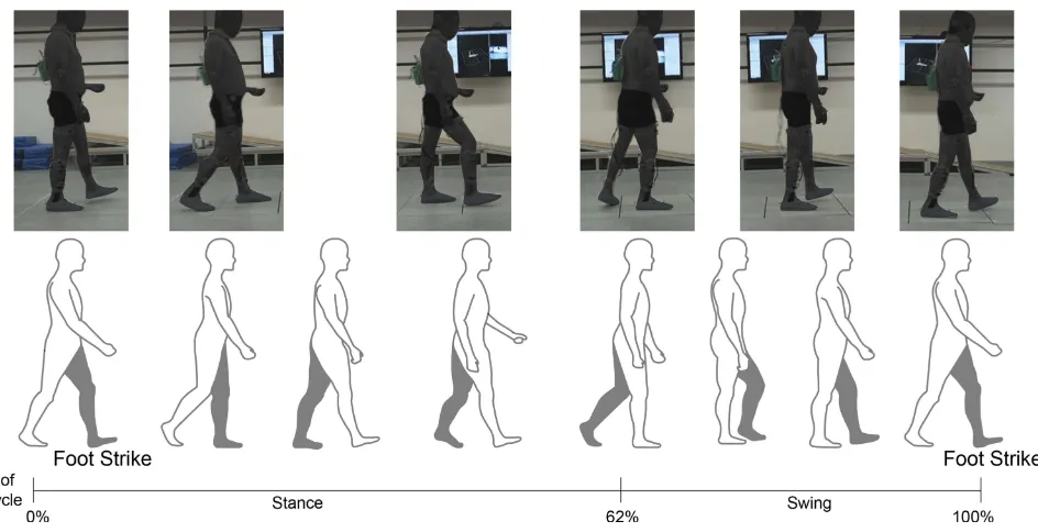
on. Each subject walked 6 m one way (Figure 2) with house
shoes on, while wearing no AFO (WI), while wearing an NP (Figure 1A), and while wearing an EP (Figure 1B).
The 3D motion capture for gait analysis was performed using a Plug-In Gait full body marker set. A total of 35
reflector markers (Figure 2) were attached on the subjects’
bodies. Twelve MX-T20 cameras (Vicon Inc., Oxford, UK) captured the motions at a frame rate of 100 Hz.
Data Analysis
Gait data were derived by dividing walking into cycles
based on a strike–strike motion, ie, the heel marker of the
affected side touching the ground and then touching it
again (Figure 2). During gait cycle stance, the knee
exten-sion (back knee) was analyzed primarily, as well as the
swing of the gait cycle and ankle plantar–dorsiflexion
(drop-foot).
To conduct a reliable gait analysis, the subjects per-formed at least three gait cycles under each condition.
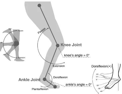
The analysis was performed for the range of the angles at the ankles and knees, and the maximum ankle
dorsi-flexion, plantarflexion, kneeflexion, and extension values
were obtained (Figure 3).
Table 1Subject Characteristics
Participants Sex Time Since Stroke (Months)
Daily Use of AFOs Paretic Side Positive History of Falls (Yes/No)
Type of Stroke
1 M 80 No Right N Ischemic
2 M 130 No Right Y Ischemic
3 F 12 No Right Y Hemorrhagic
4 M 72 Partially Right N Ischemic
5 M 82 No Right Y Ischemic
6 M 129 No Left Y Ischemic
7 M 115 No Left Y Ischemic
8 M 60 Partially Left N Hemorrhagic
9 M 24 Constantly Left N Hemorrhagic
10 M 72 Constantly Left N Hemorrhagic
Figure 1Ankle–foot orthosis (AFO) types: (A) elastic band-type AFO, (B) hard plastic material-type AFO.
Clinical Interventions in Aging downloaded from https://www.dovepress.com/ by 118.70.13.36 on 20-Aug-2020
ANOVA analysis (post examination: Tukey’s b) was performed to check whether the differences in averages
were significant (p < 0.005) because there were three
independent variables (WI, NP, and EP) affecting four dependent variables (the maximum angles of the ankles
[dorsiflexion, plantarflexion] and the maximum angles of
the knees [flexion, extension]).
To determine the correlation between ankle plantar–
dorsiflexion and kneeflexion–extension, a correlation
ana-lysis (Pearson's correlation coefficient) was performed
between the four dependent variables.
Results
The knee and ankle joint angle variables in the gait cycle showed similar trends for all three types. During stance of the affected side, the maximum knee extension and ankle
dorsiflexion were revealed, whereas during swing, the
maximum kneeflexion and plantar flexion were revealed.
The Ankle Angle Range
During dorsiflexion, the maximum angle was 10.51°
(±5.31°) in the NP condition, followed by 9.45° (±6.39°)
for WI and 6.84° (±6.22°) for EP. The results of dorsifl
ex-ion showed significant differences between all conditions
(p<0.005;Figure 4A).
In contrast, during plantarflexion, the maximum angle
was 7.12° (±7.35°) in the EP condition, followed by 5.03° (±9.32°) for WI and 1.77° (±8.17°) for NP. The results of
plantarflexion also showed significant differences between
all conditions (p<0.005;Figure 4A).
The Knee Angle Range
Duringflexion, the maximum angle was 18.51° (±8.28°) in
the NP condition, followed by 16.14° (±10.03°) for EP and
15.28° (±8.60°) for WI. Significant differences (p<0.005)
were found between the NP condition and the other
con-ditions, but no significant difference (p=0.194) was found
between WI and EP (Figure 4B).
During extension, the maximum angle was 7.30° (±11.48°) in the EP condition, followed by 7.11° (±10.95°)
for WI and 2.99° (±10.66°) for NP. Significant differences
(p<0.005) were found between the NP and other conditions,
but no significant difference (p=0.832) was found between
WI and EP conditions (Figure 4B).
Correlation Analysis
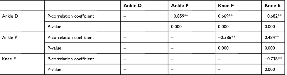
The correlation analysis showed a strong negative
correla-tion between the opposite movements (plantar–dorsiflexion
andflexion–extension), a high positive correlation
(correla-tion coefficient: 0.669**) between ankle dorsiflexion and
kneeflexion, a high negative correlation (correlation coeffi
-cient: −0.682**) between ankle dorsiflexion and knee
extension, a low negative correlation (correlation coeffi
-cient: −0.386**) between ankle plantar flexion and knee
flexion, and a moderate positive correlation (correlation
Figure 2The gait experiment of the hemiplegic patient, one gait cycle = foot strike–strike (affected side).
Clinical Interventions in Aging downloaded from https://www.dovepress.com/ by 118.70.13.36 on 20-Aug-2020
coefficient: 0.484**) between ankle plantar flexion and
knee extension (Table 2).
Discussion
Our research showed that the NP can prevent drop-foot during walking in hemiplegia patients. They are more convenient to use than the existing AFOs, and they pro-vide better wearing sensation.
In this research, the gait analysis for hemiplegia patients was conducted under three different conditions. Kinematic analysis was performed for the ankles and knees to evaluate the effectiveness of the NP. During the kinematic analysis performed during walking, the
maxi-mum ankle dorsiflexion value increased and the maximum
ankle plantarflexion value decreased. From these results,
we could confirm the preventive effect of the NP on
drop-foot.
Further, based on the correlation between AFOs and
the knee joints, as reported in previous studies,25,26 we
confirmed the positive effect of the NP on the knee joint.
In addition, in hemiplegia patients, the extension is 0° or more because of the back-knee phenomenon that occurs when they walk, and this phenomenon is caused by muscle rigidity. Therefore, in this research, the effectiveness of AFOs for the knees was evaluated based on the maximum angle offlexion–extension.25,26Based on the results of the
evaluation, we confirmed that the use of the NP increases
the maximum knee flexion and decreases the maximum
knee extension.25 In addition, this result can be attributed
to the fact that owing to its material characteristics, the NP does not resist the angular change at the ankle excessively. Moreover, considering that the subjects in this experiment could walk independently and that more than 6 years (77.3
months), which is a sufficiently long period for them to be
Figure 3Definition of the knee and ankle joint angles.
Clinical Interventions in Aging downloaded from https://www.dovepress.com/ by 118.70.13.36 on 20-Aug-2020
accustomed to walking, had passed since the onset of their disease, we found that the NP showed better effects than the EP. The NP ensures the angular elasticity of the ankles, thus yielding positive effects. This viewpoint is similar to
the results of Toshiki’s research, which showed that
“excessive resistance to plantar flexion rather restricts
knee flexion”.25 It also agrees with Marcelo’s research,
which reported that “[a]n appropriate use of AFO leads
to increases in knee’s flexion”.26 Furthermore, based on
the results of this experiment, we can understand why the EP yielded low effects. The angular variations at the
ankles when using the EP (range: dorsiflexion–plantar
flexion) were smaller than those when using the WI. This
result is attributed to the excessive restriction imposed by the plastic material on the ankles, thus restricting
dorsi-flexion, which is a positive effect. Further, as reported by
Toshiki, excessive angular restriction on the ankles is considered the cause of ineffectiveness of the EP during
kneeflexion–extension.
In this research, a correlation analysis was conducted for
four dependent variables (ankle dorsiflexion, ankle plantar
flexion, kneeflexion, and knee extension) using Pearson’s
correlation coefficient. The results of the correlation
analy-sis showed strong negative correlations between opposite
movements. As ankle dorsiflexion increased, the ankle’s
plantarflexion decreased. This shows that negative
correla-tion was maintained, while the range of angular changes did not change much. Similarly, in the case of the knee joint, as
flexion increased, extension decreased. This represents
a negative correlation between opposite movements. In
other words, as ankle dorsiflexion and knee flexion—ie,
the positive phenomena—increase, ankle plantar flexion
(drop-foot) and knee extension (back knee)—ie, the
nega-tive phenomena—decrease.
Further, the correlation between the ankle and knee could be determined based on the results of the correlation
analysis. In particular, ankle dorsiflexion and “knee joint
flexion (0.669**)–extension (−0.682**)” showed a
high-level correlation. Similar to the abovementioned analysis results, the correlation analysis showed that the positive effects of AFOs on the ankles include a reduction in the occurrence of the back-knee phenomenon and increased
kneeflexion.
This study had some limitations. The study was per-formed with hemiplegia patients who were accustomed to some degree of walking. Therefore, our results cannot be generalized to all hemiplegia patients, and NPs may not be better for all patients.
Conclusions
The kinematic analysis of the ankles and knee joints in this
research confirmed that the elastic band-type AFO (NP)
was effective in reducing the occurrence of drop-foot and the back-knee phenomena. Many hemiplegia patients and rehabilitation therapists who use AFOs believe that increasing resistance with the use of AFOs made of plastic or similar hard materials will be effective in preventing drop-foot. Hence, even the patients who were already accustomed to walking continued to use AFOs made of hard materials despite the discomfort. However, it has Figure 4A comparison of ankle and knee joint kinematics and three conditions
(Wl, NP, and EP). (A) Ankle angle range in dorsiflexion and plantarflexion; and (B) knee angle range inflexion–extension.
Notes:In the result graph, the Y-axis represents the joint angle of the ankle and the knee, (+) area is dorsiflexion of the ankle,flexion of the knee, (–) area is plantarflexion of the ankle, extension of knee, and the white line in the box represents the mean value of the moving range. An asterisk (*) indicates a significant level ofp< 0.005.
Abbreviations: AFO, ankle–foot orthosis; WI, wearing nothing; NP, wearing
elastic band-type AFO; EP, wearing hard plastic-type AFO.
Clinical Interventions in Aging downloaded from https://www.dovepress.com/ by 118.70.13.36 on 20-Aug-2020
been proven that the NP, which was developed to over-come the issues of traditional AFOs, is more effective than an existing AFO (EP) in improving drop-foot, increasing
kneeflexion, and reducing the occurrence of the back-knee
phenomenon.
The results of the effectiveness evaluation performed in this study are consistent with those of the preceding research. Previous researchers have claimed that elastic AFOs can have more positive effects on the ankles and knees.
When AFOs are chosen, different conditions, including
the patient’s health and walking ability, should be
consid-ered. This study confirmed that the soft material (fabric)
used in the elastic band-type AFO was no less effective than the plastic material. Thus, we hope to inform hemi-plegia patients and medical personnel of the results of this
research of our finding.
Acknowledgments
This study was supported by the Korea Institute of Industrial
Technology. A study of postural stability30 with the same
subjects was published in the journal‘Clinical Interventions
in Aging’.
Disclosure
The authors report no conflicts of interest in this work.
References
1. MK Business News (South Korea). The cumulative of stroke patients and onset number of stroke in Korea.2011April 12.
2. Cohen JW, Ivanova TD, Brouwer B, et al. Do performance measures of strength, balance, and mobility predict quality of life and commu-nity reintegration after stroke? Arch Phys Med Rehabil.
2018;99:713–719. doi:10.1016/j.apmr.2017.12.007
3. Datta Gupta A, Chu WH, Howell S, et al. A systematic review: efficacy of botulinum toxin in walking and quality of life in post-stroke lower limb spasticity.Syst Rev.2018;7:1–9. doi:10.1186/s13643-017-0670-9
4. Springer S, Friedman I, Ohry A. Thoracopelvic assisted movement training to improve gait and balance in elderly at risk of falling: a case series. Clin Interv Aging.2018;13:1143–1149. doi:10.2147/ CIA
5. Ahn BR, Kang HJ, Choi EJ, et al. Correlation of the Korean version of falls efficacy scale-international with quantitative balance and gait parameters through exercise program in elderly men.Ann Rehabil Med.2019;43:195–203. doi:10.5535/arm.2019.43.2.195
6. Begg R, Galea MP, James L, et al. Real-time foot clearance bio-feedback to assist gait rehabilitation following stroke: a randomized controlled trial protocol. Trials. 2019;20:317. doi:10.1186/s13063-019-3404-6
7. Saleh MS, Rehab NI. Effect of ankle proprioceptive training on gait and risk of fall in patients with diabetic neuropathy: a randomized controlled trial.Int J Diabetes Res.2019;2:40–45.
8. Petersen W, Ellermann A, Henning J, et al. Non-operative treatment of unicompartmental osteoarthritis of the knee: a prospective rando-mized trial with two different braces—ankle–foot orthosis versus knee unloader brace.Arch Orthop Trauma Surg.2019;139:155–166. doi:10.1007/s00402-018-3040-8
9. Qiao M, Feld JA, Franz JR. Aging effects on leg joint variability during walking with balance perturbations. Gait Posture.
2018;62:27–33. doi:10.1016/j.gaitpost.2018.02.020
10. Buckon CE, Thomas SS, Jackobson-Huston S, Sussman M, Aiona M. Comparison of three ankle–foot orthosis configurations for children with spastic hemiplegia.Dev Med Child Neurol.2001;43:371–378. doi:10.1017/S0012162201000706
11. Wang C, Goel R, Rahemi H, et al. Effectiveness of daily use of bilateral custom-made ankle-foot orthoses on balance, fear of falling and physical activity in older adults: a randomized controlled trial. Gerontology.2019;65:299–307. doi:10.1159/000494114
12. Taiar R, Adel C, Belassian G, et al. Can a new ergonomical ankle– foot orthosis (AFO) device improve patients’ daily life? A preliminary study.Theor Issues Ergonom Sci.2019. doi:10.1080/ 1463922X.2019.1616332
13. Lee S-H, Choi C, Lee D, et al. A novel hinged ankle foot orthosis for gait performance in chronic hemiplegic stroke survivors: a feasibility study. Biomed Eng Lett.2018;8:301–308. doi:10.1007/s13534-018-0074-3 14. Sheng Y, Kan S, Wen Z, et al. Effect of kinesio taping on the walking
ability of patients with foot drop after stroke.Evid Based Complement Alternat Med.2019:2459852. doi:10.1155/2019/2459852
15. Liu Y, Zang X, Zhang N. Design and evaluation of a wearable powered foot orthosis with metatarsophalangeal joint.Appl Bionics Biomech.2018;9289505. doi:10.1155/2018/9289505
16. Yeung LF, Ockenfeld C, Pang MK, et al. Randomized controlled trial of robot-assisted gait training with dorsiflexion assistance on chronic stroke patients wearing ankle-foot-orthosis. J Neuroeng Rehabil.
2018;15:51. doi:10.1186/s12984-018-0394-7 Table 2Correlation Between Four Dependent Variables by Pearson’s Correlation Coefficient
Ankle D Ankle P Knee F Knee E
Ankle D P-correlation coefficient – −0.859** 0.669** −0.682**
P-value – 0.000 0.000 0.000
Ankle P P-correlation coefficient – – −0.386** 0.484**
P-value – – 0.000 0.000
Knee F P-correlation coefficient – – – −0.738**
P-value – – – 0.000
Notes:P-correlation: Pearson's correlation coefficient. An asterisk (**) indicates a significant level of p < 0.01. Abbreviations:D, dorsiflexion; P, plantarflexion; F,flexion; E, extension.
Clinical Interventions in Aging downloaded from https://www.dovepress.com/ by 118.70.13.36 on 20-Aug-2020
17. Kim JH, Sim WS, Lim SY, Won BH. Analysis of lower-body-joint angle in hemiplegic patients wearing new elastic-band-type ankle-foot ortho-sis while walking.Asian Conf Ergon Des.2014;5:901–904.
18. Kobayashi T, Leung AK, Akazawa Y, Hutchins SW. Effect of ankle-foot orthoses on the sagittal plane displacement of the center of mass in patients with stroke hemiplegia: a pilot study.Top Stroke Rehabil.2012;19:338–344. doi:10.1310/tsr1904-338
19. Danielsson A, Sunnerhagen KS. Energy expenditure in stroke sub-jects walking with a carbon composite ankle foot orthosis.J Rehabil Med.2004;36:165–168. doi:10.1080/16501970410025126
20. Ohsawa S, Ikeda S, Tanaka S, et al. A new model of plastic ankle foot orthosis (FAFO (II)) against spastic foot and genu recurvatum.Prosthet Orthot Int.1992;16:104–108. doi:10.3109/03093649209164320 21. Hachisuka K, Ogata H, Tajima F, Ohmine S. Clinical evaluations of
dorsiflexion assist controlled by spring ankle-foot orthosis for hemi-plegic patients.J UOEH.1998;20:1–9. doi:10.7888/juoeh.20.1 22. Chen CC, Hong WH, Wang CM, et al. Kinematic features of
rear-foot motion using anterior and posterior ankle-foot orthoses in stroke patients with hemiplegic gait. Arch Phys Med Rehabil.
2010;91:1862–1868. doi:10.1016/j.apmr.2010.09.013
23. Bleyenheuft C, Deltombe T, Detrembleur C. Influence of ankle-foot orthoses on kinematic segmental covariation among stroke patients. Ann Phys Rehabil Med.2013;56:3–13. doi:10.1016/j.rehab.2012.11.001
24. Yamamoto S, Miyazaki S, Kubota T. Quantification of the effect of the mechanical property of ankle-foot orthoses on hemiplegic gait. Gait Posture.1993;1:27–34. doi:10.1016/0966-6362(93)90040-8 25. Kobayashi T, Leung AK, Akazawa Y, Hutchins SW. The effect of
varying the plantarflexion resistance of an ankle-foot orthosis on knee joint kinematics in patients with stroke.Gait Posture.2013;37:457–459. doi:10.1016/j.gaitpost.2012.07.028
26. Gatti MA, Freixes O, Fernandez SA, et al. Effects of ankle foot orthosis in stiff knee gait in adults with hemiplegia. J Biomech.
2012;45:2658–2661. doi:10.1016/j.jbiomech.2012.08.015
27. Perry J. Gait and Analysis: Normal and Pathological Function. Thorofare, NJ: Slack Inc.;1992.
28. Boudarham J, Roche N, Pradon D, Bonnyaud C, Bennsmail D, Zory R. Variations in kinematics during clinical gait analysis in stroke patients. PLoS One.2013;8:e66421. doi:10.1371/journal.pone.0066421 29. Nolan KJ, Savalia KK, Yarossi M, Elovic EP. Evaluation of
a dynamic ankle foot orthosis in hemiplegic gait: a case report. NeuroRehabilitation.2010;27:343–350. doi:10.3233/NRE-2010-0618 30. Kim JH, Sim WS, Won BH. Effectiveness of elastic band-type ankle-foot orthoses on postural control in poststroke elderly patients as determined using combined measurement of the stability index and body weight-bearing ratio. Clin Interv Aging.2015;10:1839–1847. doi:10.2147/CIA.S92888
Clinical Interventions in Aging
Dove
press
Publish your work in this journal
Clinical Interventions in Aging is an international, peer-reviewed journal focusing on evidence-based reports on the value or lack thereof of treatments intended to prevent or delay the onset of maladaptive correlates of aging in human beings. This journal is indexed on PubMed Central, MedLine, CAS, Scopus and the Elsevier
Bibliographic databases. The manuscript management system is completely online and includes a very quick and fair peer-review system, which is all easy to use. Visit http://www.dovepress.com/ testimonials.php to read real quotes from published authors.
Submit your manuscript here:https://www.dovepress.com/clinical-interventions-in-aging-journal
Clinical Interventions in Aging downloaded from https://www.dovepress.com/ by 118.70.13.36 on 20-Aug-2020