University of South Carolina
Scholar Commons
Theses and Dissertations
8-9-2014
Mechanical Properties of Diet and Its Effect on
Oral Health
Varsha Kapoor
University of South Carolina - Columbia
Follow this and additional works at:https://scholarcommons.sc.edu/etd Part of theBiology Commons
This Open Access Thesis is brought to you by Scholar Commons. It has been accepted for inclusion in Theses and Dissertations by an authorized administrator of Scholar Commons. For more information, please contactdillarda@mailbox.sc.edu.
Recommended Citation
M
ECHANICALP
ROPERTIES OFD
IET AND ITSE
FFECT ONO
RALH
EALTHby
Varsha Kapoor
Bachelor of Science George Mason University, 2010
Submitted in Partial Fulfillment of the Requirements
For the Degree of Master of Science in
Biomedical Science
School of Medicine
University of South Carolina
2014
Accepted by:
Adam Hartstone-Rose, Director of Thesis
Edie Goldsmith, Reader
Chandrashekhar Patel, Reader
Lacy Ford, Vice Provost and Dean of Graduate Studies
ACKNOWLEDGEMENTS
I would never have been able to finish my dissertation without the guidance of my
committee members, help from friends, and support from my family members. I would
like to express my deepest gratitude to my advisor, Dr. Adam Hartstone-Rose, for his
guidance throughout the process.
I would like to thank my committee members Dr. Edie Goldsmith and Dr.
Chandrashekhar Patel for their time and valuable advice.
Also special thanks goes to Tyler Antonelli and Hannah Selvey for collecting
valuable data and always being there to help. My research would not have been possible
without their help.
ABSTRACT
The predominant diet fed to captive carnivores consists of ground meat
formulated to provide full nutritional requirements. However, this ground meat diet
completely lacks the mechanical properties such as toughness, hardness and stiffness of
the foods the animals would eat in the wild. The goal of this research was to evaluate the
effect of mechanical properties of diet on oral health by comparing prevalence of
periodontal disease and calculus accumulation in wild and captive felids. One-way
ANOVA analysis of variance indicated that these differences are statistically significant
(P<.0001). The results of this study indicate that diet texture is a significant factor in oral
health of felids. Also there is a significant correlation between oral health and overall
TABLE OF CONTENTS
ACKNOWLEDGEMENTS ... ii
ABSTRACT ... iii
LIST OF TABLES ... vi
LIST OF FIGURES ... vii
Chapter 1: Introduction & Background ... 1
1.1. Background ... 2
1.2. Experimental Findings ... 3
1.3. Dental Anatomy ... 4
1.4. Dental Formulae of Felids ... 5
1.5. Dental Calculus ... 6
1.6. Periodontal Disease ... 7
1.7. Effects of aging on oral disease... 9
1.8. Specific Aims and Hypotheses ... 10
Chapter 2: Methods ... 13
Sample ... 14
2.1. Calculus Scoring ... 16
2.2. Periodontal Disease Scoring... 19
2.4. Statistical Analysis ... 23
Chapter 3: Results ... 25
Initial Observations ... 26
3.1. Effect of Captivity Status on Periodontal Disease and Calculus ... 26
3.2. Correlation of Oral Health to Overall Health ... 28
3.3 Comparison of Posterior versus Anterior Dentition ... 30
3.4. Influence of Sex and Species on Oral Health ... 31
3.5. Presence and Absence of Calculus and Periodontal Disease ... 32
Chapter 4: Discussion ... 34
4.1. Effect of Captivity on Oral Health ... 35
4.2. Effect of oral health on overall health ... 36
4.3. Dental Health of Anterior versus Posterior Dentition ... 37
4.4. Other Findings ... 38
4.5. Limitations ... 39
4.6. Broader Impacts ... 40
LIST OF TABLES
Table 2.1: Focal Sample ... 22
Table 2.2 Other preliminary Sample ... 22
Table 2.3: Calculus Index ... 25
Table 2.4: Periodontal Index Score ... 27
Table 2.5: Marker Key For All Graphs ... 31
Table 3.2: One-way ANOVA Analysis of Oral Health and Captivity Status ... 35
Table 3.3: Descriptive statistics for RMA regressions against the PC2 scores ... 37
Table 3.4: Descriptive statistics for RMA regressions against anterior dentition ... 39
Table 3.5: Presence and absence of Calculus and Periodontal in wild Felids ... 40
LIST OF FIGURES
Figure 1.1: The Mandibular Tooth Row of Panthera tigris ... 14
Figure 1.2: Intraoral Bisecting-angle Dental Radiograph of a Dog ... 16
Figure 1.4: Severe Calculus on Maxillary Premolars of a Juvenille Tiger ... 18
Figure 2.1: Sectioning of Mandibular (a) and Maxillary (b) teeth ... 24
Figure 2.2: Comparison of calculus score ... 26
Figure 2.3: Comparison of Periodontal Disease... 28
Figure 2.4: PCA output with second principal component against third ... 30
Figure 3.1: Periodontal and Calculus Scores versus Captivity Status ... 36
Figure 3.2: Periodontal and Calculus Scores versus PC2 ... 38
CHAPTER 1
The composition of the diet of captive carnivores is of great interest to animal
curators, keepers and veterinarians. However, that interest has focused almost entirely on
the nutritional composition of the diet (Haberstroh et al., 1984) and there is little
consideration given to the mechanical properties such as toughness, hardness and
stiffness of the foods. While the incorporation of bones as dietary supplements is a
growing trend for captive carnivores, the predominant diet that captive carnivores are fed
consists of ground meat (Haberstroh et al., 1984). While this diet contains all of the
nutrients found in a natural diet, it does not simulate the ingestive or masticatory
challenges that wild carnivores face. In particular, the lack of bone and connective tissue
manipulation in the captive diet may lead to unnatural dental health issues (Skibiel,
Trevino, & Naugher, 2007). Numerous studies in domestic animals, as reviewed in
Watson (1994) and Logan (2006) have demonstrated that mechanical attributes of the diet
appear to affect calculus buildup and oral health in general. Thorough examination of
diets of captive felids and their wild conspecifics, with a focus on the mechanical
properties of the diet and their effects on the oral health remains to be carried out.
In this study, I will investigate oral health of captive and wild felids by comparing
degree of calculus build-up and periodontal disease and finally correlate these markers of
oral health to a proxy of overall health.
Background
Several factors may affect the pathogenesis of dental abnormalities such as
2003). My research will focus on the nutrition aspect and how diet affects the oral health
in captive versus wild felids. Studies have shown that the mechanical properties of diet
may have a distinct effect on an animal’s dental health and cranial morphology (O'Regan
& Kitchener, 2005).
Experimental Findings
A study conducted on dogs and cats showed that food composition and texture
could directly affect the oral health through maintenance of tissue integrity, stimulation
of salivary flow and cleaning of the tooth surfaces by appropriate abrasive action
(Watson, 1994). The texture of food has been long thought to affect the oral health of
dogs and cats (Logan, 2006). Several studies have proven that excessive calculus
accumulation; gingivitis and periodontal disease have been noted in animals on a soft
diet. A study conducted by (Vosburgh, 1982) on timber wolves showed that diet texture
is a significant factor in the oral health of these captive wolves. The wolves fed a hard,
dry diet exhibited significantly lower levels of plaque accumulation than those fed a soft,
moist, meat-based diet (Vosburgh, 1982). Dry diet is associated with less gingival
pathology because the soft textured diet, does not provide adequate abrasive action to
clean the teeth (Haberstroh et al., 1984).
Another study conducted on ferrets showed the importance of diet and texture on
oral health. Ferrets were used for this study because they exhibit similar patterns of
plaque and calculus accumulation leading to periodontal disease as seen in other domestic
carnivores. The findings showed that ferrets kept on a soft diet such as bread and milk
prevented periodontal disease and even caused a decrease in calculus accumulation
(Verstraete, 2003). Numerous other studies conducted on different types of animals
showed similar relationship between diet and oral health. Since my study is based on
analyzing periodontal disease and calculus build-up in captive and wild felids it is
important to have a basic understanding of dental terminologies and general anatomy of
teeth.
Dental Anatomy
Although the teeth of felids can vary in shape, size and functions, the constituents
and structure of all teeth are similar. A tooth comprises of crown, which is the anatomical
area of teeth usually covered by enamel, and roots, which is covered by the cementum.
The junction where the enamel of the crown and the cementum of the root converge is
called the cementoenamel junction (CEJ). (Logan, 2006)
The periodontium provides the support necessary to maintain healthy functions of
teeth. It consists of gingiva, periodontal ligament, alveolar bone and cementum (Mariotti,
2007). The gingiva is the soft tissue oral mucosa that covers the alveolar bone of the jaws
and surrounds the necks of the teeth (Mariotti, 2007) . In healthy mammals, normal
gingiva covers the alveolar bone and tooth root to a level just below the area where
enamel meets the root surface (Niemiec, 2008a). The periodontal ligament is composed
of a complex vascular and highly cellular connective tissue that surrounds the tooth root
and connects it to the inner wall of the alveolar bone (Newman, 2006). The alveolar bone
surrounds the teeth and it is a tooth dependent structure; it creates the socket that the
It is formed with the eruption of the teeth and generally reabsorbed when the teeth is
removed (Gioso & Carvalho, 2005).
Problems with the periodontium arise once accumulation of food particles and
bacteria form a substance called plaque that sticks to the surface of the tooth (Gioso &
Carvalho, 2005). Plaque is a microbial biofilm, consisting of an organized community of
cooperating microorganism on surface of the teeth (Albuquerque et al., 2012). If plaque is
not removed by mechanical cleaning, minerals in the saliva harden the plaque into dental
calculus. As plaque and calculus spread under the gingival line it causes irritation to the
gingiva and leads to an inflammatory condition known as gingivitis (Campbell, 2007).
Gingivitis is the mildest form of periodontal disease and it does not affect the underlying
supporting structures of the teeth and is usually reversible (Pihlstrom, Michalowicz, &
Johnson, 2005). This early stage of inflammation, if left untreated, will lead to
periodontal disease (Gioso & Carvalho, 2005) .
Dental Formulae of Felids
A typical felid has a total of thirty teeth. Generally their dental formulae is written
as I = 3/3 C=1/1 P=3/2 M=1/1 where I, C, P and M stand for Incisors, Canines, Premolars
and Molars respectively and the number on either side of the “/” stand for the number of
teeth of that kind in each upper and lower quadrant respectively (Wiggs & Bloom, 2003).
The generalized Felids dental row (Figure1.1) can be broken into four functional units:
Figure.1.1: The Mandibular Tooth Row of Panthera tigris USNM 253289
Dental Calculus
Dental calculus can be defined as calcified or calcifying deposits that are attached
to the surface of teeth (Newman, 2006) . Calculus can be classified into two groups
depending on the location. Supra-gingival calculus is located above the gingival margin
and sub-gingival calculus located below the gingival margin (Roberts-Harry &
Clerehugh, 2000). Calculus or tartar are usually formed as a result of the calcification of
dental plaque or biofilm (Busscher, 2004).
According to Vosburgh (1982), plaque a precursor of calculus tends to
accumulate at the opening of salivary duct near the maxillary premolar or where the
tongue does not regularly contact the tooth surface. This could explain why the front of
Calculus is mostly made up of inorganic compound, consisting of crystalline salts
and the organic portion of dental calculus consists of proteins, carbohydrates and lipids.
A layer of active plaque usually covers calculus, plaque comprises of living bacteria and
as the active layers of plaque mineralizes, and new calculus is formed. Calculus
formation results in displacing epithelium around the gum line, allowing bacteria from
the plaque to reach the alveolar bone. (Greene, Kuba, & Irish, 2005)
Periodontal Disease
In veterinary medicine periodontal disease is the most prevalent disease in
domestic carnivores and is found in approximately 80% of dogs aged 2 years or older
(Niemiec, 2008b). Periodontal diseases are pathological processes that affect the tissues
surrounding the teeth. Previous names for periodontal diseases includes gum disease,
pyorrhea, and periodontitis (Mariotti, 2007). Periodontal disease (PD) can be referred as a
group of inflammatory diseases caused by bacterial plaque in the periodontium
(Albuquerque et al., 2012).
Periodontal disease can be broadly divided into two groups, gingivitis and
periodontitis. Gingivitis is the reversible stage of periodontal disease and can be treated
with thorough plaque removal whereas periodontitis is more severe (Logan, 2006).
Periodontitis can be referred to as the destructive form of periodontal disease and it is
characterized by several alterations in periodontal tissues such as inflammation and loss
of the periodontal attachment (Mariotti, 2007). The hallmarks for periodontitis are
alveolar bone loss, periodontal pocketing, gingival inflammation and attachment loss
Plaque deposits present in cases of untreated gingivitis will form calculus. As this
calculus builds under the gingiva, the bacteria in the sub-gingival plaque set in motion a
cycle of damage to the supporting tissues around the tooth. Once periodontal disease
begins, pockets will form where the teeth meet the gingiva and bone fostering bacterial
growth. This growth can lead to bone loss, tissue destruction or tooth loss. (Campbell,
2007)
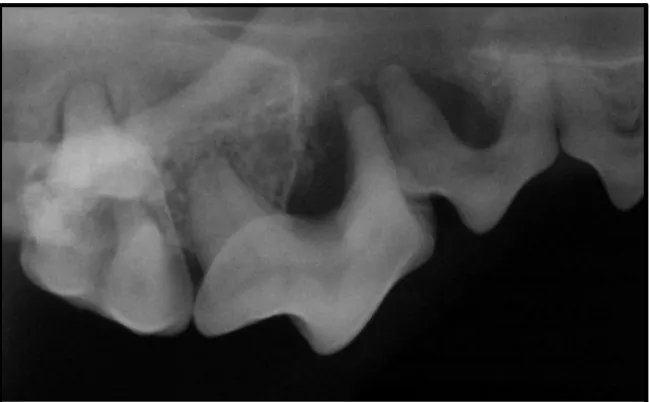
Figure 1.2: Intraoral Bisecting-angle Dental Radiograph of a Dog modified from (Niemiec, 2008b).
This clinical radiograph of the maxillary right premolars reveals severe
periodontium loss and reabsorption of alveolar bone associated with periodontal disease. This is indicative of severe periodontal disease (Niemiec, 2008b) .
For this study we used skulls of captive and wild felids to score for periodontal
disease and dental calculus. Since periodontal disease affects the alveolar bone (Figure
1.2 and 1.3), skulls were scored for periodontal disease by measuring the distance the
the alveolar bone, refer to Chapter II for a detailed scoring technique which was used to
measure dental calculus and periodontal disease.
Although a combination of dental plaque and dental calculus is the primary cause
of periodontal disease, we should keep in mind several additional factors that may
contribute to periodontal disease such as malocclusions, absence of oral hygiene, teeth
crowding and genetics (Albuquerque et al., 2012).
Effects of aging on oral disease
When comparing wild and captive felids we should keep in mind how age affects
calculus and periodontal disease. Improved veterinary care in zoos has resulted in an
increased longevity in captive felids (Longley, 2011). According to several sources
(Albuquerque et al., 2012; Gawor et al., 2006; Patterson, Neiburger, & Kasiki, 2003;
Sone, Koyasu, & Oda, 2004; Watson, 1994) the prevalence and severity of periodontal
disease is age related. One of the limitations of this study was that we did not know the
age of our specimen when scoring for periodontal disease and dental calculus.
One might argue our results to be skewed due to age restriction but a study by
Gawor (2006) on the influence of diet on oral health of cats and dogs showed that even
after they adjusted for age, the mean oral health index was significantly higher in cats and
dogs fed soft food compared with those fed dry or mixed food. These results clearly
indicate that feeding a dry food diet has a positive influence on oral health (Gawor et al.,
Furthermore, we have found clearly young specimens with advanced oral health
diseases (Fig. 1.4) demonstrating that although age might be a confounding factor in this
study it is not necessarily the main correlate of dental disease.
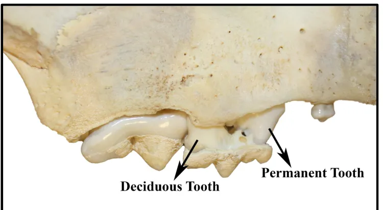
Figure 1.3: Severe Calculus on Maxillary Premolars of a Juvenille Tiger. Catalog number: USNM 396272
Note the deciduous premolar being replaced by the
permanent premolar. This specimen was studied for graphical purposes though only adult specimens are included in the statistical analyses.
Specific Aims and Hypotheses
The overall goal of this study is to assess the effect of mechanical properties of
diet on oral health. This goal is achieved by determining if differences in periodontal
disease and dental calculus exist between wild and captive felids. Multiple hypotheses
Aim 1: To determine if the incidence of dental calculus and periodontal disease is
higher in captive felids.
Studies on various animals as discussed in this paper, have shown that excessive calculus
accumulation and periodontal disease have been noted in animals on a soft diet.
Experimental Hypothesis 1: Because the predominant diet that captive carnivores are fed
consists of ground meat (Haberstroh et al., 1984), it is predicted that captive felids will
have higher incidence of dental calculus and periodontal disease compared to the wild
felids.
Aim 2: To determine the correlation between oral health and overall health.
Since gingivitis and early stages of periodontal disease is both curable and preventable,
evidence showing that it is an independent risk factor leading to other systemic diseases
and condition would be of great importance in public health (Campbell, 2007).
Experimental Hypothesis 2: Higher PC2 scores, which is a measure of skull deformity is
correlated to higher periodontal disease and dental calculus.
Aim 3: To determine prevalence of calculus and periodontal disease in the posterior
versus anterior dentition.
Carnivores display a diverse array of teeth, all of which are presumed to be adapted for
certain functions, such as slicing flesh, killing its prey, cracking into bones and chewing
(VanValkenburgh, 1996). Van Valkenburgh (1996) describes the general dentition of
carnivores as being comprised of three regions: the grasping incisors, penetrating canines
interactions between the anterior and posterior teeth. Since the function differs in anterior
versus posterior teeth, will this affect the degree of periodontal disease and calculus
buildup? When scoring the skulls at the museum, we noticed higher incidence of calculus
on the posterior teeth, when compared to the anterior teeth and an opposite trend was
observed when scoring for periodontal disease.
Experimental Hypothesis 3: Higher prevalence of calculus will be observed in the
posterior teeth and higher prevalence of periodontal disease will be observed in the
CHAPTER 2
Sample
The present study will focus primarily on lions (Panthera leo) and tigers
(P. tigris) (Table 2.1) though data were also collected on other carnivores as well
(Table 2.2) for future analysis.
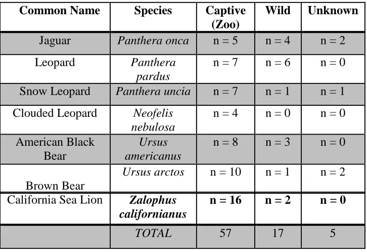
Table 2.1: Focal Sample (N = 83)
Common Name Species Captive
(Zoo)
Wild Unknown
Lion Panthera leo n = 22 n = 25 n = 0
Tiger Panthera tigris n = 23 n = 13 n = 0
TOTAL 45 38 83
Table 2.2 Other preliminary Sample (N = 133)
Common Name Species Captive (Zoo)
Wild Unknown
Jaguar Panthera onca n = 5 n = 4 n = 2
Leopard Panthera pardus
n = 7 n = 6 n = 0
Snow Leopard Panthera uncia n = 7 n = 1 n = 1
Clouded Leopard Neofelis nebulosa
n = 4 n = 0 n = 0
American Black Bear
Ursus americanus
n = 8 n = 3 n = 0
Brown Bear
Ursus arctos n = 10 n = 1 n = 2
California Sea Lion Zalophus californianus
n = 16 n = 2 n = 0
For the sake of this paper, “wild” refers to individuals who did not live in
captivity and “captive” refers to individuals who resided in zoos or animal rescue
facilities at the time of their deaths. Although their captivity status at birth is
unknown for most of the specimens, since importation of wild animals has
historically been rare, we believe that most if not all were born in captivity. In
this study, only adult specimens were used. Samples were obtained from
collections at the American Museum of Natural History (AMNH) the Smithsonian
(USNM) and the research collection of Dr. Hartstone-Rose (University of South
Carolina School of Medicine). These specimens originated at the Bronx Zoo,
Central Park Zoo, New York Zoo, New York Zoo Society, New York Zoo
Gardens, New York Park Commission, National Zoological Park (Smithsonian),
Toledo Zoological Society, Academy of Natural Science, Barnum and Bailey
Circus, Prospect Park Zoo, and the Carolina Tiger Rescue.
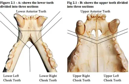
All specimens were evaluated and scored for calculus and periodontal
disease by carefully examining each skull and assessing the dental arcade in six
sections upper and lower anterior teeth (canines and incisors), left and right cheek
teeth (premolars and molars). Figure 2.1 depicts how the mandibular (2.1 – A)
and maxillary (2.1 – B) teeth were categorized into six sections and each of the
six regions was scored for calculus and periodontal disease. After dividing the
upper and lower teeth into six sections, each section was given a score of 0 to 5
for calculus build-up and periodontal disease according to the index provided in
Figure 2.1: Sectioning of Mandibular (a) and Maxillary (b) teeth.
In this study, the score of any gi
index (Table 2.3 and Table 2.
the six sections as a whole rather than scoring for each individual tooth.
eliminates the discrepancies that may arise due to
teeth.
2.1. Calculus Scoring
To score for dental calculus we observed amounts of calculus covering the crown
of all teeth and each of the six sections were scored according to the calculus index
(Table 2.3).
Figure 2.1: Sectioning of Mandibular (a) and Maxillary (b) teeth.
In this study, the score of any given section in the calculus and periodontal
2.4) tend to reflect the amalgamated score for each of
rather than scoring for each individual tooth. This
eliminates the discrepancies that may arise due to individual missing or damaged
To score for dental calculus we observed amounts of calculus covering the crown
of all teeth and each of the six sections were scored according to the calculus index periodontal
4) tend to reflect the amalgamated score for each of
This
individual missing or damaged
To score for dental calculus we observed amounts of calculus covering the crown
Table 2.3: Calculus Index
Calculus Scores
Observations
0 No evidence of calculus (Figure 2.2-A)
1 1 – 10% of the crown of the teeth is covered by calculus (Figure 2.2-B) 2 10 – 25% of the crown of the teeth is covered by calculus (Figure 2.2-C) 3 25 – 50% of the crown of the teeth is covered by calculus (Figure
2.2-D).
4 50 – 75% of the crown of the teeth is covered by calculus and/or minor caries (Figure 2.2-E).
5 75 – 100% of the crown of the teeth is covered by calculus OR thickened calculus and/or major caries (Figure 2.2-F).
Figure 2.2 illustrates varying degrees of calculus build-up for the lower left cheek
teeth. A score of 0 as seen in figure 2.2-a shows no evidence of calculus. Figure 2.2-b we
see about 10 percent of the crown covered with calculus in the lower left molar. Figure
2.2-c received a score of 2 because approximately 15 percent of the lower left molar was
covered with calculus. In figure 2.2-d we see about 50 percent of the lower left pre-molar
covered with calculus. Figure 2.2-e received a score of 4 because of the visible calculus
on the lower left premolar and caries seen in the molar. Finally figure 2.2-f received a
score of 5 because of thick calculus surrounding the molar and major caries observed in
the lower left pre-molar. On the right side of each figure we see a magnified view of the
2.2. Periodontal Disease Scoring
Skulls were scored for periodontal disease by measuring the distance the
alveolar ridge degraded from its original position combined with abscess scoring
technique. This scoring technique relies on the size of any holes in the alveolar
bone created by abscess damage, and a score for the entire skull may be based on
the number of abscess damaged sites. Below is the index used to score for
periodontal disease (Table 2.4).
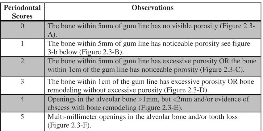
Table 2.4: Periodontal Index Score
Periodontal Scores
Observations
0 The bone within 5mm of gum line has no visible porosity (Figure
2.3-A).
1 The bone within 5mm of gum line has noticeable porosity see figure
3-b below (Figure 2.3-B).
2 The bone within 5mm of gum line has excessive porosity OR the bone
within 1cm of the gum line has noticeable porosity (Figure 2.3-C).
3 The bone within 1cm of the gum line has excessive porosity OR bone
remodeling without excessive porosity (Figure 2.3-D).
4 Openings in the alveolar bone >1mm, but <2mm and/or evidence of
abscess with bone remodeling (Figure 2.3-E).
5 Multi-millimeter openings in the alveolar bone and/or tooth loss
Figure 2.3 illustrates varying stages of periodontal disease in the left posterior
mandibular dentition. A score of 0 as seen in figure 2.3-a, shows no evidence of
periodontal disease because of no visible porosity and the alveolar bone is within 5mm of
gum line. In figure 2.3-b there are noticeable porosity in the alveolar bone around the
premolar and molar teeth. Figure 2.3-c received a score of 2 because of excessive and
noticeable porosity along the two premolars and molar teeth. In figure 2.3-d we see the
alveolar bone within 1 cm of the gum line and has excessive porosity. Figure 2.3-e
received a score of 4 because of the openings in the alveolar bone >1mm, but <2mm
surround the premolars. Finally figure 2.3-f received an over all score of 5 for the lower
left check teeth because of multi-millimeter openings in the alveolar bone and tooth loss.
2.3. Principle Component Analyses (PCA)
Principle component analyses is a statistical procedure that applies data to a new
coordinate system, which separates the data that differs the most as the first principal
component and the subsequent variation as second principal component, third principal
component and so on. Hence PCA is used to show the strongest factor driving the
variation across a population. An important component of my research was to correlate
oral health to overall health. This was accomplished by taking advantage of another study
that was conducted in the research lab of Dr. Adam Hartstone-Rose where the cranial
morphology of wild versus captive felids were compared.
The cranial morphology study examined the effect of differences in mechanical
geometric morphometric examination of their skulls and analyzed these samples with
PCA to statistically discern the results across a sample of eighty-one specimens, with
each specimen being comprised of forty-three landmarks. According to the PCA the
second greatest source of variation (PC2) across the population was driven by captivity as
seen in figure 2.4 (Hartstone-Rose, Selvey, Villari, Atwell, & Schmidt, 2014). For this
study we compared the PC2 scores to periodontal disease and calculus build-up to see if
the variables were correlated. Table 2.5 shows the markers that were used to generate all
graphs.
Figure 2.4. PCA output with second principal component against third.
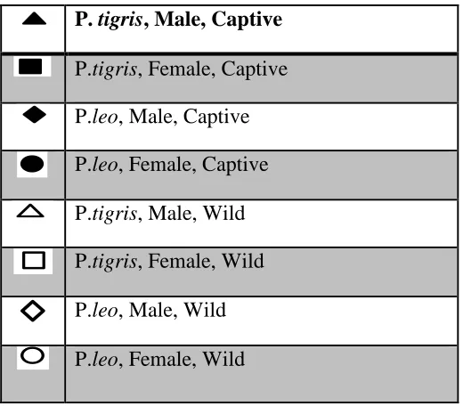
Table 2.5: Marker Key For All Graphs
P. tigris, Male, Captive
P.tigris, Female, Captive
P.leo, Male, Captive
P.leo, Female, Captive
P.tigris, Male, Wild
P.tigris, Female, Wild
P.leo, Male, Wild
P.leo, Female, Wild
2.4. Statistical Analysis
JMP statistical software was used to analyze our data. We used mean and
maximum values of periodontal disease and calculus build-up to look for general trends
such as: how the mean and maximum values change with captivity statuses, PC2 scores
and regions of the teeth. We used t-test analysis and compared the captivity statuses to
mean periodontitis score, maximum periodontitis scores, mean calculus scores and
maximum calculus score to determine if captive felids have higher periodontal and
captive felids. P-value was used to determine if the zoo animals had statistically
significant higher periodontal disease than wild animals.
The main assumption used throughout our results section is that the
samples have been independently drawn from their populations. The null hypothesis is
CHAPTER 3
Initial Observations
When scoring the skulls we observed, that the occurrence and magnitude of
periodontal and calculus scores were much higher in captive carnivores compared to the
wild carnivores. We also noticed higher calculus scores on the posterior teeth when
compared to the anterior teeth and saw an opposite trend for periodontal disease.
2.1. Effect of Captivity Status on Periodontal Disease and Calculus
To test our hypothesis, captive carnivores will have higher incidence of calculus
and periodontal disease we performed a t-test analyses comparing the mean and
maximum periodontal and mean and maximum calculus scores relative to captivity
statuses. On our x-axis we have wild and zoo felids and on our y-axis we have
periodontal and calculus scores. Box plots in red are used for identifying outliers and for
comparing distributions. Mean Periodontal Disease, Maximum Periodontal Disease,
Mean Calculus and Maximum Calculus (table 3.2) all significantly separate captive and
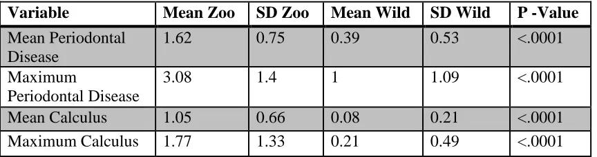
wild populations. Table 3.2 shows that captive felids have significantly higher (P <
.0001) mean periodontal disease, maximum periodontal disease, mean calculus and
Table 3.2: One-way ANOVA Analysis of Oral Health and Captivity Status.
Variable Mean Zoo SD Zoo Mean Wild SD Wild P -Value
Mean Periodontal Disease
1.62 0.75 0.39 0.53 <.0001
Maximum
Periodontal Disease
3.08 1.4 1 1.09 <.0001
Mean Calculus 1.05 0.66 0.08 0.21 <.0001 Maximum Calculus 1.77 1.33 0.21 0.49 <.0001
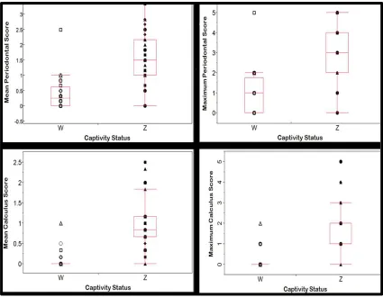
Figure 3.1: The mean and maximum periodontal score and mean and maximum
calculus score statistically separates captive and wild carnivores. On the right side of the
graph we have the captive felids (Z) and on the left we have the wild (W) felids. The box
plot in red shows the average of mean periodontal disease for wild and captive felids. The
average values of mean and maximum periodontal disease for wild felids = 0.39 +/- 0.53,
1 +/- 1.09 and captive felids = 1.62 +/- 0.75, 3.08 +/- 1.4 respectively. The average values
of mean and maximum dental calculus for wild felids = 0.08 +/- 0.21, 0.21 +/- 0.49 and
captive felids = 1.05 +/- 0.66, 1.77 +/- 1.33 ( see Table 3.2). The P value of <.0001
suggest that zoo animals have significantly higher mean and maximum periodontal
Figure 3.1: Periodontal and Calculus Scores versus Captivity Status. Symbols described in Table 3.1.
2. 2. Correlation of Oral Health to Overall Health
In order to find a correlation of oral health to overall health, we compared PC2
scores (measure of skull deformity or captivity coefficient) to mean/maximum
periodontal disease, and mean/maximum calculus scores by using bivariate analysis on
JMP statistical software. On our x-axis we have PC2 scores of wild and captive tigers and
lions and on the y-axis we have mean and maximum periodontal disease and dental
calculus. For this research it was difficult to determine which of the two variables (PC2
errors we used Reduced Major Axis regression (RMA) instead of Ordinary Least Square
(OLS) since RMA incorporates an assumption that there is error in x, which is our
independent variable. Table 3.3 outlines the descriptive statistic for mean/maximum
periodontal and calculus scores versus PC2 and shows that the relationship between oral
health and overall health is statistically significant P <. 05. Higher PC2 scores, which is a
measure of skull deformity is correlated to higher periodontal disease and dental calculus.
Table 3.3: Descriptive statistics for RMA regressions against the PC2 scores.
y-variable Slope y-intercept r Lower CL Upper CL P-Value
Mean Periodontal 34.04 1.09 0.42 18.22 63.61 >0.0006 Maximum
Periodontal
61.84 2.19 0.48 37.52 101.94 >.0001
Mean Calculus 26.65 0.63 0.41 13.84 51.33 >0.0009 Maximum
Calculus
49.03 1.11 0.33 19.59 122.71 >0.0076
Figure 3.2: A bivariate fit of mean and maximum periodontal score and mean
and maximum dental calculus score against PC2 (captivity coefficient) shows a positive
slope. This positive slope shows that there is a direct relationship between PC2 scores
and oral health. The relationship between the mean and maximum periodontal score and
Figure 3.2: Periodontal and Calculus Scores versus PC2. Symbols described in Table 3.1.
3.3 Comparison of Posterior versus Anterior Dentition
Bivariate analysis on JMP statistical software was used to analyze the correlation
between posterior and anterior dentition (see Figure 3.3). P<.0001 suggest that the
relationship between posterior and anterior teeth are statistically significant and they are
highly correlated. Positive slope suggest that there is a direct relationship between the
two regions. Since the slope for mean posterior calculus (1.64) is greater than 1, we can
anterior teeth and a slope of 0.70 (see Table 3.4) suggest that there will be higher
prevalence of periodontal disease in the anterior teeth.
Table 3.4: Descriptive statistics for RMA regressions against anterior dentition
y-variable Slope y-intercept R2 Lower CL Upper CL
P-Value
Mean Posterior Periodontal
Score
0.70 -0.24 0.38 0.50 0.98 <.0001
Mean Posterior Calculus Score
1.64 0.14 0.35 1.14 2.366 <.0001
Figure 3.3: Anterior versus Posterior Teeth. Symbols listed in Table 2.5.
3.4. Influence of Sex and Species on Oral Health
In order to study the influence of sex and species on periodontal and calculus
scores, we performed a one-way ANOVA analysis on JMP statistical software. Species
(Tiger versus Lion), sex (Male versus Female) and captivity (Wild versus Captive) were
on our y-axis. When analyzing all eight variables, the p-value for almost all our responses
were greater than .05 suggesting that these variables are not statistically significant. The
only response that was significant (p-value = 0.038) was the analysis of maximum
periodontal disease by species. The analysis showed that Tigers have slightly higher
maximum periodontal score. This variation could be due to random chance. To conclude
tigers have slightly high maximum periodontal disease but all the other variables score
equally across sexes and species.
3.5. Presence and Absence of Calculus and Periodontal Disease
A simple yet important analysis of this research was to calculate the total
percent of wild and captive specimens that have zero or minor (a max score of
zero or one) calculus and periodontal disease.
Table 3.5: Presence and Absence of Calculus and Periodontal in Wild Felids.
Variables % Score of 0 % Score between 0-1
Mean Periodontal Score 36% 89%
Maximum Periodontal Score 36% 75%
Mean Calculus Score 82% 96%
Maximum Calculus Score 82% 96%
Table 3.6: Presence and Absence of Calculus and Periodontal in Captive Felids.
Variables % Score of 0 % Score between 0-1
Mean Periodontal Score 3% 12%
Maximum Periodontal Score 3% 17%
Mean Calculus Score 3% 54%
In (Table 3.5 and 3.6) we can see that a score of zero or one is more common in
wild felids when compared to captive felids meaning that felids in wild have lower
CHAPTER 4
Effect of Captivity on Oral Health
The goal of this research was to evaluate the effect of mechanical properties of diet
on oral health by comparing wild and captive felids. Several studies showed that
consistency of the diet plays a major role in the occurrence of periodontal disease and
calculus accumulation.
The result of my research indicates that diet and texture plays a significant role in
the oral health of captive lions and tigers. Our first aim was to determine if the incidence
of dental calculus and periodontal disease was higher in captive felids. Since captive
carnivores in the zoo are fed a soft diet and if captive felids have higher incidence of oral
disease, we could correlate soft diet to dental health. As expected when comparing wild
and zoo felids, we see higher incidence of maximum/mean periodontal and calculus
scores in captive tigers and lions (Table 3.2). Figure 3.1 clearly shows that the average
periodontal and calculus scores were much higher in captive felids when compared to
their wild counterparts.
The results of this research supports our first hypothesis, captive felids on soft diet,
have higher incidence of dental calculus and periodontal disease when compared to the
wild felids. This is probably due to lack of abrasive action that usually accompanies
chewing on bones or connective tissues. Also soft diets tend to produce more bacterial
plaque than firm diets resulting in an increased calculus accumulation and eventually
Periodontal Disease is not the only complication that can arise due to lack of
mechanical properties of diet. Lack of texture in diets can lead to malocclusion, which
can have serious consequences for an animal’s overall health and oral health.
Malocclusion could lead to animals unable to graze effectively or catch and kill its prey.
Also mechanical properties of an animal’s food may have a distinct effect on its cranial
morphology (O'Regan & Kitchener, 2005).
Effect of oral health on overall health
The possibility of damage to other organs and tissues, as a consequence of
periodontal disease has been a major topic of study for a long time. Although changes in
nutritional aspect of the diet have arguably improved the health of cats and dogs,
periodontal disease remains a serious problem (Gawor et al., 2006). Periodontal disease is
estimated to affect approximately 75% of the people in the United States, and 20 to 30%
of the adults suffer moderate to severe forms of periodontal disease (Campbell, 2007).
Historically dental caries and periodontal disease have been considered the most
important global oral health burdens (Petersen, 2008). Hence it is important to study the
pathogenesis of periodontal disease and its effect on overall health.
Scientists and dental professionals have long suspected associations between oral
health and systemic health. In people, an association has been established between
periodontal disease and diabetes, cardiovascular disease and adverse pregnancy (Logan,
2006). Although the nature of the relationship is not fully understood, researches in this
field clearly point to a connection between periodontal and systemic health (Campbell,
mean/maximum periodontal and calculus scores to the cranial deformation axis from our
lab’s other study that correlated with the captivity status of these same specimens (PC2
scores). The results of that comparison showed that there is a direct relationship between
cranial deformation and oral health. An increase in cranial deformation scores is
correlated to an increase in periodontal disease and calculus accumulation (Table 3.3 and
Figure 3.2).
The results of this study supports our second hypothesis, higher cranial deformation
scores is correlated to higher periodontal disease and dental calculus and the correlation
is statistically significant (P<.0001), indicating that skull abnormality is related to
periodontal disease. There are many risk factors associated with periodontal disease.
Progression to periodontitis can be prevented by proper oral hygiene and effective plaque
control. Although there is no direct evidence linking periodontal disease to overall health,
researches continues to confirm that periodontitis is strongly associated with other
systemic conditions (Campbell, 2007).
Dental Health of Anterior versus Posterior Dentition
Wild and captive felids depend on teeth for survival. Van Valkenburgh (1996)
described the general dentition of felids as being comprised of three regions, the grasping
incisors, penetrating canines and food processing cheek teeth. Different teeth of
carnivores provide different functions. The incisors are used for grasping and tearing, the
canines are used for capturing and killing preys and the premolars and molars are used
primarily for grinding and chewing (Logan, 2006). Since the functions of these teeth
Bivariate analysis was used to test if variation exists in posterior versus anterior
dentition of captive and wild lions and tigers due to differences in functions of the teeth.
Our results support our third hypothesis, proving that the relationship between posterior
and anterior teeth are statistically significant and they are highly correlated P<.0001
(Table 3.4). The slope in figure 3.3 showed that calculus buildup was greater in the
posterior teeth when compared to the anterior teeth. The opposite trend was seen for
periodontal disease, higher prevalence of periodontal disease was noted in the anterior
teeth when compared to the posterior teeth. Since carnivores primarily use their anterior
teeth for grasping and tearing, this action may damage the gingiva, leading to
inflammation and if not treated could lead to periodontal disease, this could explain why
periodontal disease is more prevalent on the anterior teeth. Higher prevalence of calculus
accumulation on the posterior teeth could be due to lack of regular tongue contact on the
tooth surface. Also plaque tends to accumulate at the openings of salivary duct near the
maxillary premolar (Vosburgh, 1982).
Other Findings
One-way ANOVA analysis was performed in order to study the influence
of sex and species (lions and tigers) on periodontal and calculus scores. The
results of this study showed that the influence of sex and species on oral health
were not statistically significant (P>.05). Sex and species had no effect on the oral
health of wild and captive felids and there were no significant correlation. The
only response that was significant (p-value = 0.038) was the analysis of maximum
explained earlier could be due to random chance. To conclude tigers have slightly
higher maximum periodontal disease when compared to lions but overall, the
other variables score equally across sexes and species.
Another important findings of this research were to calculate the total
percent of wild and captive specimens that have zero or minor (a max score of
zero or one) calculus and periodontal disease. Table 3.5 and 3.6 shows that 3% of
captive specimens have zero mean calculus and mean periodontal disease
respectively, compared to 82% and 36% for those same metrics for wild
specimens. When considering specimens with only the most mild evidence of
both metrics (scores of 0 or 1), then the difference is more stark: 66% and 17%
for maximum calculus and periodontal disease scores in captive animals versus
96% and 75% for the same metrics in the wild specimens."
Limitations
One of the limitations of this study was that we did not know the exact age of our
specimen when scoring for periodontal disease and dental calculus, especially knowing
the age of wild felids was challenging. We learned that improved veterinary care in zoos
has resulted in an increased longevity in captive felids and according to several studies,
the prevalence and severity of periodontal disease is age related (Longley, 2011).
Although Figure 1.4 depicts severe calculus on a juvenile Tiger suggests that age might
Another potential caveat is that some zoos may practice regular dental care, which
may result in some captive carnivores to have lower prevalence of periodontal disease
and calculus accumulation. Also overcrowding of teeth may increase accumulation of
calculus and require additional care to maintain healthy gingiva.
We should also keep in mind that there are other factors that can affect periodontal
disease such as malocclusions, absence of oral hygiene, diet, environment and genetics
(Albuquerque et al., 2012).
Broader Impacts
The results of the research should give an indication of which physical aspects of
diets are most important to oral health. This study could lead to recommendations for
improvement in captive animal diets from a mechanical perspective. Management of
dental disorders provides animals with a higher quality of life, extends their life span and
improves breeding capabilities (van Foreest, 1993).
Most importantly this is a study that shows, yet again, another link between oral
health and overall health (as represented by the cranial deformation score).
Understanding the possible relationships between periodontal health and other systemic
REFERENCES
Albuquerque, C., Morinha, F., Requicha, J., Martins, T., Dias, I., Guedes-Pinto, H., . . .
Viegas, C. (2012). Canine periodontitis: The dog as an important model for
periodontal studies. The Veterinary Journal, 191(3), 299-305. doi:
http://dx.doi.org/10.1016/j.tvjl.2011.08.017
Busscher, H. J. (2004). A surface physicochemical rationale for calculus formation in the
oral cavity. Journal of Crystal Growth, 261(1), 87-92. doi:
http://dx.doi.org/10.1016/j.jcrysgro.2003.09.012
Campbell, E. (2007). It's more than the mouth: the effects of periodontal disease on
systemic health. Dental assistant (Chicago, Ill. : 1994), 76(3), 26-28, 30-21.
Fagan, D. A. (1980). Diet consistency and periodontal disease in exotic carnivores.
American Association of Zoo Veterinarians Annual Proceedings, 1980, 34-37.
Gawor, J. P., Reiter, A. M., Jodkowska, K., Kurski, G., Wojtacki, M. P., & Kurek, A.
(2006). Influence of diet on oral health in cats and dogs. J Nutr, 136(7 Suppl),
2021s-2023s.
Gioso, M. A., & Carvalho, V. G. G. (2005). Oral Anatomy of the Dog and Cat in
Veterinary Dentistry Practice. Veterinary Clinics of North America: Small Animal
Greene, T. R., Kuba, C. L., & Irish, J. D. (2005). Quantifying calculus: A suggested new
approach for recording an important indicator of diet and dental health. HOMO -
Journal of Comparative Human Biology, 56(2), 119-132. doi:
http://dx.doi.org/10.1016/j.jchb.2005.02.002
Haberstroh, L. I., Ullrey, D. E., Sikarski, J. G., Richter, N. A., Colmery, B. H., & Myers,
T. D. (1984). Diet and Oral Health in Captive Amur Tigers
(Panthera-Tigris-Altaica). Journal of Zoo Animal Medicine, 15(4), 142-146. doi:
10.2307/20094710
Hartstone-Rose, A., Selvey, H., Villari, J., Atwell, M., & Schmidt, T. (2014). The
Morphological Effects of Captivity; how the cranial shape of large felids is
affected by their captivity status.
Logan, E. I. (2006). Dietary influences on periodontal health in dogs and cats. Vet Clin
North Am Small Anim Pract, 36(6), 1385-1401, ix. doi:
10.1016/j.cvsm.2006.09.002
Longley, L. (2011). A review of ageing studies in captive felids. International Zoo
Yearbook, 45(1), 91-98. doi: 10.1111/j.1748-1090.2010.00125.x
Mariotti, A. (2007). Periodontal Diseases. In S. J. Enna & D. B. Bylund (Eds.), xPharm:
The Comprehensive Pharmacology Reference (pp. 1-5). New York: Elsevier.
Newman, M. G. (2006). Carranza's Clinical Periodontology (10th Edition ed.): Saunders
Niemiec, B. A. (2008a). Oral Pathology. Topics in Companion Animal Medicine, 23(2),
59-71. doi: http://dx.doi.org/10.1053/j.tcam.2008.02.002
Niemiec, B. A. (2008b). Periodontal Disease. Topics in Companion Animal Medicine,
23(2), 72-80. doi: http://dx.doi.org/10.1053/j.tcam.2008.02.003
O'Regan, H. J., & Kitchener, A. C. (2005). The effects of captivity on the morphology of
captive, domesticated and feral mammals. Mammal Review, 35(3-4), 215-230.
doi: 10.1111/j.1365-2907.2005.00070.x
Patterson, B. D., Neiburger, E. J., & Kasiki, S. M. (2003). Tooth breakage and dental
disease as causes of carnivore-human conflicts. Journal of Mammalogy, 84(1),
190-196. doi: 10.1644/1545-1542(2003)084<0190:tbadda>2.0.co;2
Petersen, P. E. (2008). Oral Health. In H. K. Heggenhougen (Ed.), International
Encyclopedia of Public Health (pp. 677-685). Oxford: Academic Press.
Pihlstrom, B. L., Michalowicz, B. S., & Johnson, N. W. (2005). Periodontal diseases. The
Lancet, 366(9499), 1809-1820. doi:
http://dx.doi.org/10.1016/S0140-6736(05)67728-8
Roberts-Harry, E. A., & Clerehugh, V. (2000). Subgingival calculus: where are we now?
A comparative review. Journal of Dentistry, 28(2), 93-102. doi:
http://dx.doi.org/10.1016/S0300-5712(99)00056-1
Skibiel, A. L., Trevino, H. S., & Naugher, K. (2007). Comparison of several types of
enrichment for captive felids. Zoo Biology, 26(5), 371-381. doi:
Sone, K., Koyasu, K., & Oda, S. (2004). Dental and skull anomalies in feral coypu,
Myocastor coypus. Archives of Oral Biology, 49(10), 849-854. doi:
10.1016/j.archoralbio.2004.02.015
Van Foreest, A. W. (1993). Veterinary dentistry in zoo and wild animals.
VanValkenburgh, B. (1996). Feeding behavior in free-ranging, large African carnivores.
Journal of Mammalogy, 77(1), 240-254. doi: 10.2307/1382725
Verstraete, F. J. M. (2003). Advances in diagnosis and treatment of small exotic mammal
dental disease. Seminars in Avian and Exotic Pet Medicine, 12(1), 37-48. doi:
http://dx.doi.org/10.1053/saep.2003.127877
Vosburgh, K. M. (1982). A Soft Versus Hard Diet and Oral Health in Captive Timber
Wolves (Canis-Lupus). Journal of Zoo Animal Medicine, 13(3), 104-107. doi:
10.2307/20094590
Watson, A. D. (1994). Diet and periodontal disease in dogs and cats. Aust Vet J, 71(10),
313-318.
Wiggs, R. B., & Bloom, B. C. (2003). Exotic placental carnivore dentistry. Veterinary
Clinics of North America: Exotic Animal Practice, 6(3), 571-599. doi: