DOI: 10.1534/genetics.110.119917
Refinement of Tools for Targeted Gene Expression in Drosophila
Barret D. Pfeiffer,
1Teri-T B. Ngo, Karen L. Hibbard, Christine Murphy, Arnim Jenett,
James W. Truman and Gerald M. Rubin
Janelia Farm Research Campus, Howard Hughes Medical Institute, Ashburn, Virginia 20147 Manuscript received June 14, 2010
Accepted for publication July 30, 2010
ABSTRACT
A wide variety of biological experiments rely on the ability to express an exogenous gene in a transgenic animal at a defined level and in a spatially and temporally controlled pattern. We describe major improvements of the methods available for achieving this objective in Drosophila melanogaster. We have systematically varied core promoters, UTRs, operator sequences, and transcriptional activating domains used to direct gene expression with the GAL4, LexA, and Split GAL4 transcription factors and the GAL80 transcriptional repressor. The use of site-specific integration allowed us to make quantitative comparisons between different constructs inserted at the same genomic location. We also characterized a set of PhiC31 integration sites for their ability to support transgene expression of both drivers and responders in the nervous system. The increased strength and reliability of these optimized reagents overcome many of the previous limitations of these methods and will facilitate genetic manipulations of greater complexity and sophistication.
T
HE ability to express a gene of interest in a spatially restricted manner in a transgenic animal has greatly contributed to the use of Drosophila in a wide variety of biological studies. In conjunction with RNA interference and proteins that have been engineered to alter or report cell function in specific ways, directed gene expression enables the precise manipulation of the function of single cells or cell types, as well as their visualization. This ability is essential in cases such as the nervous system, where understanding function requires probing the structure and activity of individual, identified cells.We previously described an approach for identifying a large set of enhancers that can each reproducibly drive expression of a reporter gene in a distinct, small subset of cells in the adult central nervous system (Pfeiffer et al. 2008). In the course of that work, we became aware of many limitations of current methods that employ the yeast GAL4 (Brandand Perrimon1993; Duffy2002) and bacterial LexA (Lai and Lee 2006) transcription factors to drive gene expression in Drosophila. In this article, we report our efforts to systematically improve these widely used methods.
The GAL4 transcriptional activator from Saccharomy-ces cerevisiae functions in Drosophila (Fischer et al. 1988), and the GAL4/UAS binary expression system
(UAS denotes a GAL4 binding site) has become a powerful and widely used tool for directed gene expres-sion (Brandand Perrimon1993; Duffy2002). In such two component systems, one transgenic construct drives the expression of a site-specific transcriptional activator and a second construct contains its binding sites positioned upstream of a responder gene. We evaluated many factors that affect the pattern and strength of GAL4-driven expression that had not been well charac-terized previously, including codon usage, transcrip-tional terminator, and activation domain. In addition, we show how varying the number of UAS sites or in-cluding introns or post-transcriptional regulatory ele-ments in the UTRs influences the expression level of the target gene and the perdurance of its product. These manipulations allow the GAL4/UAS system to be tuned to adjust expression to desired levels.
The repressor LexA is a regulator of the SOS response to DNA damage inEscherichia coli(Walker1984). LexA is a 202-amino-acid protein consisting of a DNA-binding domain and a dimerization domain and binds as a dimer with varying affinities to single or multiple copies of gene-specific LexA DNA-binding motifs (LexAop) found upstream of its target genes (Littleand Mount 1982; Butalaet al. 2008). Fusing a C-terminal activation domain derived from GAL4 or VP16 (Sadowski et al. 1988) to LexA allows it to drivein vivotranscription of reporter transgenes in Drosophila whose promoters contain LexAop motifs (Szutsand Bienz2000; Laiand Lee2006). Use of LexA/LexAop as a complementary binary system in conjunction with GAL4/UAS has proven useful in a variety of applications such as mosaic Supporting information is available online athttp://www.genetics.org/
cgi/content/full/genetics.110.119917/DC1.
Available freely online through the author-supported open access option.
1Corresponding author: Janelia Farm Research Campus, Howard Hughes Medical Institute, Ashburn, VA 20147.
E-mail: [email protected]
synaptic partners (GRASP) (Feinberg et al. 2008; Gordonand Scott2009), and intersectional strategies for refining gene expression patterns (Shang et al. 2008). However, we found that the published LexA drivers tend to be less effective than GAL4 as transcrip-tional activators. Moreover, even in the absence of the LexA protein, there is detectable expression from the colE1-derived LexA binding sites used in the LexAop from Lai and Lee (2006). In this report we describe LexA drivers containing either the more potent ex-tended GAL4 or human p65 (Schmitzand Baeuerle 1991) activation domains and a new reporter of LexA activity that uses LexA-binding sites from sulA (Cole 1983), another LexA target gene. Unlike a reporter containing colE1-derived LexA-binding sites, one de-rived fromsulA has no apparent leak in the adult or larval CNS when assayed by histochemical methods.
The DNA-binding and transcription-activating func-tions of GAL4 are accomplished through different functional domains of the protein that can be separated into distinct polypeptides (Brent and Ptashne1985; Keeganet al. 1986). When association of the separated domains is promoted by protein–protein interactions, the activity of GAL4 as a transcription factor is restored; this is the basis of the two-hybrid method for screening for protein–protein interactions (Fieldsand Song1989). Luanet al. (2006) demonstrated that it is possible to drive GAL4’s DNA-binding and activation domains separately, under different enhancers, thereby restricting reporter expression to the overlap of the two patterns, where the complete GAL4 is reconstituted by means of a leucine zipper attached to each domain (Split GAL4). However, the expression levels obtained with Split GAL4 were greatly reduced from those obtained with the same enhancers driving intact GAL4, even when the strong VP16 activation domain was used. We improved the efficacy of this method, primarily by the use of the p65 activation domain.
Another strategy for refining expression patterns in GAL4 lines is targeted suppression of GAL4 activity in a subset of cells comprising the pattern by expression of GAL80 (Leeand Luo1999); GAL80 binds to GAL4 and neutralizes its transcription-promoting activity (Yun et al. 1991; Travenet al. 2006). The successful imple-mentation of this strategy requires that GAL80 be expressed at a sufficiently high level ( Johnston and Hopper1982; Salmeronet al. 1989). We wanted to be able to use GAL80 to suppress GAL4-driven transcrip-tion when GAL4 and GAL80 are driven by enhancers of similar strength. To this end, we generated and tested vectors expressing a version of a GAL80 optimized for Drosophila codon usage and varied both the copy number and the presence of two post-transcriptional regulatory elements: an intron and the woodchuck post-transcriptional regulatory element (WPRE). The WPRE has been shown to increase protein expression from
We also tested mutant versions of GAL80 that were reported to provide greater suppression of GAL4 ac-tivity in yeast.
The ability to repeatedly target the same genomic sites using PhiC31 integrase (Grothet al. 2004) is a major advance in that it enables the experimenter to control for the influence of local genomic environment on transgene expression (Lewis1950; Wilsonet al. 1990). However, the many locations where theattPdocking site has been inserted vary themselves in their local environ-ments, and it is necessary to test each site for each desired property. We assayed 16 genomicattPdocking sites to identify sites that showed minimal expression in the adult nervous system when a basal enhancer trap vector was inserted, allowed an exogenous enhancer to drive strong expression, and allowed a UAS construct to respond strongly to the presence of GAL4. Further, by using site-specific integration of transgenes (Groth et al. 2004), we were able to directly compare constructs without the variation that results from insertion at different genomic locations (Spradling and Rubin 1983).
Taken together, the efforts described in this article have generated and characterized a new set of vectors and methods that allow unprecedented control over the expression of exogenous genes in Drosophila. These vectors are modular to facilitate further improvements and allow the addition of enhancer fragments in a reproducible orientation by site-specific recombination using Gateway technology (Invitrogen, Carlsbad, CA). When integrated into well-characterized genomic loca-tions, this approach maximizes the consistency and predictability of the resultant expression patterns.
MATERIALS AND METHODS
Codon optimization and gene synthesis: Codon optimiza-tion was performed using Gene Designer (Villaloboset al. 2006; DNA 2.0). DNA 2.0 (Menlo Park, CA) synthesized the DNA.
Construction of GAL4, LexA, Split GAL4, and GAL80 vectors:Standard molecular biology methods were used and constructs were sequence verified prior to injection into flies. Restriction enzymes and mung bean nuclease were from New England Biolabs (Ipswich, MA). PCR amplifications were performed withPfuUltraHigh-Fidelity DNA polymerase (Stra-tagene, La Jolla, CA). Vectors, maps, and sequences are avail-able from Addgene (http://www.addgene.org/pgvec1).
(gift of Gary Struhl, Columbia University, New York) as a 59 -KpnI to 39-HindIII fragment. A multistep cloning process was used to construct the codon-optimized GAL4d variant. First, the codon-optimized GAL4 DNA-binding domain (DBD) (amino acids 1–147) and the activation domain II (ADII) (amino acids 768–881) were PCR amplified from a codon-optimized GAL4 gene. Second, the PCR amplicons were cloned as a triple ligation into pBDP (Pfeiffer et al. 2008) 59-EcoRI to 39-NotI. The resultant vector, pBDP236, includes a BamHI linker between the GAL4 DBD and ADII protein domains. Finally, the codon-optimized GAL4d was excised from pBDP236 as a 59-KpnI to 39-HindIII fragment and cloned into pBPGUw. Codon-optimized GAL4Tp65 and GAL4TVP16
were made by first liberating GAL4 DBD from pBDP236, as a 59-KpnI to 39-BamHI fragment, and then cloning it as a fusion to codon-optimized p65 (amino acids 283–551) or VP16 (amino acids 413–490) activation domains. GAL4Tp65 was
cloned 59-KpnI to 39-PmeI into pBPLexATp65Uw from which
LexATp65 had been removed. GAL4TVP16 was cloned by
excising GAL4Tp65 from pBPGAL4.2Tp65Uw as aHindIII
fragment and replacing it with GAL4TVP16.
Construction of LexA vectors: LexATVP16 was PCR
am-plified from pBS_LexATVP16_SV40 (Laiand Lee2006) and cloned 59-KpnI to 39-HindIII into pBPGUw to create pBPLexAT
VP16Uw. Codon-optimized LexA transgenes were cloned in pBPGUw as described above. LexA vectors pBPnlsLexAT
GADflUw and pBPnlsLexATp65Uw contain an N-terminal
nuclear localization signal consisting of 11 amino acids (residues 125–135) derived from SV40 large T antigen (Smithet al. 1985).
Construction of Split GAL4 vectors: Design of the Split GAL4 vectors has been described (Luanet al. 2006). However, we included codon-optimized GAL4 DNA-binding domain and p65 activation domain and corrected the Y/N mutation at amino acid 36 of the leucine zipper domain in the VP16AD and GAL4AD versions of Luan et al. (2006) to create pBPZpGAL4DBDUw and pBPp65ADZpUw.
Construction of GAL80 vectors:Codon-optimized GAL80 was cloned 59-KpnI to 39-HindIII into pBPGUw and pBPGAL4. 2Uw-2 to create pBPGAL80Uw-1 and pBPGAL80Uw-5. The DrosophilaMyosin heavy chain intron 16 was PCR amplified from pJFRC2 (see below) and cloned 59-KpnI to 39-NheI in pBPGAL80Uw-1, making pBPGAL80Uw-3. The WPRE was PCR amplified from FLEX vector DNA (Atasoyet al. 2008) with flankingHindIII sites and cloned into pBPGAL80Uw-1 and pBPGAL80Uw-3 to generate pBPGAL80Uw-2 and pBPGAL80Uw-4, respectively. pBPGAL80Uw-6 was generated in the same fash-ion as pBPGAL80Uw-4, but using pBPGAL80Uw-5 instead of pBPGAL80Uw-1 as the starting vector.
Construction of pJFRC reporter vectors:pJFRC-MUH was created in a multistep cloning process. First, pBDP (Pfeiffer et al. 2008) was cut withBglII, treated with DNA polymerase, and religated. Next, two copies of 5XUAS sites were PCR amplified from pUASBP (Horne-Badovinac and Bilder 2008) as 59-NotI/HindIII to 39-AvrII and 59-AvrII to 39-NheI/ BamHI and cloned, as a triple ligation, 59-NotI to 39-BamHI into pBDP. The resultant plasmid, pBDP-10XUAS, was digested 59 -NheI to 39-BamHI and ligated with a PCR-amplifiedhsp70basal promoter that included flanking 59-AatII and 39-BglII sites from pUASBP, generating pBDP-MUH. Mung bean nuclease was used to destroy theNotI site in pBDP-MUH. Finally, the SV40 terminator and multiple cloning site (MCS) were ex-tracted from pUASBP as aBglII toFseI fragment and cloned into pBDP-MUH, yielding pJFRC-MUH.
Construction of vectors pJRC1–pJFRC8: mCD8TGFP was
removed from pUAST (Leeand Luo1999) as aXhoI toXbaI fragment and then cloned into pJFRC-MUH to make pJFRC1. To generate pJFRC2, the DrosophilaMyosin heavy chainintron
16 was PCR amplified to include splice site consensus sequen-ces using Phusion High-Fidelity polymerase (FINNZYMES, Espoo, Finland) fromy; cn bw spDNA (Adamset al. 2000) and cloned as a 59-BglII to 39-NotI fragment into pJFRC1. Promoters containing anhsp70basal promoter and one or three UAS sites were cloned into pJFRC2 59-HindIII to 39-BglII to yield pJFRC3 and pJFRC4, respectively. Digestion of pJFRC2 withAvrII and NheI and subsequent ligation with thehsp70 promoter and intron removed 5 copies of the UAS site, generating pJFRC5. A modular cassette containing 10 additional UAS copies was cloned in pJFRC2 as a 59-AatII to 39-NheI fragment, yielding pJFRC7, followed by a subsequent digest withNheI andSpeI to yield pJFRC6. pJFRC8 was made by cutting pJFRC7 with HindIII, blunting with DNA polymerase, digesting withZraI, and finally cloning as a blunt end 20XUAS fragment intoZra I-cut pJFRC7.
Construction of pJRC9–pJFRC11 tandem vectors: To construct pJFRC9, pJFRC2 was first cut withHindIII and made blunt using DNA polymerase, followed byPmeI digestion and gel extraction. The resultant fragment was cloned into pJFRC2, which was cut with FseI and treated with DNA polymerase; products were screened for orientation. pJFRC10 and pJFRC11 were created in a multistep process. First, pJFRC2 was cut with FseI, followed by ligation with a synthesized gypsy-insulated spacer of 2.8 kb, generating pJFRC2-INS. pJFRC2-INS was cut withPmeI and ligated with aHindIII (blunt) toPmeI-cut pJFRC2 fragment (see above). Orientation was determined via PCR and restriction digest.
Construction of pJFRC12–pJFRC14: DNA encoding the first 85 amino acids of DrosophilaSrc oncogene at 64B(Src64B) was PCR amplified from P{UAS-myr-mRFP} (gift of Henry Chang, Purdue University, West Lafayette, IN) as a 59-XhoI to 39-BamHI fragment and cloned into pJFRC2, replacing the mCD8 membrane tag. Next, a Drosophila codon-optimized GFP utilizing the same F64L and S65T mutations as in the published version (Leeand Luo1999) was cloned 59-BamHI to 39-XbaI, creating pJFRC12. Codon-optimized GFP from pJFRC12 was generated by PCR amplification and used to replace the myrTGFP transgene in pJFRC12 as a 59-XhoI to 39
-XbaI fragment, making pJFRC13. Next, the WPRE was PCR amplified from FLEX vector DNA and cloned as a XbaI fragment into pJFRC13, yielding pJFRC14.
Construction of pJRC15–pJFRC18: A modular cassette, containing eight LexA-binding sites from thesulA operator and an hsp70 basal promoter, was cloned in pJFRC1 as a 59-HindIII to 39-BglII fragment yielding pJFRC18. Five LexA-binding sites were PCR amplified from pJFRC18-8XLexAop2-mCD8TGFP with the inclusion of a 39-SpeI restriction site.
Next, the PCR amplicon was cloned 59-NheI to 39-AatII into pJFRC18 to generate pJFRC15. pJFRC16 was made as follows: pJFRC18 vector was digested withHindIII andZraI to liberate a fragment containing eight LexA-binding sites; the fragment was gel extracted and theHindIII sticky ends were filled in; the HindIII (blunt) toZraI fragment was cloned into anAvrII-cut 8XLexAop2-mCD8TGFP that was made blunt with DNA
polymerase, creating pJFRC16. pJFRC17 was generated in a similar fashion, but using pJFRC15 as template DNA for all steps.
Developmental Studies Hybridoma Bank, Iowa City, IA), rabbit anti-GFP IgG (1:1000; no. A11122, Invitrogen), and 3% NGS in PAT. After four 30-min washes in PAT, samples were incubated overnight at 4°in a secondary antibody solution containing Alexa Fluor 568 goat anti-mouse IgG (1:1000, Invitrogen), Alexa Fluor 488 goat anti-rabbit IgG (1:1000, Invitrogen), and 3% NGS in PAT. After at least four 30-min washes in PAT, samples were rinsed in PBS and mounted in Vectashield mounting medium (Vector Labs, Burlingame, CA) within multiwell silicone adhesive spacers (Grace Bio-labs, Bend, OR). Samples were covered with a no. 1.5 glass coverslip before imaging.
Immunolabeled adult brains were imaged with a Model 510 laser scanning confocal microscope (Zeiss, Thornwood, NY) un-der 203 magnification. Twelve-bitz-stack images were scan-ned at 1-mm section intervals with a resolution of 102431024 pixels.Z-projection images were converted from 12-bit to 8-bit, inverted, and rotated using macros written for Fiji (http:// pacific.mpi-cbg.de/wiki/index.php/Main_Page). Image pro-cessing macros are available upon request.
Larval dissection and histochemistry: Larval tissues were dissected in PBS and fixed for 1–2 hr in 4% paraformaldehyde at room temperature. After multiple rinses in PBS with 1% Triton X-100 (PBS-TX), tissues were preblocked in 2% NGS in PBS-TX for 30 min and then incubated overnight at 4°with mouse anti-neuroglian (1:50 BP-104; Developmental Studies Hybridoma Bank) and rabbit anti-GFP IgG (1:1000) in PBS-TX. After multiple rinses in PBS-TX, tissues were incubated overnight in the cocktail of fluorescent secondary antibodies described above. Nervous systems were then washed two to three times in PBS-TX, mounted on polylysine (Sigma, St. Louis)-coated coverslips, dehydrated through a graded etha-nol series, cleared in xylene, and mounted in DPX Mountant (Sigma).
Immunolabeled larval nervous systems were imaged under 403magnification as a 233 array of tiled stacks. Each stack was scanned as an 8-bit image with a resolution of 5123512 and az-step interval of 2mm. For a given series, all images were taken at the same gain settings.
Quantitative reverse transcriptase PCR:Flies were anesthe-tized with CO2 and then frozen on dry ice. Heads were removed with a scalpel and immediately placed in Eppendorf tubes containing TRIzol reagent (Invitrogen) on ice. Heads were homogenized in TRIzol using a pellet pestle (Kontes, Vineland, NJ) and RNA was extracted according to Invitrogen instructions. RNA concentration was read using a NanoDrop ND-1000 spectrophotometer (Nano Drop Technologies, Wil-mington, DE). RNA integrity was confirmed on a nondenatur-ing agarose gel. Reverse transcription was performed on 1mg of RNA treated with gDNA Wipeout Buffer using the Quanti-tect Reverse Transcription kit (QIAGEN, Valencia, CA) in a final volume of 20ml. PCR was carried out using 2.5ml of the reverse transcription reaction and 15 ml of Brilliant SYBR Green QPCR Master Mix (Stratagene) in a total volume of 30ml. Cycling conditions were as follows: 95°for 10 min followed by 40 cycles at 95°for 30 sec, 55°for 1 min, and 72°for 30 sec.
QRT–PCR reactions were run in the Stratagene Mx3005P and analyzed with the accompanying MxPro software. Experi-ments included no-reverse-transcriptase controls for each template and no-template controls for each pair of primers. Percentage of error was determined by dividing the upper and lower relative quantity (RQ) value limits by the reported value and multiplying by 100. The given error is the largest resultant percentage of error from all samples. Primers used were as follows: GFP, CM29F 59-ATTGGCGATGGCCCTGTCCT-39and CM30R 59-GTTCATCCATGCCATGTGTAATCC-39; and Dro-sophila melanogaster beta-Tubulin at 56D(NM_079071) (normal-izer), CM57F 59-ATCCCGCCCCGTGGTCTG-39 and CM59R 59-AAAGCCTTGCGCCTGAACATAGC-39.
Drosophila stocks: Flies were reared at 25°and raised on standard cornmeal/molasses food. In addition to the materials presented in this study we used three published transgenic flies: y w; UAS-mCD8TGFP; 1 and y w; Pin/CyO;
UAS-mCD8TGFP (Lee and Luo 1999) andw; Sp/Cyo; LexAop-rCD2TGFP (Laiand Lee2006).
RESULTS
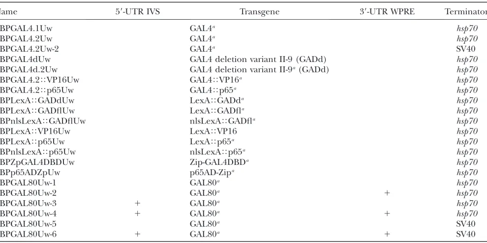
Optimization of vectors for GAL4 expression: We examined the effects of altering the sequence of the GAL4 gene and the UTR sequences that flank it in commonly used vectors. Figure 1 diagrams the structure of the vector we used to test these variants and Table 1 lists the resultant constructs. Codon-optimizing genes for ex-pression in heterologous hosts can improve levels of protein expression (Gustafssonet al. 2004). We found that using a variant of the yeast GAL4 gene that had been optimized for both Drosophila codon-usage and translation-initiation sequence increased levels of GAL4-driven GFP by50% (compare Figure 2B with 2C).
lacks the salivary gland background (Pfeifferet al. 2008). If indeed theP-element vector carries a cryptic salivary gland enhancer, it seems to have been lost in our constructs.
Most GAL4 constructs carry thehsp7039-UTR, which contains degradation sequences that cause rapid turnover in non-heat-shock conditions (Petersenand Lindquist 1989). Replacing the hsp70 transcriptional terminator used in our GAL4 vectors with the SV40 UTR is expected to result in greater mRNA stability, consistent with the increased expression levels we observed (Figure 2F). It may also result in perdurance of expression from earlier developmental stages, one explanation for the extra cells observed when using CRM R9C11-GAL4-SV40 (Figure 2F). Similar results were obtained using enhancers R9C11, R9B05, and R9D11 (supporting information, Figure S1, and Pfeifferet al. 2008).
Choice of activation domain can strongly affect levels of transcription (Ptashne 1988; Melcher 2000). We compared the GFP expression patterns driven by GAL4 constructs with four different activation domains: (1) GAL4; (2) GAL4 deletion variant II-9 (GAL4d, GAL4 amino acid residues 1–147 fused by a small linker with 768–881; Maand Ptashne1987a), which has been used in LexA (Lai and Lee 2006) and Split GAL4 vectors (Luanet al. 2006); (3) herpes simplex virus protein VP16
(Sadowskiet al. 1988); and (4) human p65 (Schmitz and Baeuerle1991), which has been successfully used to drive transcription of reporter transgenes in Dro-sophila as part of GeneSwitch (Osterwalderet al. 2001; Romanet al. 2001). Figure 3 shows expression driven by these GAL4 variants. R9C11-GAL4d (Figure 3B) drove GFP at a greatly reduced level, relative to intact GAL4 (Figure 3A). Conversely, replacing the GAL4 activation domain with either VP16 (Figure 3C) or p65 (Figure 3D) resulted in severalfold increases in GFP mRNA; the observed expression patterns were also broader, pre-sumably because weakly expressing cells became visible. Although GAL4Tp65 yielded the highest transcription levels, it reduced transformant viability. Cytotoxicity has been previously reported when GAL4 is expressed at very high levels (Kramerand Staveley2003).
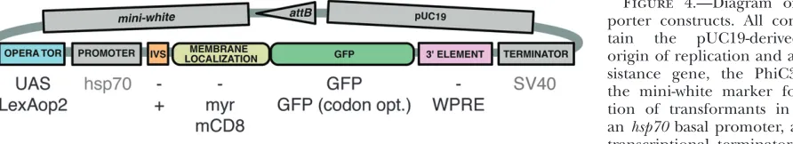
Improved UAS reporter constructs:Using a modular backbone (Figure 4), we generated a series of UAS-mCD8TGFP reporter constructs (Table 2) and used them to determine the effects on GFP expression of (1) the choice of basal promoter, (2) the presence of an intron in the 59-UTR, (3) the number of UAS sites, (4) the copy number of the reporter construct, (5) the nature of the protein localization signals fused to the GFP protein, and (6) the presence of the WPRE reg-ulatory element in the 39-UTR .
TABLE 1
New GAL4, LexA, Split GAL4, and GAL80 vector backbones used in this study
Name 59-UTR IVS Transgene 39-UTR WPRE Terminator
pBPGAL4.1Uw GAL4a hsp70
pBPGAL4.2Uw GAL4a hsp70
pBPGAL4.2Uw-2 GAL4a SV40
pBPGAL4dUw GAL4 deletion variant II-9 (GADd) hsp70
pBPGAL4d.2Uw GAL4 deletion variant II-9a(GADd) hsp70
pBPGAL4.2TVP16Uw GAL4TVP16a hsp70
pBPGAL4.2Tp65Uw GAL4Tp65a hsp70
pBPLexATGADdUw LexATGADda hsp70
pBPLexATGADflUw LexATGADfla hsp70
pBPnlsLexATGADflUw nlsLexATGADfla hsp70
pBPLexATVP16Uw LexATVP16 hsp70
pBPLexATp65Uw LexATp65a hsp70
pBPnlsLexATp65Uw nlsLexATp65a hsp70
pBPZpGAL4DBDUw Zip-GAL4DBDa hsp70
pBPp65ADZpUw p65AD-Zipa hsp70
pBPGAL80Uw-1 GAL80a hsp70
pBPGAL80Uw-2 GAL80a 1 hsp70
pBPGAL80Uw-3 1 GAL80a hsp70
pBPGAL80Uw-4 1 GAL80a 1 hsp70
pBPGAL80Uw-5 GAL80a SV40
pBPGAL80Uw-6 1 GAL80a 1 SV40
BP plasmid vector backbones are derived from pBPGUw (Pfeifferet al.2008) and contain the pUC19-derived bacterial origin of replication and ampicillin resistance gene, the PhiC31attBsite, the mini-white marker for identification of transformants in Drosophila, and the DSCP basal promoter. For more details see Figure 1 and Addgene plasmid 17575. Abbreviations: U, DSCP basal promoter; w, mini-white marker; nls, nuclear localization signal; IVS, intervening sequence within the 59-UTR; WPRE, a woodchuck hepatitis virus post-transcriptional regulatory element within the 39-UTR; TERMINATOR, the transcriptional termi-nator; and pBP, plasmid BP backbone.
a
We tested two basal promoters, the Drosophila syn-thetic core promoter (DSCP) (Pfeifferet al. 2008) and thehsp70basal promoter (Brandand Perrimon1993) to determine the levels of expression they gave when paired with UAS sites in the presence or absence of GAL4. The DSCP contains a large set of core promoter elements to promote robust expression with a broad range of enhancers that bind different activator pro-teins, which themselves might have specific preferences
for one or more of these promoter motifs (Pfeifferet al. 2008). While the DSCP promoter works well with a variety of enhancers, we found that in the specific case of GAL4-driven UAS expression, thehsp70basal promoter yielded twofold higher expression levels than the same construct built with the DSCP, while still displaying nearly undetectable leak in the absence of GAL4 (data not shown). We therefore used thehsp70basal promoter in subsequent UAS constructs.
terminators on GAL4-driven GFP transgene ex-pression. Adult brains are shown after immunos-taining to reveal GFP expression. (A) As a control for transcription of the UAS-mCD8TGFP
reporter construct (Lee and Luo1999) in the absence of a GAL4 driver, as well as for the back-ground of the immunohistochemistry proce-dure, UAS-mCD8TGFP was crossed to theattP2
site with no integrated construct. (B–F) The CRM R9C11 fragment (Pfeiffer et al. 2008) was used to drive GAL4 expression in constructs that are integrated into theattP2site and crossed to the UAS-mCD8TGFP reporter. (B) The GAL4
gene from Brandet al.(1994), which contains 45 bp of thehsp70 59-UTR and transcriptional ter-minators from both the GAL4 gene and the hsp70gene. CRM R9C11 drives expression prom-inently in the antennal mechanosensory and mo-tor center (AMMC) and mushroom body (MB). (C) GAL4.1, the same construct as in B, but with a GAL4 coding sequence optimized for Drosoph-ila codon usage. (D) GAL4.2, the same construct as in C, but with the 45-bphsp7059-UTR and yeast transcriptional terminator removed. (E) Same as D, but with GAL4 deletion variant II-9 (pMA236; Ma and Ptashne 1987a) replacing the full-length GAL4 gene. (F) Same as D, but with the SV40 terminator replacing the hsp70 terminator. Relative quantities of GFP mRNA expression levels as measured by QRT– PCR in homogenates of heads of each genotype relative to the control (calibrator) in A, which was arbitrarily set at 1.0, are in-dicated in the top right of each panel. Assays were done in triplicate except for F, which was done in duplicate; error was within 9% of reported values.
Figure 3.—Activation domain choice has a large effect on GAL4-driven levels. Drosophila adult brains were immunostained for GFP after crossing drivers to the UAS-mCD8TGFP
re-porter, as in Figure 2. All GAL4 constructs are di-rected by CRM R9C11, integrated intoattP2, and contain an hsp70 terminator. (A) GAL4 from pGawB (Brand and Perrimon 1993). (B) GAL4d, containing the GAL4 deletion variant II-9 (pMA236; Ma and Ptashne 1987a). The 45-bp segment of hsp70 59-UTR has been re-moved, but the yeast transcriptional termi-nator from the GAL4 gene is present. (C) GAL4.2TVP16, containing a fusion of the
GAL4 DNA-binding domain to the VP16 activa-tion domain. The entire coding region has been optimized for Drosophila codon usage and the hsp7059-UTR and yeast transcriptional termina-tor have been removed. (D) GAL4.2Tp65, as
in C, but with the activation domain from p65. Relative GFP QRT–PCR values, standardized toattP2crossed to UAS-mCD8TGFP
Intervening sequences (IVS, introns) placed in the 59-UTR of genes can boost expression by promoting steady-state export of spliced mRNAs to the cytoplasm (Huangand Gorman1990; Dunckeret al. 1997; Zieler and Huynh 2002). We added a 67-bp intron from DrosophilaMyosin heavy chain(Mhc IVS16) to pJFRC1, our 10XUAS-mCD8::GFP vector to make pJFRC2. GAL4-driven GFP mRNA levels from pJFRC2 were el-evated 20% compared to the same construct without the intron (data not shown). We also tested four other introns: synthetic intron IVS8 (Invitrogen),whiteintron 2 (Leeand Carthew2003),ftzintron (Niet al. 2009), and a hybrid intron (Choi et al. 1991). As none were substantially better than Mhc IVS16 in terms of
expres-sion levels and background (data not shown), we chose to use Mhc IVS16 in subsequent constructs.
pJFRC2 contains 10 copies of an optimized GAL4 DNA-binding site, the ‘‘ScaI 17-mer’’ (Websteret al. 1988), and drives GFP expression more than twofold higher than the UAS-mCD8TGFP construct described by Lee and Luo (1999) that contains 5 sites (compare Figure 5B with 5C). We varied the number of UAS sites from 5 to 40 and assayed levels of GFP expression driven from R9C11-GAL4 (Figure 5). Maximal GFP expression was obtained with 20 sites (pJFRC7; Figure 5E). Similar results were obtained by Ni et al. (2008) with RNAiNotchhairpin-induced phenotypes. Finally, while inclusion of gypsy insulators has been shown to boost transgene expression (Marksteinet al. Figure 4.—Diagram of pJFRC re-porter constructs. All constructs con-tain the pUC19-derived bacterial origin of replication and ampicillin re-sistance gene, the PhiC31 attB site, the mini-white marker for identifica-tion of transformants in Drosophila, anhsp70basal promoter, and an SV40 transcriptional terminator. The vector backbone is modular to allow for many possible combinations: gray shading indicates components that were held constant, while the colored elements were varied between the constructs we describe in this report. Examples of some of these alternatives are listed below colored elements; see text for more details.
TABLE 2
New reporter transgenes used in this study
Name Tandem construct Operator 59-UTR IVS Reporter 39-UTR WPRE
pJFRC-MUH 10XUAS
pJFRC1 10XUAS mCD8TGFP
pJFRC2 10XUAS 1 mCD8TGFP
pJFRC3 1XUAS 1 mCD8TGFP
pJFRC4 3XUAS 1 mCD8TGFP
pJFRC5 5XUAS 1 mCD8TGFP
pJFRC6 15XUAS 1 mCD8TGFP
pJFRC7 20XUAS 1 mCD8TGFP
pJFRC8 40XUAS 1 mCD8TGFP
pJFRC9 TH1 10XUAS 1 mCD8TGFP
10XUAS 1 mCD8TGFP
pJFRC10 THS2 10XUAS 1 mCD8TGFP
10XUAS 1 mCD8TGFP
pJFRC11 TTS3 10XUAS 1 mCD8TGFP
10XUAS 1 mCD8TGFP
pJFRC12 10XUAS 1 myrTGFPa
pJFRC13 10XUAS 1 GFPa
pJFRC14 10XUAS 1 GFPa 1
pJFRC15 13XLexAop2 mCD8TGFP
pJFRC16 16XLexAop2 mCD8TGFP
pJFRC17 26XLexAop2 mCD8TGFP
pJFRC18 8XLexAop2 mCD8TGFP
Janelia Farm Reporter Construct ( JFRC) backbones are derived from pBDP (Pfeifferet al.2008) and con-tain the pUC19-derived bacterial origin of replication and ampicillin resistance gene, the PhiC31attBsite, and the mini-white marker for identification of transformants in Drosophila. For more details see Figure 4 and Addgene plasmid 17566. In addition, all vectors also contain a basal promoter derived fromhsp70and an SV40 transcriptional terminator (Brandand Perrimon1993). Abbreviations: IVS, intervening sequence within the 59-UTR; and WPRE, a woodchuck hepatitis virus post-transcriptional regulatory element within the 39-UTR.
a
2008), we did not observe notable increases from the inclusion of these elements in pJFRC2 or pJFRC12 when integrated inattP2(data not shown).
We also tested expression driven from tandem con-structs, containing two copies of pJFRC2 in a single insert (Figure 6; Table 2), as the ability to drive multiple UAS constructs without complicated genetics can be helpful. A tandem construct with two copies of the internal components of pJFRC2 in a tail-to-head orien-tation, pJFRC9 (Figure 6D), gave more expression than pJFRC2 (Figure 6B), but less than pJFR7 (Figure 6C), which has the same number of UAS sites upstream of a single GFP gene. The addition of a 2.8-kb gypsy in-sulated spacer between the tandem copies of these same components in pJFRC10 (Figure 6E) increased expres-sion to above that of pJFRC7. The relative orientation of the tandem genes was significant; a tail-to-tail construct with a spacer, pJFRC11 (Figure 6F), gave the highest level of GFP expression. Both the spacer and a tail-to-tail orientation appear to be required to obtain the expected twofold increase over pJFRC2. While helpful in increas-ing expression levels, such tandem constructs will likely have their greatest utility when used to direct the ex-pression of two different responder genes under the control of the same GAL4 driver.
For many experiments, it is desirable to generate the same level of reporter expression while utilizing en-hancers of very different strengths. For example, de-creasing the number of UAS sites when using a strong enhancer-GAL4 driver will result in lower levels of reporter expression. We sought to evaluate our ability to modulate expression levels by varying the strength of the activation domain of the GAL4 driver and the
number of UAS sites carried by the reporter construct (Figure 7). mCD8TGFP driven from a single UAS site was almost undetectable, even with our strongest GAL4 driver, GAL4Tp65 (Figure 7C). The expression levels obtained increased with UAS number from 3 to 20 and with activation domain strength (Figure 7, D–R). Thus, using a single enhancer, we can obtain a range of expression levels from undetectable to undesirably high and cytotoxic, by simply modulating the strength of the activation domain and UAS responder. The toxicity ap-parent with the GAL4Tp65 driver and reporter con-structs with $10 UAS sites (Figure 7, L, O, and R) appears to result largely from reporter protein expres-sion levels, as this same driver with a 3X or a 5XUAS reporter results in labeled cells of apparently normal morphology (Figure 7, F and I; also seeFigure S2).
Depending on the experiment, it may be desirable to target the reporter protein to either membrane or cytoplasmic sites. pJFRC2 employs the first 222 amino acids of the mouse CD8 protein to target GFP to the plasma membrane as first described by Lee and Luo (1999). We also evaluated a second method of targeting proteins to the membrane,N-myristoylation (Resh1999); pJFRC12 employs the first 85 amino acids encoded by the DrosophilaSrc oncogene at 64B, Src64B, which has been used previously in Drosophila to target proteins to the membrane (Mausset al. 2009). Myristoylation improved signal strength; with 10XUAS, theN-myristoylated GFP reporter (pJFRC12; Figure 7, M–O) expresses GFP at levels similar to our 203 UAS-mCD8TGFP reporter (pJFRC7; Figure 7, P–R).
Signal strength observed with our cytoplasmic GFP reporter (pJFRC13; Figure 7, S–U) was weak compared Figure 5.—Increasing the number of GAL4 DNA-binding sites boosts GFP levels. Drosophila adult brains were immunostained for GFP after crossing R9C11-GAL4 to mCD8TGFP reporters
with 5 to 40 UAS sites. With the exception of the UAS-mCD8TGFP construct of Lee and Luo(1999), which is a P-element insertion on the second chromosome, all constructs are inte-grated into attP2. (A) attP2 (no GAL4 driver) crossed to pJFRC2 (negative control). (B–F) R9C11-GAL4 crossed to reporters containing (B) 5, (C) 10, (D) 15, (E) 20, and (F) 40 UAS sites. We also tested a 5XUAS construct in the pJFRC backbone (pJFRC5; see Table 2), which gave similar results to UAS-mCD8TGFP (data
not shown). Relative GFP QRT–PCR values, nor-malized to a level of 1.0 for attP2 3 UAS-mCD8TGFP (see Figure 2A), are indicated in
to that obtained with an equivalent membrane-targeted GFP construct (pJFRC2; Figure 7, J–L). However, inclusion of the woodchuck hepatitis virus post-transcriptional regulatory element (WPRE) (Zuffereyet al. 1999) in the 39-UTR (pJFRC14; Figure 7, V–X) substantially increased GFP levels, making them comparable to those seen with pJFRC2. However, the addition of the WPRE did not cause a notable increase in GFP expression from either the myristoylated or the mCD8 membrane-targeted reporters (data not shown).
Improved LexA drivers and operators: Laiand Lee (2006) showed that a C-terminal fusion with either the GAL4 activation domain II (GADd, residues 768–881 of GAL4) or the more potent VP16 activation domain from herpes simplex virus allows LexA-driven transcription of reporter transgenes in a GAL80-sensitive or -insensitive manner, respectively. We sought to improve the potency of these published LexA drivers by fusing LexA with two alternative activation domains. First, we used an ex-tended version of the GAL4 activation domain, GADf l (residues 148–881 of GAL4), which includes activation domains I and II (Maand Ptashne1987a). Second, we replaced VP16 with the human p65 activation domain. Also, because LexA is a prokaryotic protein and is not specifically targeted to the nucleus in eukaryotic cells (Brentand Ptashne1984), we reasoned that a nuclear localization signal (NLS) might increase its ability to promote transcription.
We constructed R9C11 enhancer-directed LexA drivers with a codon-optimized GADd, VP16 (codon
optimiza-tion of VP16 did not significantly improve LexA-driven transgene expression; data not shown), codon-optimized GADfl, or codon-optimized p65, with or without an NLS (Table 1). We compared the patterns and strength of expression observed when these constructs drove LexAop-rCD2TGFP (Lai and Lee2006). LexA drivers containing GADfl (Figure 8D) or p65 (Figure 8G) activation domains drove GFP expression at higher levels than GADd (Figure 8C) or VP16 (Figure 8F). In all cases, we observed the expected expression pattern, based on R9C11-GAL4 driving mCD8TGFP (Figure 8B). Surprisingly, expression levels of GFP driven from LexATGADfl were only slightly lower than those ob-tained with LexATp65. Although a rigorous quantitative comparison cannot be made, as the GFP reporter constructs used differ in structure and genomic loca-tion, the level of LexAop-rCD2TGFP expression driven from LexATGADfl seemed comparable to that seen with R9C11-GAL4 driving the mCD8TGFP reporter of Leeand Luo(1999). Further, as expected, tub-GAL80 (Leeand Luo1999) was able to suppress GFP expression from LexATGADfl (data not shown). Addition of a nuclear localization signal to the LexATGADfl (Figure 8E) or the LexATp65 (Figure 8H) proteins modestly increased their efficacy.
LexA protein binds, with various affinities, to a 20-bp motif found in the promoters of .20 E. coli genes (Schnarr et al. 1991; Wade et al. 2005). Drosophila LexA reporters (Laiand Lee2006) have used the colE1-binding motif because it was found to have one of the Figure6.—Tandem reporters can be used to increase output. (A) Diagram of reporter con-structs. Note variation in UAS copy number (in-dicated in the blue box), tandem orientation (indicated by the arrows), and presence of a gypsy-insulated spacer of 2.8 kb (INS). The syn-thesized spacer was designed by independently randomizing a minimum of five times (Shuffle program GCG Version 11.1; Accelrys, San Diego) a sequence derived from kanamycin CDS (base pairs 1–810) and a sequence derived from the E. colilacZ CDS (base pairs 799–2000) and then fusing these randomized sequences. The spacer was then flanked on either end with 424 bp from the 59-UTR of gypsy (base pairs 647–1074) that contains 12 binding sites for thesu(Hw)protein (Marlor et al.1986; Spanaet al. 1988). (B–F) Drosophila adult brains immunostained for GFP after crossing R9C11-GAL4 driver to indi-cated reporter. All constructs integrated into attP2. (B and C) Controls showing driver pat-tern with (B) 10 UAS copies (pJFRC2) or (C) 20 copies (pJFRC7). (D) A tandem 10XUAS-mCD8TGFP reporter in a tail-to-head
orienta-tion (pJFRC9). (E) As in D, but with inclusion of a gypsy-insulated 2.8-kb spacer between in-serts (pJFRC10). (F) As in E, but reporters are inserted tail to tail (pJFRC11). Relative GFP QRT–PCR values, standardized toattP23UAS-mCD8TGFP, are indicated in the top right corners. Assays were done in
Figure7.—Tuning expression levels by varying the strength of the activation domain and UAS responder. Drosophila adult brains were immunostained for GFP. With the exception of the 53UAS-mCD8TGFP (Leeand Luo1999) construct all transgenes are integrated intoattP2. CRM R9B05 was used to drive three GAL4 variants: standard GAL4 (as used in the constructs described by Pfeifferet al.2008), GAL4.2TVP16, or GAL4.2Tp65. These three GAL4 drivers were crossed to different responders as in-dicated, which vary in number of UAS sites, localization tag, and inclusion of a WPRE: (A–C) The 1XUAS-mCD8TGFP (pJFRC3).
10XUAS-highest affinities for LexA (Ebinaet al. 1983). Although thecolE1sequence permits robust transgene expression in Drosophila, these LexA reporters allow low levels of expression, or leak, in the absence of LexA, presumably due to affinity of endogenous transcription factors for the colE1-derived LexA-binding sites (Figure 9A and data not shown). In an attempt to identify other LexA-binding sites that would support strong expression in the presence of LexA, but not show any expression in its absence, we compared leak and levels of GFP induction from reporter transgenes containing sites from colE1 (Ebinaet al. 1983),sulA(Cole1983),umuDC(Kitagawa et al. 1985; Perryet al. 1985), or a synthetic 22-bp lexA operator (Brent and Ptashne 1984). The latter two operator sequences did not perform well in our assays and were dropped from further studies (data not shown). Moreover, the publishedcolE1LexA-binding sites showed strong leak in both larval and adult brains when used to drive mCD8-GFP under the DSCP promoter in attP2 (data not shown). Thus, we focused on sulA-derived LexA-binding sites for subsequent constructs. We also varied the number of binding sites, which we expected would influence levels of transgene expression on the basis of studies with the GAL4/UAS system (this study; Ni et al. 2008). We built mCD8TGFP reporters containing 8, 13, 16, or 26 LexA DNA-binding sites from the sulA operator and found the 133 version to give optimal expression with minimal leak (Figure 9 and data not shown). This reporter (pJFRC15) reproduced the
ex-pected expression pattern for three different enhancer– LexA constructs (Figure 9).
Refinement of expression patterns using Split GAL4: Luan et al. (2006) used GADd and VP16 as activation domains in their implementation of the Split GAL4 system. However, the low levels of reconstituted GAL4 expression obtained limit the utility of this intersectional approach. We tested whether the GADfl and p65 activa-tion domains, which we have shown drive much higher expression levels than the GADd and VP16 domains when fused to the LexA DNA-binding domain, could similarly improve expression in the Split GAL4 approach. We used enhancer R20B05 to drive either GADfl or p65 Split GAL4 activation domains. These were combined with the GAL4 DBD driven under either R35B08 or R50B06 and their ability to drive pJFRC2 was assayed. The p65 domain performed well. Both R35B08Gal4DBD\R20B05p65ADand R50B06Gal4DBD \ R20B05p65AD resulted in robust and specific GFP expression in the predicted cells, where the expression patterns of the two parent enhancers over-lapped; expression outside the overlap was not detected (Figure 10). In contrast, the GADfl domain performed poorly; neither R35B08Gal4DBD \ R20B05GADfl nor R50B06Gal4DBD\R20B05GADflyielded detectable GFP ex-pression (data not shown).
Luan et al. (2006) reported instances where Split GAL4 intersections showed expression in cells not observed in either parent pattern and we also observed this phenomenon, to varying extents, in many of the Figure8.—Improved LexA drivers with GADf l and p65 activation domains. Drosophila adult brains were immunostained for GFP after crossing LexA drivers to a published LexAop-rCD2TGFP (Laiand Lee2006). All GAL4 and LexA constructs are di-rected by CRM R9C11 and integrated intoattP2. (A)attP2(no LexA driver) crossed to published LexAop-rCD2TGFP (negative
control; note ‘‘leak’’ expression in the lamina). (B) R9C11-GAL4 with UAS-mCD8TGFP. (C and D) R9C11-LexA drivers containing
GAL4 activation domain variants GADd (C) or GADfl (D), crossed to rCD2TGFP. (E) As in D, but with a nuclear localization signal
(nls). (F and G) R9C11-LexA drivers with GAL80-insensitive activation domains VP16 (F) or p65 (G), crossed to rCD2TGFP. (H)
As in G, but with an nls. Relative GFP mRNA levels as measured by QRT–PCR, standardized toattP23UAS-mCD8TGFP (set as
1.0), are indicated in the top right of each panel. Assays were done in duplicate; error was within 14% of reported values.
mCD8TGFP (pJFRC2). (M–O) The 10XUAS-myrTGFP (pJFRC12): myristoylated, codon-optimized GFP. (P–R) The
20XUAS-mCD8TGFP (pJFRC7). (S–U) The 10XUAS-GFP (pJFRC13): untagged (cytoplasmic), codon-optimized GFP. (V–X) As in S–U,
Split GAL4 intersections we examined in the adult brain (A. Nern, B. D. Pfeifferand G. M. Rubin, unpublished results). One possible explanation is that stronger activation domains were used in the Split GAL4 con-structs than in the parent lines; we have shown that both the VP16 and the p65 activation domains drive broader transcription than the GAL4 activation domain (Figure 7). We therefore made additional attempts to develop Split GAL4 reagents, employing the GADf l domain. Since the GADfl peptide is approximately seven times
larger than GADd, GADf l may introduce steric hin-drance or other deleterious protein–protein interac-tions. However, our attempts to address this problem by increasing the length of the polyglycine linker connect-ing the leucine zipper to the GADfl domain from 20, 40, or 80 residues were unsuccessful (data not shown). In summary, when using the p65 activation domain, the Split GAL4 intersectional method performs well in a majority of cases. However, it is important to verify the resultant expression pattern by direct assays.
Figure9.—A LexA operator containing 13 binding sites fromsulAprovides robust and nonleaky expression. Drosophila adult brains were immunostained for GFP. With the exception of LexAop-rCD2TGFP (Laiand Lee2006) all constructs are integrated intoattP2. (A)attP2(no LexA driver) crossed to LexAop-rCD2TGFP (negative control; note ‘‘leak’’ expression in the lamina
indicated by arrowheads). (B)attP2(no LexA driver) crossed to pJFRC15-13XLexAop2-mCD8TGFP (pJFRC15; negative control,
Subtraction of GAL4 expression by GAL80:One of the features of the modular system of enhancers and vectors we are generating (Pfeiffer et al. 2008 and this study) is that, once the pattern of expression produced by an enhancer has been established, we expect to be able to use that enhancer to drive another protein in the same pattern. This assumption is particularly important for experiments using GAL80 to refine the patterns of GAL4-driven expression, as it has not been feasible to assay GAL80 expression directly. For the results to be predictable, the vectors used to express the two proteins must differ as little as possible in sequences that affect expression patterns. Moreover, GAL80 must be expressed at levels similar to or higher than GAL4, as it acts by directly binding to GAL4.
We first tested two enhancers that drive GAL4 in overlapping patterns and then moved one of the enhancers to a GAL80 vector. When these GAL4 and GAL80 constructs were present in the same fly, we obtained the expected pattern in which GAL4 activity is detected only in those cells that do not overlap between the patterns (Figure 11).
In the experiment described above the GAL80 vector used a 39-UTR from Simian virus 40 (SV40), which had been used in previous GAL80 constructs (Leeand Luo 1999; Stoleruet al. 2004; Susteret al. 2004). However, after observing the strong effect on expression of in-cluding the SV40 UTR in GAL4 vectors (Figure 2 and also seeFigure S1), we realized that it would be important Figure10.—Refinement of GFP expression using Split GAL4. Drosophila third instar central nervous systems were immunos-tained for GFP. All constructs are integrated intoattP2. (A)attP2(no GAL4 driver) crossed to pJFRC2-10XUAS-IVS-mCD8TGFP
to use the same 39-UTR in both our GAL4 and GAL80 vectors.
Thus, we generated a series of vectors for the expression of GAL80 that contain either the SV40 or the hsp70 39-UTR (Table 1) and tested them with the enhancer R11F05 (Figure 12). R11F05 directs reporter expression to100 sensory neurons that project into the ventral nerve cord of the third instar larva. The constructs were integrated into two genomic docking sites: attP2, on the third chromosome, which shows robust expression with GAL4 drivers, andattP40, on the second chromosome, which supports weaker expres-sion (see below). A single copy of R11F05-GAL80-SV40 in attP2, but not attP40, reduced GFP expression
generated by R11F05-GAL4 (in attP2) and UAS-mCD8TGFP (Leeand Luo1999) to undetectable levels (Figure 12, B and C). The increased suppression seen when the GAL80 construct is inserted in attP2 is presumably a simple reflection of the increased levels of transcription seen from constructs inserted at this genomic docking site compared with attP40. We also generated a tandem construct with two copies of R11F05-GAL80-SV40; when integrated in either attP40 orattP2, this construct was able to suppress GFP expres-sion to undetectable levels (data not shown).
When we substituted thehsp70UTR for the SV40 UTR in the GAL80 vector, suppression levels decreased (Figure 12, D and E). This decrease allowed us to test refinement of a GAL4 pat-tern using GAL80. Dis-sected third instar central nervous systems were im-munostained for GFP (green) and neurotactin (magenta). All GAL4 and GAL80 constructs are inte-grated inattP2; the reporter is a P-element insertion of UAS-mCD8TGFP on the
second chromosome (Lee and Luo1999). (A) R20B05 drives expression in the im-mature neurons of the op-tic lobes (OL) and in all of the lineages of second-ary neurons in the central brain and ventral nerve cord in third instar larvae. (B) A single optical section of the region indicated by the blue line in A at the level of the intermedi-ate commissures. R20B05 drives GFP expression in both anterior and posterior commissures of thoracic segments T1–T3 and ab-dominal segment A1: ante-rior commissures are indicated by solid yellow ar-rowheads. (C) As in B, but only the green channel (anti-GFP) is shown. (D) R15E07 drives expression in most of the secondary lineages in the central brain and VNC, with the notable exception of the neuroblast lineages whose axon bundles comprise the anterior intermediate commissure in the thoracic segments. It does not drive optic lobe expression. (E) A single optical section of the region indicated by the blue line in D. Note that R15E07 drives GFP expression in only the posterior commissures; the anterior commissures lacking GFP expression are in-dicated by open yellow arrowheads. (F) As in E, but only the green channel (anti-GFP) is shown. (G) R15E07-GAL80-SV40 (attP40) crossed to w; UAS-mCD8TGFP; R20B05-GAL4. As predicted, GFP expression is now restricted to the optic lobes (OL), a few
other methods for raising levels of GAL80 expression. We explored the use of two post-transcriptional regula-tory elements thought to act by increasing RNA trans-port from the nucleus to the cytoplasm, rather than mRNA stability: intron 16 (IVS) from DrosophilaMyosin heavy chain, and the WPRE (Zuffereyet al. 1999). One
or both elements were added to R11F05-GAL80-hsp70 (Table 1) and then assayed as above (Figure 12, F–I). Addition of either IVS or WPRE to R11F05-GAL80-hsp70 increased the extent of GFP suppression. When both were added, a single copy of GAL80 was able to reduce GAL4-driven GFP expression to almost undetectable Figure12.—GAL80 sup-pression of GAL4 is impro-ved with post-transcriptional regulatory elements (intron and WPRE). Drosophila third instar central nervous sys-tems were immunostained for GFP. Enhancer R11F05 was used to drive different GAL80 constructs. These constructs were then tested against their parent enhan-cer using a recombinant reporter line of R11F05-GAL4 (attP2) and a P -element insertion of UAS-mCD8TGFP on the third
chromosome. Both GAL4 and GAL80 constructs use anhsp70terminator, unless otherwise noted. GFP expres-sion was assayed after cross-ing R11F05-GAL4 (attP2) UAS-mCD8TGFP to (A)
Can-ton S(positive control), show-ing GFP expression in a subset of sensory neurons that project into the ven-tral nerve cord, and (B) R11F05-GAL80-SV40 (no post-transcriptional regula-tory elements), integrated in attP2. The inset at the bottom right shows a por-tion of the VNC at higher gain. (C) As in B, but inte-grated inattP40.The inset at the bottom right shows a portion of the VNC at higher gain. (D) R11F05-GAL80, integrated into attP2. A small number of neurons in the pattern es-cape suppression (express GFP). (E) As in D, but inte-grated into the weaker site attP40. Note increased inci-dence of neurons that es-cape suppression. (F–I) Inclusion of post-transcrip-tional regulatory elements to R11F05-GAL80 increases the level of GFP suppression: (F) R11F05-GAL80 with a WPRE, inattP40; (G) R11F05-GAL80 with an IVS, inattP40; (H) R11F05-GAL80 with both IVS and WPRE, inattP40; (I) R11F05-GAL80 with both IVS and WPRE, inattP2.Weak background GFP expression from GAL4-independent expression from the UAS-mCD8TGFP reporter was present
levels when inserted inattP40 and suppressed it com-pletely when inserted inattP2. Thus, R11F05-IVS-GAL80-WPRE-hsp70suppresses GAL4 at least as well as the SV40 version. We expect the addition of IVS and WPRE would further enhance GAL4 suppression when added to a GAL80-SV40 construct, as might be required when using a weak enhancer to drive GAL80 expression; for this reason we constructed pBPGAL80Uw-6 (Table 1).
Nogiand Fukasawa(1984) cloned and sequenced mutants of GAL80 (GAL80s) that suppress GAL4 in both
inducing and noninducing conditions inS. cerevisiae. One of these mutants, GAL80s-1, contains a
glycine-to-arginine substitution at amino acid 323, which is
thought to give it a higher affinity for GAL4 (Salmeron et al. 1990). We thought this mutation might also increase its GAL4 suppression in Drosophila, so we generated lines in which the R11F05 enhancer drives GAL80s-1or a
triple-mutant GAL80 containing theS-0,S-1, andS-2mutations, with or without the addition of IVS and WPRE. These GAL80s
constructs varied in their ability to suppress GAL4-driven GFP expression, but none was substantially better than 11F05-IVS-GAL80-WPRE-hsp70 (data not shown).
Assaying position effects at genomic attP docking sites: To generate complex genotypes it will be necessary to have severalattPsites with similar and favorable pro-uated genomic attP sites. The indicated 16 PhiC31 genomic attP integration sites were assayed for four properties: (1) expression in the adult nervous system when an enhancer trap vector was inserted, (2) expression from an exoge-nous enhancer, (3) expres-sion from a UAS construct responding to a GAL4 driver, and (4) transgene in-tegration rate. Sites shown in red meet all four criteria, while those shown in blue performed well with UAS reporter constructs, but not with enhancer-GAL4 drivers. Sites shown in black were rejected. See text for details. GenomicattP site references: attP1, attP2, attP3, attP18, and attP40 (Groth et al. 2004; Markstein et al. 2008); attP16 (Markstein et al. 2008); VK00005, VK00016, VK00026, and VK00027(Venkenet al.2006); and su(Hw)attP1, su(Hw)attP2, su(Hw)attP4, su(Hw)attP5, su(Hw)attP6, and su(Hw)attP8(Ni et al. 2009; this study).
perties. To identify such a set, we began with 16 PhiC31 genomicattPdocking sites (Figure 13) and assayed their ability to support (1) expression in the adult nervous system when an enhancer trap vector was inserted, (2) expression driven by an exogenous enhancer, and (3) expression from a UAS construct responding to a GAL4 driver. We tested for criterion 1 by constructing an en-hancer detector (O’Kane and Gehring 1987; Bellen et al. 1990), pBDPGAL4Uw, and integrating it into each of these sites. We crossed lines containing pBDPGAL4Uw with UAS-mCD8TGFP (Leeand Luo1999) and assayed for GFP in the adult brain (data not shown). In four cases, strong position effects were observed, while the remain-ing 12 candidateattPsites showed minimal or no detect-able expression of GFP in the adult brain. We confirmed these results using the stronger GFP reporter pJFRC2 (see Figure S3).
We then tested for the ability of these 12 selectedattP sites to allow robust expression of GAL4 driven by an enhancer and a strong core promoter, the DSCP pro-moter (Pfeiffer et al. 2008). Of the 12 sites, 2 gave integrants at low efficiency rates and were not analyzed further (Figure 14). We integrated three GAL4 drivers, R9C11, R9B05, and R9D11 (Pfeifferet al. 2008), which have different expression patterns, into the remaining 10 sites and crossed them to pJFRC2 (in attP2) to measure reproducibility and fidelity of expression be-tween sites (Figure 14; also seeFigure S4andFigure S5). Five sites (labeled in red in Figure 13) were identified as superior on the basis of their displaying little modifica-tion of the expression patterns observed when the three
test drivers were inserted in attP2, a site known to support robust expression (Markstein et al. 2008; Pfeifferet al. 2008).
Chromatin environment can influence transgene expression unpredictably, and thus the same genomic landing sites might not work well for both enhancer-driven GAL4 expression and UAS reporters. We there-fore also evaluated the 12 sites for their ability to support expression from an integrated UAS construct in response to GAL4. We crossed pJFRC2 integrated into each of the 12 differentattPgenomic sites to three GAL4 drivers (all in attP2), see above, to measure pattern fidelity and reliability of expression relative to the same construct inserted inattP2. Reproducibility and fidelity of pJFRC2 expression patterns were consistent across 11 of the 12 insertion sites (Figure 15; also seeFigure S6andFigure S7). Unlike enhancer–GAL4 constructs, the UAS trans-genes seem more refractory to chromatin influence and thus offer a larger choice for genomic integration sites, an observation also reported by Bischofet al. (2007). As summarized in Figure 13, by screening 16 differentattP docking sites we were able to identify 5 sites (labeled in red) that meet all three of our criteria as well as 6 additional sites (labeled in blue) that appear suitable for UAS–effector constructs but not for enhancer–GAL4 constructs.
whether this common background was caused by the insulators themselves, we assayed the ability of the gypsy insulator alone to drive transcription of GAL4. We found that the lateral horn expression seen in docking sites su(Hw)attP1, su(Hw)attP2, su(Hw)attP5, su(Hw) attP6, and su(Hw)attP8 is reproduced when the in-sulator itself is used as an enhancer (data not shown). In contrast to the GAL4 drivers, UAS effectors in-tegrated into these sites lacked the lateral horn back-ground and showed reproducible expression patterns relative toattP2. However, we did not see a significant boost in reporter gene expression levels when either a UAS or GAL4 was inserted in insulated genomic attP sites.
DISCUSSION
Many experiments in biological research rely critically on the ability to express exogenous proteins or RNAs in transgenic animals in a manner that is regulated for level, timing, and cell type. Methods that seek to attain that goal in D. melanogaster have been developed in a number of laboratories. Although widely used, the characteristics and limitations of these methods have generally not been critically evaluated and are often not appreciated by end users. In the studies we report here, we have attempted to assess different methods, un-derstand the variables that affect their performance, and use that knowledge to improve them.
We first studied the GAL4/UAS binary system (Brand and Perrimon 1993; Duffy 2002). Modifications in codon usage, activation domains, transcriptional termi-nators, and other features of the UTRs can result in substantial changes in both strength and pattern of expression. Varying the GAL4 activation domain has the single biggest effect on GAL4-driven expression, in terms of both expression level and number of cells observed in the pattern. The choice of GAL4 transcrip-tional terminator can also effect roughly twofold changes in reporter levels and alter the observed expression pattern, most likely by modifying the half-life of the GAL4 mRNA. We also systematically charac-terized the effects of promoters, introns, number of UAS-binding sites, tandem copies of UAS constructs, 39-post-transcriptional modifiers, and protein tags on GFP expression levels from UAS reporter constructs. These data should facilitate optimal construct design for most future GAL4/UAS applications.
The ability to use distinct binary systems to indepen-dently target different cells or tissues in the same animal has many applications. The bacterial repressor LexA (Brent and Ptashne 1981; Little et al. 1981) binds DNA sequences distinct from those recognized by GAL4 and has been successfully used in diverse eukaryotic systems such as yeast (Estojaket al. 1995), Drosophila
and Parinov 2008). However, the utility of LexA/ LexAop as a second binary system in Drosophila has been limited by the relative leakiness of the colE1-derived binding sites and weakness of the drivers compared to GAL4/UAS. We replaced the published GAL4 deletion (GADd) and VP16 activation domains with an extended GAL4 activation domain (GADf l) and the human p65 activation domain. We also screened LexA-binding sites from a variety of LexA target genes and found that 13 copies of a binding site derived from thesulAgene produced robust reporter expression in the presence of LexA, with no expression detectable by histochemical methods in its absence. However, sensi-tive assays using flippase suggest there may still be a very low level of leak under some circumstances (A. Nern, B. D. Pfeifferand G. M. Rubin, unpublished results). Used together, our new LexA drivers and reporter constructs produce similar expression levels and signal-to-noise ratios to those of the GAL4/UAS system.
Potteret al. (2010) reported development of a third binary system for use in Drosophila based on the qa gene cluster ofNeurospora crassa(Gileset al. 1991). This system shows great promise, but the apparent toxicity of the Q transcription factor (Potteret al. 2010) currently limits its widespread application. Once this issue is addressed, and with the improvements to the LexA system we report here, there will be three independent binary transcriptional activation systems available for use in Drosophila.
Even when single enhancers are used to drive GAL4 expression, few resultant patterns will be limited to a single cell type. In many cases, more restricted expres-sion will be desired for use in behavioral, anatomical, or developmental studies. Using Split GAL4 to restrict expression to the overlap between two ‘‘parent’’ en-hancers is one attractive option (Luan et al. 2006). However, the low level of expression obtained using the reconstituted GAL4 limited its utility. We were able to increase expression levels significantly by replacing the VP16 activation domain with the stronger activation domain from the human p65 protein.
the stoichiometric requirement for GAL80. In cases where GAL80 is driven from a strong promoter, such as
a-1 tubulin, a single transgene is sufficient to suppress GAL4-mediated transcription (Leeand Luo 1999; Vef et al. 2006); however, we found that when GAL4 and GAL80 are driven under the same enhancer, a single copy of GAL80 may be insufficient, especially when the vector utilizes the same, mRNA-destabilizing hsp70 39 -UTR as used in the GAL4 constructs. This problem can be fixed by using two copies of GAL80, either by generating a stock bearing GAL80 in two locations or by building a tandem construct with two copies of GAL80 in one insertion. Alternatively, the efficacy of a single copy of GAL80 can be improved by optimizing the codon usage of the GAL80 gene and including two post-transcriptional regulatory elements thought to increase the efficiency of mRNA transport, an intron and the WPRE. These changes allowed us to completely suppress GAL4 activity with a single copy of GAL80 when the two genes were expressed from the same enhancer in vectors designed to maximize the concurrence of the expression of the two proteins.
The local chromatin environment at the site of trans-gene insertion can alter both the pattern and the level of transgene expression (Spradling and Rubin 1983; Hiromiet al. 1985; Kirkpatricket al. 1994). Markstein et al. (2008) used luciferase to quantify such position effects in 20 genomicattPdocking sites and found a wide range of basal and inducible levels of expression. In addition, leakiness and inducibility varied not only across insertion loci, but also between tissues. These effects are not surprising, considering that chromatin changes are involved in tissue-specific gene regulation (Schulzeand Wallrath2007; Girtonand Johansen 2008). Thus, identification of suitable genomic docking sites relies on empirical testing in the tissues and developmental stages of interest. We assayed for posi-tion effects affecting adult brain GFP expression in 16 attPgenomic docking sites. Although there is no single ideal locus for all transgenes, we identified 5 sites that show reproducible levels of expression for enhancer– GAL4 constructs and 11 for UAS-mCD8TGFP. As far as we have tested, theseattPsites work well for other driver and responder combinations. These selected sites are also all permissive to integration, making them efficient for the production of transgenic lines.
In the course of this work we identified two areas that require further technology development. First, in a significant minority of driver lines, transgene expression is stochastic; that is, not all cells of a given type express the transgene, and the precise cells showing expression vary from animal to animal. We do not know the underlying cause of this variation, but we speculate that it most likely relates to the difficulty in overcoming chromatin blocks to initiating transcription. This phe-nomenon has been observed in a wide variety of trans-genic systems (see, for example, Ahmadand Henikoff
2001; Skoraand Spradling2010). Detecting stochastic expression often requires careful examination and is most easily scored in lines that express in repeating patterns, for example, in a cell type that occurs in each of the 800 cartridges of the lamina or each of the segments of the larval ventral nerve cord. We observed that increasing the strength of the activation domain can reduce this variation; also, certain integration sites in the genome appear to favor more uniform expression. But we have not found a general mechanism to resolve this issue, and at this point we avoid using such lines in experiments that depend on nonstochastic expression, such as measuring the behavioral effects of inactivation of a cell class.
The second issue that has not yet been adequately addressed is intrinsic to any experiment involving ex-pression of an exogenous protein or an endogenous protein at elevated levels: such expression can perturb cell function and cell structure. These effects can be obvious, such as cell death. However, sometimes they are subtle. Here the only solution is to assay the exogenous protein for its intended effects on the cell and use only the minimal level of expression sufficient for the experiment. The tools we have presented—for example, a series of vectors with different numbers of UAS sites—will be useful in achieving the desired expression level and also in normalizing expression levels when using enhancers of different strengths.
In conclusion, we report here an extensive set of experiments in which we empirically tested a wide range of modifications to the vectors and methods commonly used to direct exogenous gene expression in Drosoph-ila. We have been able to modulate the level of trans-gene expression by varying the strength of the activation domain carried by the transcriptional activator as well as the number of copies of its binding site and other properties of the reporter construct. Additional engi-neering to increase expression levels is not warranted, as significant toxicity appears to result from the exogenous proteins at the high end of our current expression range. We also solved the problem of leakiness of the LexA operator in the absence of LexA protein and made the Split GAL4 and GAL80 intersectional strategies more robust. Given the widespread use of these methods, we expect our results will have considerable utility.