RHODES, CASSIE LEA. Microglia Colonization and Disruption in the Developing Prairie Vole Brain. (Under the direction of Dr. Heather Patisaul.)
Microglia are neural immune cells that play an important role in normal brain development, and are susceptible to disruption by environmental factors, including chemicals and pollutants. Such exposures during developmental periods have been shown to alter microglia colonization and to result in behavioral changes at juvenile and adult ages. Many of these effects are reminiscent of neurodevelopmental disorders, including autism. This study utilized the prairie vole, an established model for the study of social behaviors and disorders, to investigate long-term effects on microglia colonization following early life exposure to a known endocrine disrupting chemical (EDC), bisphenol A (BPA). In order to understand developmental disruption of microglia in this species, we first quantified microglia colonization in the normally developing prairie vole brain over time – specifically in the dentate gyrus, medial amygdala, and central amygdala at postnatal days (PNDs)0, 1, 2, 4, 7, 10, 14, and 21. To assess long term effects of
early life exposure to BPA we quantified microglia density in the central and posterodorsal medial amygdala of adult voles previously exposed to BPA at PND8-14. Results in pups revealed that colonization increased with age and in region-specific patterns, but with no sex differences. Colonization in adults also showed no sex differences and no permanent impact on microglia numbers following BPA exposure. To our knowledge these data represent the first
by
Cassie Lea Rhodes
A thesis submitted to the Graduate Faculty of North Carolina State University
in partial fulfillment of the requirements for the degree of
Master of Science
Comparative Biomedical Sciences
Raleigh, North Carolina 2018
APPROVED BY:
_______________________________ _______________________________ Dr. Heather Patisaul Dr. Nanette Nascone-Yoder
Chair of Advisory Committee
_______________________________ _______________________________ Dr. John Metizen Dr. John Godwin
BIOGRAPHY
Cassie Rhodes was born in 1981 and grew up in Rocky Point, North Carolina. After graduating first in her class, Cassie attended The University of North Carolina at Chapel Hill, where she earned a Bachelor of Arts in both Psychology and Communication Studies in 2003. While there she interned in a psychology lab focused on social cognition and schizophrenia; worked as a residence counselor supporting youths with emotional difficulties; and spent a summer as a human resources intern for the Sanford division of Newell Rubbermaid. After graduating with honors, she began work at The Link Group, a market research firm based in Atlanta, GA and Durham, NC. While with the company she oversaw tracking studies for Fortune 500 companies and conducted ad hoc national and international projects. With a heavy emphasis on
pharmaceutical clients and a lifelong interest in both health and human behavior, Cassie left the company in 2012 to begin graduate school at North Carolina State University, where she earned a Masters of Physiology in 2014. Following graduation, she remained at NC State to work in the laboratory of Dr. Chad Stahl, where she learned about the role of inflammation in
obesity and disease. The following year she joined the graduate program in Comparative Biomedical Sciences at the NC State College of Veterinary Medicine, and worked in the
ACKNOWLEDGMENTS
I would like to think the many people who have helped me complete this stage of my education. First and foremost, I would like to thank my family and friends. My parents have always supported my many interests, and without their unending love and encouragement, I would not be where I am today. A special thank you to Saundra, Wade, and little Mason who have always been, and continue to be, a source of love and support. A note of love and
remembrance to my dear friend Lindsay, who’s life and memory continues to inspire and guide me on a daily basis. And a thank you to the instructors and students of the Krav Maga
community who I’ve had the pleasure to train with and to teach in recent years, and who are a constant source of inspiration and motivation. I’d also like to thank all of the advisors and
mentors I’ve had while at NC State, including Dr. Heather Patisaul for allowing me to work in her lab and introducing me to microglia; Dr. John Meitzen for his time, attention, and unending enthusiasm; and to Drs. Nanette Nascone-Yoder, Sabrina Robertson, and John Godwin for their guidance and support. I’d also like to thank Dr. Chad Stahl for his time, support, and advice; Dr.
James Petitte for his sincere and thoughtful insight; and Dr. Sam Jones for creating such a supportive and student oriented environment. And a special thank you Dr. Robert Kelly and the GAANN Fellowship for allowing me to be part of such a wonderful group of people, and for the support, both financial and otherwise. Last, but certainly not least, thank you to Andrea Vogel, Megan Serr, Sheryl Arambula, and Caroline Leitschuh for their friendship and perspective. My
sincerest thanks to each and everyone one of you, and to everyone else who has helped me throughout my time at NC State. The education I’ve received goes well beyond the classroom
TABLE OF CONTENTS
LIST OF TABLES ... vi
LIST OF FIGURES ... vii
Introduction ... 1
Methods and Materials: Experiment 1: Microglia Colonization of the Postnatal Vole Brain ... 7
Animal Care ... 7
Tissue Collection and Preparation ... 8
Immunocytochemistry ... 8
Quantification ... 9
Statistical Analysis ... 11
Experiment 2: Impact of Postnatal BPA Exposure on Adult Vole Microglia ... 12
Animal Care ... 12
Tissue Collection and Preparation ... 13
Immunocytochemistry ... 13
Quantification ... 14
Statistical Analysis ... 15
Results: Experiment 1: Microglia Colonization of the Postnatal Vole Brain ... 15
Dentate Gyrus ... 15
Medial Amygdala ... 16
Central Amygdala ... 16
Experiment 2: Impact of Postnatal BPA Exposure on Adult Vole Microglia ... 17
Medial Amygdala (posterodorsal nucleus) ... 17
Central Amygdala ... 17
LIST OF TABLES
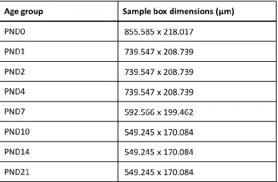
Table 1 Sample box sizes for the medial amygdala (MeA) in prairie vole pups by
LIST OF FIGURES
Figure 1 Microglia sample areas in the developing prairie vole brain ... 24 Figure 2 Microglia colonization in the developing prairie vole brain ... 25 Figure 3 Microglia in the brains of adult prairie voles that were postnatally
Introduction
Microglia are the resident immune cells of the central nervous system, and quickly respond to pathogenic insult and injury in the brain. However, they are also important for normal development. Specifically, microglia are involved in synaptic pruning, phagocytosing of dead or dying cells, and supporting the development of new neurons (Lenz & McCarthy, 2015; Morgan et al., 2010; Salter & Stevens, 2017; Vargas, Nascimbene, Krishnan, Zimmerman, & Pardo, 2005). Such activities are key parts of neural development, and make proper function of these cells an important part of forming a normal, healthy brain. In turn, disruption of such functions could result in abnormal development such as altered numbers of neurons or synapses, which have been associated with neurodevelopmental disorders like autism
(Courchesne et al., 2011; Hutsler & Zhang, 2010; Kim et al., 2017; Qiu, Aldinger, & Levitt, 2012; Schumann & Amaral, 2006). Interestingly, postmortem studies of patients with
neurodevelopmental disorders have shown increased microglia density and activity (Nelson & Lenz, 2017), suggesting microglia involvement, though not necessarily as a cause. Additionally,
there is evidence that one factor in the occurrence of these types of disorders is developmental exposure to endocrine disrupting chemicals (EDCs) (Bilbo, Block, Bolton, Hanamsagar, & Tran, 2017; Goines & Ashwood, 2013), some of which may alter microglia specifically. Previous work in our lab suggests that bisphenol A (BPA) may be one such chemical (Rebuli, Gibson, Rhodes, Cushing, & Patisaul, 2016). Therefore, the purpose of this study was to examine first a normal
study of sociality and social disorders, including autism spectrum disorder (ASD). In doing so, we hope to similarly establish the prairie vole as a neurotoxicological model, and to increase its utility as a tool for understanding toxicological factors at play in such disorders.
Unlike peripheral tissue macrophages which derive from the bone marrow and appear much later, microglia are born from the yolk sack and arise very early in embryonic
development. In rodents, microglia colonization begins around embryonic day 8-10 (Bilbo & Schwarz, 2012; Lenz & McCarthy, 2015). And while there is variation by brain region, overall microglia colonization appears to increase with age throughout the pre- and postnatal period (Harry & Kraft, 2012; Schwarz, Sholar, & Bilbo, 2012). However, colonization also appears to differ by sex. Specifically, at postnatal day (PND)4 male rats have significantly more microglia than females in a number of brain regions, including the dentate gyrus (DG) of the
hippocampus and the amygdala. Yet in these same brain regions at PNDs 30 and 60 females have more microglia than males, indicating a sex reversal at juvenile and adult ages (Schwarz et al., 2012). Further, microglia morphology appears to progress with age from a more amoeboid
phenotype during embryonic and early postnatal days, to a more ramified appearance with long processes in adulthood (Harry & Kraft, 2012; Schwarz et al., 2012). Though not entirely clear, these phenotypes may be associated with varying degrees of phagocytic activity. Specifically, amoeboid cells, characterized by large cell bodies and few processes, are more motile and phagocytic, and resemble the dominant morphology of microglia following tissue
overall, morphology appears to vary by sex, with males having more amoeboid cells at PND4,
and females having more cells with an “activated” or non-ramified morphology at PND30 and 60 (Schwarz et al., 2012).
Such sex differences, both in number and morphology, may provide a clue to the sex biases seen in disorders like autism that are much more prevalent in males and have early postnatal onsets (Hanamsagar & Bilbo, 2016; Lai, Lombardo, & Baron-Cohen, 2014).
Specifically, greater microglia colonization and higher numbers of amoeboid cells during the postnatal time period may suggest a higher degree of reactivity among males early in life (Hanamsagar & Bilbo, 2016). If this is the case, microglia in males may be more susceptible to disruption by exogenous factors during this time simply because they are more primed to produce an immune response. Studies of pathogenic insult support this idea of sex-biased immune activation, as bacterial infection at PND4 causes long term changes in microglial function in male rat pups, but not in females. These changes are in turn linked to altered behavior in adulthood, as well as to altered cytokine expression in the brain (Bilbo & Schwarz,
2012). Additionally, both altered cytokine levels and early life infection of varying types have been associated with an increased risk of ASD (Goines & Ashwood, 2013). But, importantly, infection later at PND30 does not result in the same long-term changes (Bilbo & Schwarz, 2012; Schwarz et al., 2012). Together, such research suggests the early postnatal period may be a window during which the brain is particularly sensitive to disruption of immunological pathways
Exposure to EDCs has also been linked to neurodevelopmental disorders, including autism (Bilbo et al., 2017; Goines & Ashwood, 2013). Like infections, developmental exposure to some of these chemicals has been shown to alter microglia in a sex-specific manner. For example, gestational exposure to diesel exhaust particles (DEP), a major component of roadway exposures to air pollution, resulted in altered microglia morphology, as well as increased
inflammatory cytokine protein, but only in the embryonic brains of male mice (Bolton et al., 2017), suggesting either activation or delayed maturation. Additionally, these effects were dependent on toll-like receptor (TLR)4, a well-known pattern recognition receptor (PAMP) that recognizes signs of pathogenic invaders, including lipopolysaccharide (LPS), a component of bacteria known to activate microglia. Interestingly, prenatal exposure to DEP has also been shown to result in vulnerability to behavioral deficits in adult offspring overall, but especially in males (Bolton, Auten, & Bilbo, 2014; Bolton et al., 2013). Further, research in general has linked ASD to several environmental chemicals, including not only air pollution (Roberts et al., 2013; Volk, Lurmann, Penfold, Hertz-Picciotto, & McConnell, 2013), but also polychlorinated biphenyls
(additives in industrial oils and lubricants that persist in the environment), polybrominated diphenyl ethers (flame retardants), and various pesticides (Goines & Ashwood, 2013). Such evidence suggests that exposure to environmental chemicals during development can alter microglia via disruption of immune pathways, which may in turn have permanent behavioral and cognitive effects, potentially including those seen in neurodevelopmental disorders.
the majority of the US population, including children and adolescents (Geens et al., 2012). Detection in the young is of special concern considering that detectable urinary levels in children have been linked to hyperactivity, increased anxiety, and deficits in executive function (Hong et al., 2013). Similarly, animal models have shown that pre- and postnatal BPA exposure can result in behavioral changes as juveniles and adults, including increased anxiety,
hyperactivity, and altered social interactions (Cox, Gatewood, Howeth, & Rissman, 2010; Patisaul & Bateman, 2008; Sullivan et al., 2014). Such behavioral changes are reminiscent of neurodevelopmental disorders like autism, which is characterized by deficits in social
communication and interaction, and restricted or repetitive behaviors, but also often includes anxiety (42-56% of cases) and attention deficit hyperactivity disorder (ADHD, 28-44%) (Lai et al., 2014). BPA is also a xenoestrogen, and thus suspected of causing adverse effects by mimicking estrogen, a hormone to which microglia are known to be sensitive (Lenz & McCarthy, 2015). In rats for instance, treatment of females with a male-typical dose of estradiol immediately after birth results in complete masculinization of microglia, both in increased numbers and increased
amoeboid morphology (Lenz, Nugent, Haliyur, & McCarthy, 2013). Such disruption suggests that developmental exposure to estrogenic substances like BPA may be of particular concern, and previous work in our lab has hinted that early postnatal exposure may in fact increase microglia colonization in the prairie vole brain (Rebuli et al., 2016). Here we build on that work by expanding our investigation of microglia colonization in those same animals, as well as
impacts of EDC exposure during that time of dynamic change. In characterizing postnatal microglia colonization in the brain of prairie voles, we focused on the dentate gyrus and amygdala, two areas in which microglia colonization has been well studied in other rodent models (Bolton et al., 2017; Rebuli et al., 2016; Schwarz et al., 2012). We hypothesized that colonization patterns during development would be similar to those seen in rats, including region-specific colonization, increasing colonization with age, and the exhibition of sex differences - specifically higher levels of colonization in males immediately after birth and higher colonization in females by weaning at PND21. In examining the long-term effects of postnatal exposure to BPA on microglia colonization, we focused on the amygdala of adult prairie voles, a brain region that may undergo an abnormal pattern of postnatal development in autism (Munson et al., 2006; Schumann & Amaral, 2006; Schumann et al., 2004; Sparks et al., 2002). Because previous research suggested a potential increase in the most medial aspects of this region following postnatal BPA exposure (Rebuli et al., 2016), we focused specifically on the posterodorsal portion of the medial amygdaloid nucleus (MePD). Additionally, because our
previous focus was limited to the more ventral nuclei of the amygdala, we also expanded our investigation to include the nearby central amygdala (CeA). For both areas, we hypothesized that exposure during a critical developmental period would result in altered microglia colonization patterns that would persist into adulthood. Additionally, based on subsequent findings showing sex and dose specific behavioral changes in these same animals (Sullivan et al.,
Methods and Materials
Experiment 1: Microglial Colonization of the Postnatal Vole Brain
Animal Care
All work was done according to the applicable portions of the Animal Welfare Act and the U.S. Department of Health and Human Services Guide for the Care and use of Laboratory Animals. All animal care was approved by the North Carolina State University Institutional Animal Care and Use Committee (IACUC) and the resident veterinarian. All prairie voles for experiment 1 were generously contributed from a colony maintained by Lisa McGraw at North Carolina State University, and housed in the North Carolina State University Biological
Resources Facility (Vogel, Patisaul, Arambula, Tiezzi, & McGraw, 2018). The room was temperature, humidity, and light-controlled (22C, 30% humidity, 12-hour light: 12-hour
darkness cycle with lights on 6AM-6PM). Animal housing consisted of polysulfone cages with corn cob bedding and crinkle nest enrichment, and food and water provided ad libitum. Diet consisted primarily of high fiber rabbit chow (Laboratory Rabbit Diet HF 5326, LabDiet, Missouri, USA) mixed with a small amount of rodent chow (Laboratory Rodent Diet 5001,
LabDiet, Missouri, USA), as well as occasional supplementation with timothy hay and alfalfa cubes (PicoLab Certified LabDiet Timothy Hay Cubes, PicoLab 5LRT). All water was reverse osmosis purified and provided in glass water bottles.
Due to the pair-bonding habits of prairie voles, all male/female pairs in the colony were placed together in the same cage and then remained co-housed indefinitely. When litters were
Cages were checked daily, and offspring age was determined based on the day of litter discovery, which was considered the day of birth or PND0.
Tissue Collection and Preparation
Animals younger than PND10 were sacrificed by rapid decapitation. Animals PND10 or older were first anesthetized with carbon monoxide, then sacrificed by rapid decapitation. All brains were removed immediately after decapitation and immersion fixed in 4%
paraformaldehyde (~20 mL) for 24 hours with gentle agitation at 4C. Brains were then
transferred to 30% sucrose in 4% paraformaldehyde for another 24 hours at 4C, and then to
30% sucrose in 0.02M potassium phosphate-buffered saline (KPBS) for a final 24 hours at 4C.
All brains were then flash frozen on powdered dry ice, wrapped in foil and stored at 80C. Sex
was determined on the day of sacrifice via internal examination of reproductive organs. The brains were then sliced into 50 µm serial sections on a freezing sliding microtome and stored in antifreeze (30% ethylene glycol, 5% glycerol, 30% sucrose in 0.1 M sodium phosphate buffer, with 10 g/L polyvinylpyrrolidone) at 20C until use.
Immunocytochemistry
Immunolabeling was conducted as we have done previously (Rebuli et al., 2016) with minor modifications to enhance tissue permeability. Sections were washed in a series of six 10-minute incubations in KPBS to remove the antifreeze, then incubated for 15 10-minutes in a
KPBS with 2% goat serum and 0.5% Triton-X for 30 minutes. All sections were then incubated in primary antibody solution consisting of rabbit anti-Iba-1 (Wako, 019-19741) at 1:10,000 in KPBS with 2% goat serum and 0.3% Triton-X overnight at 4°C with gentle agitation. Iba-1 (ionized calcium-binding adaptor molecule) was targeted due to its specificity to microglia and its
reliable expression regardless of microglia state. Sections then underwent six 10-minute washes in KPBS, and a 90-minute incubation in biotinylated goat anti-rabbit secondary antibody
(Jackson Immuno Research Laboratories, 111-065-144) at 1:200 in KPBS with 2% goat serum and 0.3% Triton-X. After another six 10-minute KPBS washes, tissue was incubated in an ABC (avidin/biotin complex) solution for 90 minutes (Vectastain Elite ABC HRP Kit from Vector Laboratories (PK-1600)), followed by a series of six 5-minute washes in KPBS. Sections were
then exposed for approximately 5 minutes to a DAB (3,3’-diaminobenzidine) solution with nickel enhancement (DAB Peroxidase (HRP) Substrate Kit with Nickel, Vector Laboratories, SK-4100). After chromagen labeling, all tissue was thoroughly rinsed in KPBS before being mounted on pre-cleaned Fisherbrand Superfrost Plus Microscope slides, allowed to dry, and then
dehydrated through a series of successive incubations: 2 minutes in 70% ethanol, a second 2-minute round in 70% ethanol, 2 2-minutes in 95% ethanol, 2 2-minutes in 100% ethanol, and incubation in xylene for 24 hours. Upon completion, the slides were coverslipped with DPX mountant and allowed to dry.
Quantification
Microsystems, Wetzlar, Germany) and quantified with the thresholding tool at 10x on the MCID Core image software program (MCID Core 7.0, InterFocus Imaging Ltd., Cambridge, England). Because an atlas has not yet been published for the prairie vole brain, all regions were identified according to the Developing Mouse Brain Atlas (Paxinos, 2007). For all regions, density was used as the endpoint (immunolabeled cells/ µm2 sampled area).
Dentate Gyrus
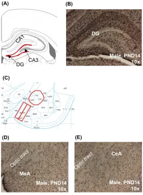
For each animal, one section approximately 4.35 mm to 5.07 mm from the most rostral section of the PND6 series was selected for quantification (Figure 1B) (Paxinos, 2007). The granular layer of the DG was outlined (Figure 1A), and the area quantified (µm2) using the MCID software. All immunolabeled cells within the boundary of the outlined region were then
counted by hand.
Medial Amygdala
One section approximately 4.95 mm from the most rostral section of the PND6 series was selected for each animal (Figure 1D) (Paxinos, 2007). Using MCID Core software, two rectangular frames were placed medially to laterally, just lateral to the optic tract (Figure 1C). Box sizes differed slightly by age to account for brain growth, and extended from the ventral edge of the MeA to just ventral to the central amygdala (see Table 1 for standardized frame
Immunolabeled cells were identified and quantified in each box using the thresholding tool. To ensure reproducibility, this procedure was independently performed by two different observers. The values from each observer were then averaged together to obtain the final cell counts and densities. Total cell count and sampled area was calculated by summing the cell counts and areas of the two boxes.
Central Amygdala
Sections selected for the MeA were also used for quantification of the CeA (Figure 1E). For each, the CeA as outlined by hand (i.e. a standardized sampling area was not used) (Figure 1C). Border identification was conservative; with the outline always falling just within the discernable edge of the CeA so as to not risk capturing any area outside of the CeA. Quantification of area and cell numbers was done with MCID as described for the MeA.
Statistical Analysis
All results were analyzed by two-way ANOVA (Sigmaplot 11.0) with sex and age as factors. Because prior work in rats has shown sex differences in microglial colonization, to ensure no potential age dependent sex differences were missed, all results were subsequently analyzed within age, even if no significant sex by age interaction was detected by the two-way ANOVA. Significant main effects (p≤0.05) were followed up with a protected Fisher’s least
by ANOVA. In these limited cases, unplanned poshoc analysis was conducted using a simple t-test and are specifically identified as unprotected post-hoc t-tests in the results.
Experiment 2: Impact of Postnatal BPA Exposure on Adult Vole Microglia
Animal Care
The animals used for experiment 2 were also used in previously published studies, and all details regarding animal husbandry and dosing for these prairie voles are detailed in that prior work (Rebuli et al., 2016; Sullivan et al., 2014). Briefly, all prairie voles for the present study were obtained via collaboration with Bruce Cushing, and originated from wild stock from Urbana, Illinois. The founders and subsequent colony were housed in an AAALAC approved facility at the Northeast Ohio Medical University at the University of Akron. Briefly, all animals were orally exposed to BPA PNDs8 – 14, which has previously been identified as a crucial sociosexual developmental window in prairie voles (Kramer, Perry, Golbin, & Cushing, 2009) and a point at which microglial colonization has been shown to be particularly dynamic in other
rodent species (Rebuli et al., 2016; Schwarz et al., 2012). Three doses of BPA were used: 5 µg/kg body weight (bw), 50 µg/kg bw (the oral reference dose established by the EPA), and 50 mg/kg bw (the EPA established lowest observed adverse effect dose) (Corrales et al., 2015). All doses were based upon the average weight of all pups on PND8 and dissolved in 2.7 g of
hydroxypropyl β cyclodextrin (Pharm Grade) in 10-mL 0.9% NaCl. A fourth group was exposed
Tissue Collection and Preparation
The tissue for experiment 2 was the same tissue used in a previous study, the collection and preparation of which has been described previously (Rebuli et al., 2016). Briefly, all animals were sacrificed between PND60 and PND90 via buprenorphine and ketamine-xylazine
overdose. Brains were removed and immersion fixed in 4% paraformaldehyde. Tissue was then shipped in cryoprotectant to the Patisaul lab at North Carolina State University. Brains were then flash frozen and stored at -80C. All tissue was coronally sliced into 35 µm sections on a
frozen sliding microtome.
Immunocytochemistry
As described in Rebuli et al (2016), and similar to what was done in experiment 1 (above), six consecutive sections per animal collectively containing the DG and amygdala were immunolabeled for Iba1 and developed with DAB as the chromagen. Only sections that
demonstrated visibly stained cells throughout, indicating complete tissue penetration, were used for quantification and analysis of microglia in this study. All sections were stained for the
ionized calcium-binding adaptor molecule (Iba-1) using standard immunolabeling protocols in our laboratory (Patisaul, Todd, Mickens, & Adewale, 2009). Rabbit anti-Iba1 (Wako Chemicals,
Quantification
Labeled microglia were imaged and quantified using the same approach and tools described in experiment 1. All regions were identified with the assistance of a Rat Brain Atlas (Paxinos & Watson, 2007). All region identification and counting was conducted by a
researcher blind to exposure groups.
Medial Amygdala (posterodorsal nucleus)
One section containing a midlevel section of the MePD (Bregma -2.76 through -3.00) was selected for each animal (Figure 3C). The sample area was defined using a standardized oval shaped frame (520 µm x 127 µm) the same width and height of the rectangular frames previously used for counting microglia in the amygdala of these animals (Figure 3A) (Rebuli et al., 2016). The shape of the frame was changed from a rectangle because it specifically fits within the boundaries of the adult MePD, as seen in previous work (Cao & Patisaul, 2013). For each animal, the oval frame was placed laterally along the optic tract. Microglia density within
the framed area was quantified using the thresholding tool of the MCID Core Image software as described for the MeA and CeA in experiment 1.
Central Amygdala
The same sections selected for the MePD were also used to quantify the CeA (Figure 3F).
Statistical Analysis
All results were analyzed by two-way ANOVA (Sigmaplot 11.0) with sex and BPA
exposure as factors. Because these regions have known sexual dimorphisms, to minimize risk of type 2 error, the data were then analyzed within sex, even if no main effect of sex was found.
Any significant main effects or interactions detected were followed up by Fisher’s least
significant differences (LSD) post-hoc test to evaluate pair-wise differences (p≤0.05).
Results
Experiment 1: Microglial Colonization of the Postnatal Vole Brain
Dentate Gyrus
Microglia colonization in the DG rapidly increased in the first two postnatal weeks (Figure 2A). Density was initially very low at PND0 through PND2 but began to show marked increases in both sexes at PND4 (Figure 2B). Two-way ANOVA revealed a significant effect of age (F(7,80)=61; p<0.001)), but not sex (F(1,80)=0.095; p=0.76) or a significant interaction with
sex (F(7,80)=0.708; p=0.67). Overall colonization increased significantly between PND4 and PND7 (p<0.001), and again between PND7 and PND10 (p<0.001) (Figure 2A). Unprotected t-test results also show that each sex alone saw increased colonization at these ages (PND4-7 males p<0.04, females p<0.002; PND7-10 males p<0.002, females p<0.001). The difference between males and females was largest at PND14 with density higher in males (Figure 2B), but an
Medial Amygdala
In the MeA, microglia density was greater in the first few postnatal days compared to the DG (Figure 2C). In both sexes, colonization increased significantly between PNDs 10 and 14 (p<0.001) (Figure 2D). Overall, two-way ANOVA revealed a main effect of age (F(7,77)=31; p<0.001) and sex (F(1,77)=5; p=0.03) with numbers significantly higher in males compared to females. There was, however, no significant sex by age interaction (F(7,77)=0.88; p=0.53). As in the DG, the largest difference between males and females occurred at PND14 (Figure 2D), with density higher in males. In the MeA, an unprotected t-test identified this difference as
statistically significant (p≤0.007).
Central Amygdala
The pattern of microglia colonization was similar to the MeA with density in the first few
postnatal days higher than in the DG (see Fig. 2C). A main effect of age (F(7,74)=51; p≤0.001)
was identified but not sex (F(1,74)=2; p=0.18), nor a significant interaction between age and sex
(F(7,74)=1; p=0.4). The first significant postnatal increase came earlier than in the MeA, however, occurring between PNDs7 and PND10 (p<0.001) (Figure 2E). Density also increased significantly between PNDs10 and PND14 (p<0.001). The most noticeable differences between males and females occurred at PNDs 14 and 21, with density higher in males at both points (Figure 2F), but unprotected t-tests did not find either to be statistically significant (PND14,
Experiment 2: Impact of Postnatal BPA Exposure on Adult Vole Microglia
Medial Amygdala (posterodorsal nucleus)
There were no significant differences in microglia colonization of the MePD in adult prairie voles postnatally exposed to BPA (Figure 3B). A two-way ANOVA showed no effect of sex (F(1,65)=2; p=0.17) or exposure (F(3,65)=1; p=0.66), and no significant sex by exposure
interaction (F(3,65)=0.4; p=0.76). In all exposure groups colonization appeared slightly higher among females, but even unprotected t-tests showed no significant differences. This included the gap between males and females within the 5µg/kg treatment group, which appeared to be largest, but an unprotected t-test showed no significant difference (p=0.14).
Central Amygdala
As in the MePD, there were no significant differences in microglia colonization of the CeA in adult voles postnatally exposed to BPA (Figure 3E). A two-way ANOVA showed no effect of sex (F(1,90)=2; p=0.13) or exposure level (F(3,90)=0.056; p=0.98), and no significant sex by
exposure interaction (F(3,90)=1.1; p=0.35). The male-female gap was most noticeable within the 50-mg exposure group, but the results of an unprotected t-test did not reach statistical significance (p=0.06).
Discussion
immediately after birth vs. low but noticeably higher colonization in the MeA and CeA at the same time points. This is similar to the much sparser colonization seen in the DG relative to the amygdala in early postnatal rat brains (Schwarz et al., 2012). Additionally, while microglia numbers increased with age in all regions of the prairie vole brain, the timing of those increases varied, with density in the DG climbing significantly within the first postnatal week, while the MeA and CeA did not see significant increases until the second postnatal week. However, colonization in both amygdalar subregions remained higher relative to the DG throughout the ages examined. This consistently higher density in the amygdala compared to the DG is also similar to reported colonization patterns in rats (Schwarz et al., 2012). Such comparable
patterns of colonization by age and region suggest a similar trajectory of microglia colonization across species.
Despite this generally congruous timeline with other species, predicted sex differences in microglia colonization were mostly absent in the developing prairie vole brain. Specifically, colonization did not vary significantly by sex at any age for any brain region. There were also no
significant differences in microglia colonization by sex in either of the amygdala subregions (MePD or CeA) of adult control animals, suggesting the lack of sex differences in amygdala subregions of PND0-PND21 animals persists into adulthood. These findings are in direct contrast to studies in rats showing higher microglia numbers in the DG and amygdala of males in early postnatal periods, but higher numbers in females by juvenile and adult ages. However,
we did find a main effect of sex in the MeA, where overall male prairie vole pups had a significantly higher density of microglia than females. While this failed to hold true with the smaller sample sizes of any particular age alone, it could point to a more general sex difference in that subregion. But overall, the consistent lack of sex differences in microglia density at any postnatal age and in adult controls suggests microglia colonization in the prairie voles deviates from other species. A lack of sex differences in cell numbers, however, does not rule out the possibility of sex differences by cellular morphology.
Our study also differed from research examining rats in our approach to quantification of microglia. While we used a combination of manual and computer assisted threshold to determine microglia cell counts and density (µm2 / sample area), previous studies have used unbiased stereology (Bolton et al., 2017; Schwarz et al., 2012), a method generally considered more precise and with the ability to assess the morphology of individual cells. Additional research using this other approach would therefore allow for not only a better head-to-head comparison of microglia numbers across species, but also a look at potential morphological
differences in the prairie vole across age and sex. Importantly, we have shown in rats that a lack of sex differences in total microglia colonization does not necessarily indicate a lack of
morphological sex differences (Rebuli et al., 2016). Specifically, even if total microglia counts are the same between male and female prairie voles in a given brain region, colonization in those same regions could still vary by the number of amoeboid, ramified, and intermediate cells
approach provides a valuable initial look at microglia in the developing prairie vole brain, investigating colonization by morphology is an important next step for a more complete picture.
Our work also revealed no significant differences in microglia colonization by exposure in amygdalar subregions of adult brains following postnatal exposure to BPA. Previous work in our lab has shown that the amygdala may be vulnerable to BPA exposure (Arambula, Jima, & Patisaul, 2018), and thus an important target for continued research. And previous work with these animals in particular has suggested potential subregional susceptibility (Rebuli et al., 2016), which led us to focus on the subregions reported herein. However, overall density of microglia colonization in the MePD and CeA showed no significant effects of exposure, suggesting total numbers were not impacted. But as shown by our previous research in rats, exposure both to BPA and to estradiol can significantly alter microglia morphology without necessarily altering microglia number (Rebuli et al., 2016). Thus, as with our developmental findings, whether or not microglia morphology in these regions was affected by exposure
remains unknown, but cannot be ruled out.
Due to the timing of our microglia assessment, this study also does not eliminate the possibility of transient effects on microglia colonization at the time of BPA exposure.
Specifically, BPA may have caused changes in microglia during postnatal exposure that did not persist into adulthood. Notably, research with rats developmentally exposed to BPA until the
day of sacrifice (PND12) indicated changes in microglia numbers and morphology (Rebuli et al., 2016). And because the postnatal period constitutes a critical window, even temporary
cause permanent changes to the developing brain. This could in turn lead to altered behavior later in development and into adulthood, which, importantly, was previously reported in these same animals (Sullivan et al., 2014), even in the absence of lasting changes in microglia
colonization.
And finally, another possibility for the lack of detectable BPA related changes is that the exposure period in this study missed the window of susceptibility for microglia colonization. Importantly, our developmental data show that the PND8-14 exposure did capture periods of significant microglia increase in both the MeA (PND10-14), which included a portion of the MePD, and the CeA (PND7-14), and at least part of the PND4-10 increase in the DG. However, this exposure likely did not capture the critical window for masculinization of the prairie vole brain, which in other rodents ends during the first postnatal week (Lenz & McCarthy, 2015). Notably, it is during this first postnatal week that treating females with estradiol has been shown to result in masculinization of microglia numbers and morphology in the preoptic area (POA) (Lenz et al., 2013). And considering its estrogenic nature, BPA may share that same
window of susceptibility. Thus, despite occurring during a time of increasing colonization in the brain regions examined, BPA exposure in this study may have missed the period in which microglia are most vulnerable to its effects.
To our knowledge these data represent the first characterization of postnatal microglia colonization in the prairie vole brain. Further research is warranted to generate a more
exogenous factors. The prairie vole is an established model for the study of sociality and social deficits, including those seen in autism and other neurodevelopmental disorders. And
considering the potential role of chemical pollutants in the etiology of such disorders, they are an increasingly important tool for neurotoxicological work as well. And while this study found no evidence that BPA exposure in the second postnatal week permanently affected microglia density, other characteristics warrant investigation, as do potentially more transient effects at earlier time points.
REFERENCES
Arambula, S. E., Jima, D., & Patisaul, H. B. (2018). Prenatal bisphenol A (BPA) exposure alters the transcriptome of the neonate rat amygdala in a sex-specific manner: a CLARITY-BPA consortium study. Neurotoxicology, 65, 207-220. doi:10.1016/j.neuro.2017.10.005 Bilbo, S. D., Block, C. L., Bolton, J. L., Hanamsagar, R., & Tran, P. K. (2017). Beyond infection -
Maternal immune activation by environmental factors, microglial development, and relevance for autism spectrum disorders. Exp Neurol.
doi:10.1016/j.expneurol.2017.07.002
Bilbo, S. D., & Schwarz, J. M. (2012). The immune system and developmental programming of brain and behavior. Front Neuroendocrinol, 33(3), 267-286.
doi:10.1016/j.yfrne.2012.08.006
Bolton, J. L., Auten, R. L., & Bilbo, S. D. (2014). Prenatal air pollution exposure induces sexually dimorphic fetal programming of metabolic and neuroinflammatory outcomes in adult offspring. Brain Behav Immun, 37, 30-44. doi:10.1016/j.bbi.2013.10.029
Bolton, J. L., Huff, N. C., Smith, S. H., Mason, S. N., Foster, W. M., Auten, R. L., & Bilbo, S. D. (2013). Maternal stress and effects of prenatal air pollution on offspring mental health outcomes in mice. Environ Health Perspect, 121(9), 1075-1082.
doi:10.1289/ehp.1306560
Bolton, J. L., Marinero, S., Hassanzadeh, T., Natesan, D., Le, D., Belliveau, C., . . . Bilbo, S. D. (2017). Gestational Exposure to Air Pollution Alters Cortical Volume, Microglial
Morphology, and Microglia-Neuron Interactions in a Sex-Specific Manner. Front Synaptic Neurosci, 9, 10. doi:10.3389/fnsyn.2017.00010
Cao, J., & Patisaul, H. B. (2013). Sex-specific expression of estrogen receptors alpha and beta and Kiss1 in the postnatal rat amygdala. J Comp Neurol, 521(2), 465-478.
doi:10.1002/cne.23185
Corrales, J., Kristofco, L. A., Steele, W. B., Yates, B. S., Breed, C. S., Williams, E. S., & Brooks, B. W. (2015). Global Assessment of Bisphenol A in the Environment: Review and Analysis of Its Occurrence and Bioaccumulation. Dose Response, 13(3), 1559325815598308.
doi:10.1177/1559325815598308
Courchesne, E., Mouton, P. R., Calhoun, M. E., Semendeferi, K., Ahrens-Barbeau, C., Hallet, M. J., . . . Pierce, K. (2011). Neuron number and size in prefrontal cortex of children with autism. JAMA, 306(18), 2001-2010. doi:10.1001/jama.2011.1638
Geens, T., Aerts, D., Berthot, C., Bourguignon, J. P., Goeyens, L., Lecomte, P., . . . Covaci, A. (2012). A review of dietary and non-dietary exposure to bisphenol-A. Food Chem Toxicol, 50(10), 3725-3740. doi:10.1016/j.fct.2012.07.059
Goines, P. E., & Ashwood, P. (2013). Cytokine dysregulation in autism spectrum disorders (ASD): possible role of the environment. Neurotoxicol Teratol, 36, 67-81.
doi:10.1016/j.ntt.2012.07.006
Hanamsagar, R., & Bilbo, S. D. (2016). Sex differences in neurodevelopmental and
neurodegenerative disorders: Focus on microglial function and neuroinflammation during development. J Steroid Biochem Mol Biol, 160, 127-133.
doi:10.1016/j.jsbmb.2015.09.039
Harry, G. J., & Kraft, A. D. (2012). Microglia in the developing brain: a potential target with lifetime effects. Neurotoxicology, 33(2), 191-206. doi:10.1016/j.neuro.2012.01.012 Hong, S. B., Hong, Y. C., Kim, J. W., Park, E. J., Shin, M. S., Kim, B. N., . . . Cho, S. C. (2013).
Bisphenol A in relation to behavior and learning of school-age children. J Child Psychol Psychiatry, 54(8), 890-899. doi:10.1111/jcpp.12050
Hutsler, J. J., & Zhang, H. (2010). Increased dendritic spine densities on cortical projection neurons in autism spectrum disorders. Brain Res, 1309, 83-94.
doi:10.1016/j.brainres.2009.09.120
Kim, H. J., Cho, M. H., Shim, W. H., Kim, J. K., Jeon, E. Y., Kim, D. H., & Yoon, S. Y. (2017). Deficient autophagy in microglia impairs synaptic pruning and causes social behavioral defects. Mol Psychiatry, 22(11), 1576-1584. doi:10.1038/mp.2016.103
Kramer, K. M., Perry, A. N., Golbin, D., & Cushing, B. S. (2009). Sex steroids are necessary in the second postnatal week for the expression of male alloparental behavior in prairie voles (Microtus ochragaster). Behav Neurosci, 123(5), 958-963. doi:10.1037/a0016927 Lai, M. C., Lombardo, M. V., & Baron-Cohen, S. (2014). Autism. Lancet, 383(9920), 896-910.
doi:10.1016/S0140-6736(13)61539-1
Lenz, K. M., & McCarthy, M. M. (2015). A starring role for microglia in brain sex differences. Neuroscientist, 21(3), 306-321. doi:10.1177/1073858414536468
Lenz, K. M., Nugent, B. M., Haliyur, R., & McCarthy, M. M. (2013). Microglia are essential to masculinization of brain and behavior. J Neurosci, 33(7), 2761-2772.
doi:10.1523/JNEUROSCI.1268-12.2013
Morgan, J. T., Chana, G., Pardo, C. A., Achim, C., Semendeferi, K., Buckwalter, J., . . . Everall, I. P. (2010). Microglial activation and increased microglial density observed in the
Munson, J., Dawson, G., Abbott, R., Faja, S., Webb, S. J., Friedman, S. D., . . . Dager, S. R. (2006). Amygdalar volume and behavioral development in autism. Arch Gen Psychiatry, 63(6), 686-693. doi:10.1001/archpsyc.63.6.686
Nelson, L. H., & Lenz, K. M. (2017). The immune system as a novel regulator of sex differences in brain and behavioral development. J Neurosci Res, 95(1-2), 447-461.
doi:10.1002/jnr.23821
Palanza, P. L., Howdeshell, K. L., Parmigiani, S., & vom Saal, F. S. (2002). Exposure to a low dose of bisphenol A during fetal life or in adulthood alters maternal behavior in mice. Environ Health Perspect, 110 Suppl 3, 415-422.
Patisaul, H. B., & Bateman, H. L. (2008). Neonatal exposure to endocrine active compounds or an ERbeta agonist increases adult anxiety and aggression in gonadally intact male rats. Horm Behav, 53(4), 580-588. doi:10.1016/j.yhbeh.2008.01.008
Patisaul, H. B., Todd, K. L., Mickens, J. A., & Adewale, H. B. (2009). Impact of neonatal exposure to the ERalpha agonist PPT, bisphenol-A or phytoestrogens on hypothalamic kisspeptin fiber density in male and female rats. Neurotoxicology, 30(3), 350-357.
doi:10.1016/j.neuro.2009.02.010
Paxinos, G. (2007). Atlas of the developing mouse brain at E17.5, P0 and P6 (1st ed.). Amsterdam ; Boston: Elsevier.
Paxinos, G., & Watson, C. (2007). The rat brain in stereotaxic coordinates (6th ed.). Amsterdam ; Boston ;: Academic Press/Elsevier.
Qiu, S., Aldinger, K. A., & Levitt, P. (2012). Modeling of autism genetic variations in mice: focusing on synaptic and microcircuit dysfunctions. Dev Neurosci, 34(2-3), 88-100. doi:10.1159/000336644
Rebuli, M. E., Gibson, P., Rhodes, C. L., Cushing, B. S., & Patisaul, H. B. (2016). Sex differences in microglial colonization and vulnerabilities to endocrine disruption in the social brain. Gen Comp Endocrinol, 238, 39-46. doi:10.1016/j.ygcen.2016.04.018
Roberts, A. L., Lyall, K., Hart, J. E., Laden, F., Just, A. C., Bobb, J. F., . . . Weisskopf, M. G. (2013). Perinatal air pollutant exposures and autism spectrum disorder in the children of Nurses' Health Study II participants. Environ Health Perspect, 121(8), 978-984. doi:10.1289/ehp.1206187
Salter, M. W., & Stevens, B. (2017). Microglia emerge as central players in brain disease. Nat Med, 23(9), 1018-1027. doi:10.1038/nm.4397
Schumann, C. M., Hamstra, J., Goodlin-Jones, B. L., Lotspeich, L. J., Kwon, H., Buonocore, M. H., . . . Amaral, D. G. (2004). The amygdala is enlarged in children but not adolescents with autism; the hippocampus is enlarged at all ages. J Neurosci, 24(28), 6392-6401.
doi:10.1523/JNEUROSCI.1297-04.2004
Schwarz, J. M., Sholar, P. W., & Bilbo, S. D. (2012). Sex differences in microglial colonization of the developing rat brain. J Neurochem, 120(6), 948-963.
doi:10.1111/j.1471-4159.2011.07630.x
Sparks, B. F., Friedman, S. D., Shaw, D. W., Aylward, E. H., Echelard, D., Artru, A. A., . . . Dager, S. R. (2002). Brain structural abnormalities in young children with autism spectrum
disorder. Neurology, 59(2), 184-192.
Sullivan, A. W., Beach, E. C., Stetzik, L. A., Perry, A., D'Addezio, A. S., Cushing, B. S., & Patisaul, H. B. (2014). A novel model for neuroendocrine toxicology: neurobehavioral effects of BPA exposure in a prosocial species, the prairie vole (Microtus ochrogaster). Endocrinology, 155(10), 3867-3881. doi:10.1210/en.2014-1379
Vargas, D. L., Nascimbene, C., Krishnan, C., Zimmerman, A. W., & Pardo, C. A. (2005). Neuroglial activation and neuroinflammation in the brain of patients with autism. Ann Neurol, 57(1), 67-81. doi:10.1002/ana.20315
Vogel, A. R., Patisaul, H. B., Arambula, S. E., Tiezzi, F., & McGraw, L. A. (2018). Individual
Variation in Social Behaviours of Male Lab-reared Prairie voles (Microtus ochrogaster) is Non-heritable and Weakly Associated with V1aR Density. Sci Rep, 8(1), 1396.
doi:10.1038/s41598-018-19737-9
Volk, H. E., Lurmann, F., Penfold, B., Hertz-Picciotto, I., & McConnell, R. (2013). Traffic-related air pollution, particulate matter, and autism. JAMA Psychiatry, 70(1), 71-77.