DOI: 10.1534/genetics.109.108134
Requirement for the ERI/DICER Complex in Endogenous
RNA Interference and Sperm Development in
Caenorhabditis elegans
Derek M. Pavelec,*
,†Jennifer Lachowiec,* Thomas F. Duchaine,
‡Harold E. Smith
§and Scott Kennedy*
,1*Department of Medical Genetics and Department of Pharmacology,†Program in Molecular and Cellular Pharmacology, University of Wisconsin, Madison, Wisconsin 53706,‡McGill Cancer Center and Department of Biochemistry, McGill University, Quebec H3G 1Y6, Canada
and§National Institute of Diabetes and Digestive and Kidney Diseases, National Institutes of Health, Bethesda, Maryland 20892 Manuscript received August 3, 2009
Accepted for publication September 17, 2009
ABSTRACT
Small regulatory RNAs are key regulators of gene expression. One class of small regulatory RNAs, termed the endogenous small interfering RNAs (endo siRNAs), is thought to negatively regulate cellular transcripts via an RNA interference (RNAi)-like mechanism termed endogenous RNAi (endo RNAi). A complex of proteins composed ofERI-1/3/5,RRF-3, and DICER (the ERI/DICER complex) mediates endo RNAi processes in Caenorhabditis elegans. We conducted a genetic screen to identify additional components of the endo RNAi machinery. Our screen recovered alleles oferi-9, which encodes a novel DICER-interacting protein, and a missense mutation within the helicase domain of DICER [ DCR-1(G492R)]. ERI-9() andDCR-1(G492) animals exhibit defects in endo siRNA expression and a con-comitant failure to regulate mRNAs that exhibit sequence homology to these endo siRNAs, indicating that ERI-9 and theDCR-1helicase domain function in theC. elegansendo RNAi pathway. We define a subset of Eri mutant animals (includingeri-1,rrf-3,eri-3, anddcr-1, but noteri-9orergo-1) that exhibit temperature-sensitive, sperm-specific sterility and defects in X chromosome segregation. Among these mutants we find multiple aberrations in sperm development beginning with cytokinesis and extending through terminal differentiation. These results identify novel components of the endo RNAi machinery, demonstrate differential requirements for the Eri factors in the sperm-producing germline, and begin to delineate the functional requirement for the ERI/DICER complex in sperm development.
E
UKARYOTIC cells express a wide variety of 20–30 nucleotide small regulatory RNAs that function in a wide range of biological processes including, but not limited to, heterochromatin formation, developmental timing, defense against parasitic nucleic acids, and genome rearrangement (Lee et al.1993; Wianny andZernicka-Goetz 2000; Knight and Bass 2001; Hall et al. 2002; Mochizuki et al. 2002; Plasterk 2002;
Mochizuki and Gorovsky 2004; Verdel et al. 2004;
Camet al. 2005). Small regulatory RNAs associate with
ARGONAUTE (AGO) and PIWI proteins. Together, small regulatory RNAs and AGO/PIWI proteins seek out and regulate homologous nucleic acid sequences via a variety of mechanisms, including decreased mRNA stability, translational repression, transcriptional repres-sion, meiotic silencing of unpaired DNA, and DNA elimination (Kim2005).
One class of small regulatory RNA, termed the endogenous small interfering RNAs (endo siRNAs), was identified via biochemical purification in Caeno-rhabditis elegans(Ambroset al.2003). Endo siRNAs have
now been identified in a wide array of eukaryotic organisms, including mammals (Hamiltonet al.2002;
Llave et al.2002; Tanget al. 2003; Czechet al. 2008;
Ghildiyalet al.2008; Kawamuraet al.2008; Tamet al.
2008; Watanabeet al.2008). InC. elegans, endo siRNAs
are complementary to predicted coding and noncoding genomic sequences and map to a large number of clusters within theC. elegansgenome (Rubyet al.2006).
Several proteins that are required for the biogenesis and/or stability of a subset of the cellular endo siRNAs in C. elegans have been identified, including the exo-nuclease ERI-1, the RNA-dependent RNA polymerase (RdRP) RRF-3, ERI-3, and the Tudor-domain protein
ERI-5(collectively, ERIs) (Simmeret al.2002; Kennedy et al.2004; Duchaineet al.2006).ERI-1,RRF-3,ERI-3,
and ERI-5 coprecipitate with DCR-1, suggesting that these factors assemble into a complex with Dicer (DCR-1
inC. elegans), an RNase III enzyme that converts double-stranded RNAs (dsRNAs) to small RNAs (Duchaine Supporting information is available online athttp://www.genetics.org/
cgi/content/full/genetics.109.108134/DC1.
1Corresponding author: 2434 Genetics-Biotechnology Center Bldg., 425 Henry Mall Madison, WI 53706. E-mail: [email protected]
et al.2006). This complex of proteins has been termed the ERI/DICER complex. Animals lacking compo-nents of the ERI/DICER complex fail to express some endo siRNAs and also overexpress cellular mRNAs that exhibit sequence homology to these endo siRNAs (Duchaineet al.2006; Leeet al.2006; Asikainenet al.
2007). Consequently, endo siRNAs are postulated to initiate or perpetuate the silencing of mRNAs via a process termed endogenous RNA interference (endo RNAi) Duchaine et al. 2006; Leeet al. 2006). Finally, C. elegans lacking ERI-1, RRF-3, or ERI-3 exhibit a temperature-sensitive (ts) sterile phenotype, hinting that the ERI/DICER complex and endo siRNAs may play important roles during germline development (Kennedyet al.2004; Duchaineet al.2006).
Here we report the molecular identification and characterization of genes required for endo siRNA expression inC. elegans. We identify a subset of endo RNAi genes that are required for endo RNAi in the male germline and document a role for these genes in sperm development.
MATERIALS AND METHODS
C. elegansstrains:Bristol strainN2was used as the standard wild-type strain. See thesupporting information,File S1, for a full list of strains used in these studies.
RNAi experiments:RNAi experiments were conducted as described previously (Timmons et al. 2001). HT115 E. coli expressing dsRNA, includingsqt-3,lir-1,unc-73,cel-1,tra-2, dpy-13,lin-1,unc-22, andlin-15a, were obtained from the Ahringer RNAi library (Kamathet al.2003) and sequenced to verify their identities. lin-15b RNAi was performed as described previously (Guanget al.2008).
RNA analysis:Sequences for quantitative reverse transcrip-tase PCR (qRT–PCR) primers, Northern analysis probes and in situprobes can be found inFile S1. Total RNA samples were prepared by dounce homogenization in TRIzol solution (In-vitrogen) followed by isopropanol precipitation. Small RNAs were enriched utilizing a mirVana microRNA (miRNA) iso-lation kit (Ambion) according to the manufacturer’s protocol. For Northern analysis, 10–20mg of RNA (50 mg of RNA for ssp-16) was separated on 15% polyacrylamide (for small RNAs) or on 1.5% agarose (for mRNAs) denaturing gel, transferred to Hybond-N1
membrane (GE-Amersham) with a semi-dry appa-ratus (Hoefer), and blotted in Ultrahyb-Oligo hybridization buffer (Ambion). Strand-specific oligonucleotide probes were synthesized and labeled with [a-32P]dATP utilizing a StarFire kit
(IDT). Membranes were washed in 23SSC10.5% SDS, and signals were detected by PhosphorImager (Molecular Dynam-ics). For qRT–PCR, cDNA was generated from RNA with iScript cDNA synthesis kit (Bio-Rad) according to the manufacturer’s protocol. qPCR was performed on an iCycler machine (Bio-Rad) using iQ SYBR Green Supermix (Bio-(Bio-Rad). Whole-mountin situhybridization was performed essentially as de-scribed previously (Motohashi et al. 2006). Digoxigenin (DIG)-labeled, strand-specific probes were synthesized from full-lengthssp-16cDNA by multiple cycles of primer extension in the presence of DIG-dNTP. Fixed worms were costained with DAPI and ana-digoxigenin antibody conjugated to alkaline phosphatase (Roche).
Sperm analysis: Sperm and nuclear morphology were determined by gonad dissection into SM medium (50 mm
HEPES, 45 mmNaCl, 25 mmKCl, 5 mmCaCl2, 1 mmMgSO4, pH 7.8), supplemented with 10 mg/ml polyvinyl pyrrolidone (average molecular weight ¼ 40,000), containing DAPI (Shakesand Ward 1989). In vitro activation of spermatids was quantified after treatment with monensin at 100 nm con-centration (Sigma Chemicals) on poly-l-lysine-coated slides (Shakes and Ward 1989). In vivo activation and sperm transfer were assessed by vital staining of adult males with the fluorescent dye MitoTracker Red CMXRos (Molecular Probes) followed by mating with unstainedfem-1(hc17ts)adult hermaphrodites (Hilland L’Hernault 2001). Early germ-line development was visualized in intact young adult animals by fixation with cold methanol followed by DAPI staining. Microscopy was performed with Zeiss Axio Imager equipped for DIC Nomarski and fluorescence imaging.
RESULTS
A genetic screen identifies novel Eri genes: We previously conducted a genetic screen for regulators of RNAi that identified the geneseri-1,rrf-3,eri-3, and eri-5(Kennedyet al.2004; Duchaineet al.2006). Animals
defective for these genes fail to accumulate endo siRNAs (Ambroset al.2003; Duchaineet al.2006). In addition
to this endo siRNA defect, eri-1, rrf-3, eri-3, and eri-5
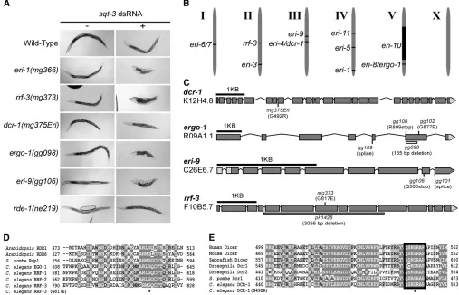
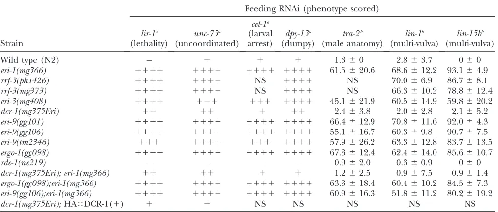
mutants respond more robustly than wild-type animals to exogenous sources of dsRNA, a phenotype referred to asenhancedRNAi(Eri) (Figure 1A and Simmeret al.
2002; Kennedyet al.2004; Duchaine et al.2006). To
explain this phenomenon, a model was proposed in which different small regulatory RNA pathways compete for limiting amounts of shared components. We have taken advantage of the Eri phenotype to screen for additional factors required for the production and stability of endo siRNAs.
We chemically mutagenized 150,000 haploid ge-nomes and screened for mutant animals that exhibit enhanced sensitivity to dsRNAs (for details of screens, seeFigure S1). We identified five new complementation groups that defined the geneseri-4,eri-8,eri-9,eri-10, and eri-11 (Figure 1B). Subsequent characterization (de-scribed below) identifiederi-4(mg375)as an allele of dcr-1anderi-8as being allelic toergo-1. For the sake of clarity, the designationsdcr-1(mg375Eri)and ergo-1 are hence-forth used where appropriate. Our screens identified a single allele oferi-4/dcr-1,mg375, and multiple alleles of eri-8/ergo-1anderi-9. These screens also recovered alleles of the previously identified eri-1, rrf-3, and eri-3genes (Figure S1). For example, we identified an allele ofrrf-3, termed rrf-3(mg373), that encodes an amino acid sub-stitution [RRF-3(G817E)] of a glycine residue that is highly conserved in related RdRPs (Figure 1, C and D). Here, we focus most of our analysis oneri-4/dcr-1,eri-8/
ergo-1, anderi-9.
Animals mutant for eri-1, rrf-3, eri-3, or eri-5 exhibit enhanced sensitivity to dsRNAs targeting a wide-array of mRNAs (Simmer et al. 2002; Kennedy et al. 2004;
Duchaine et al. 2006; Lee et al. 2006). We found that
animals likewise exhibit a generalized enhanced sensitiv-ity to RNAi (Figure 1A and Table 1). For example, hypomorphic alleles ofsqt-3trigger a dumpy phenotype (van derKeylet al.1994). Exposure of wild-type animals
to dsRNA derived from thesqt-3gene (sqt-3RNAi) fails to induce a dumpy phenotype (Figure 1A). sqt-3 RNAi, however, is sufficient to induce a dumpy phenotype in
eri-1(mg366), ergo-1(gg098), or eri-9(gg106) animals (Figure
1A).dcr-1(mg375Eri)animals exhibited an enhanced, but
less pronounced, response to sqt-3 RNAi (Figure 1A). Genetic criteria indicate that these Eri genes are not components of the class B synthetic multi-vulva pathway (Table S1), mutations of which also exhibit enhanced sensitivity to RNAi (Wang et al.2005). Rather, animals
carrying mutations foreri-1;eri-9,eri-1;ergo-1, oreri-1;dcr-1
(mg375Eri)exhibited Eri phenotypes similar to animals harboring the individual mutations, suggesting thateri-9,
ergo-1, and dcr-1(mg375Eri) are components of theeri-1
genetic pathway (Table 1).
Molecular identification of eri-4,eri-8, and eri-9:We mappederi-8to a,1.5-cM interval on chromosome V
between Y50D4C(19.99) and F52F10(18.54). The C. elegans genome encodes 27 Argonaute proteins. One of those Argonaute genes,R09A1.1, lies within this mapping interval. We sequenced R09A1.1 from DNA isolated from foureri-8alleles; each contained a unique mutation within the R09A1.1 open reading frame (Figure 1C). During the course of our studies, animals harboring a deletion within the R09A1.1 locus were shown to exhibit an enhanced RNAi phenotype and defects in endo siRNA expression (Yigitet al.2006). On
that basis, R09A1.1 was named endogenous-RNAi de-ficient Argonaute-1 (ergo-1) (Yigit et al. 2006).
eri-8(gg098) animals failed to complement ergo-1(tm1860)
animals for enhancement of RNAi (data not shown). We conclude thateri-8corresponds toergo-1.
We mappederi-9between7.4 and11.5 on chromo-some III. The ORFC26E6.7lies within this interval. Four lines of evidence indicate that eri-9 corresponds to
C26E6.7. First, like ERI-1, RRF-3, ERI-3, and ERI-5, the C26E6.7 gene product coprecipitates with DCR-1
from twoeri-9mutants and found that each had a unique mutation within the predictedC26E6.7coding sequence (Figure 1C). Third,eri-9(gg106)failed to complement the RNAi enhancement phenotype shown by a C26E6.7
deletion mutant (data not shown). Fourth, a transgene that includes the wild-typeC26E6.7gene rescued the Eri phenotype associated with eri-9(gg106) animals (Table S2). We conclude that eri-9 corresponds to C26E6.7. Database searches reveal a single homolog of ERI-9 inC. briggsae andC. remanei (data not shown). We have not detected sequence homologs of ERI-9 in other, more divergent organisms (data not shown).
We mapped the singleeri-4allele,mg375, to a,1-cM interval on chromosome III, an interval that contains
the dcr-1 gene. As DCR-1 is required for converting
dsRNA to siRNAs (a necessary prerequisite for RNAi), we did not anticipate identifying alleles of dcr-1in our screen for enhanced RNAi sensitivity. Surprisingly, sequencing of thedcr-1 locus from DNA isolated from
mg375 animals identified a mutation that encodes a
G492R substitution within the N-terminal helicase domain of DCR-1(Figure 1C). Two additional lines of evidence indicate thatmg375is a mutant allele ofdcr-1. First,mg375fails to complementdcr-1(ok247), a deletion that likely represents a null allele ofdcr-1(Knightand
Bass 2001), for sterility defects (Figure S2A, and see
below). Second, a transgene expressing full-lengthdcr-1
partially rescues the sterility and enhanced RNAi phenotypes associated withmg375hermaphrodite ani-mals (Figure S2B and Table 1). We speculate that the partial rescue ofdcr-1(mg375Eri) animals by transgeni-cally expressed dcr-1 likely reflects poor germline
expression of this transgene. We conclude thatmg375
is an allele ofdcr-1and encodes a mutant variant of DCR-1 that harbors a G492R substitution within the N-terminal helicase domain. G492 is an evolutionarily conserved residue, which suggests an important func-tion for this amino acid in RNAi-related processes (Figure 1D).
dcr-1(mg375Eri) and eri-9 animals exhibit defects in endo siRNA production and mRNA regulation: Muta-tions in previously identified Eri genes result in a failure of cells to accumulate endo siRNAs (Lee et al. 2006;
Duchaineet al.2006; Yigitet al.2006). The Argonaute
proteinNRDE-3is expressed in most, if not all, somatic tissues and escorts a subset of endo siRNAs from the cytoplasm to the nucleus. In the absence of endo siRNAs,NRDE-3resides in the cytoplasm, while, in the presence of endo siRNAs, NRDE-3 localizes to the nucleus (Guang et al. 2008). Thus, the subcellular
distribution of NRDE-3 is reflective of endo siRNA abundance in somatic cells. To begin to assess whether
eri-9ordcr-1(mg375Eri)regulate endo siRNA expression,
we examined the subcellular distribution ofNRDE-3in
eri-9()ordcr-1(mg375Eri)animals. In contrast to
wild-type controls, where NRDE-3 was predominantly nu-clear,NRDE-3localized predominantly to the cytoplasm
in eri-9() and dcr-1(mg375Eri) animals (Figure 2A).
These results suggest that wild-type eri-9and dcr-1 are required for expression of the majority of endo siRNAs that associate withNRDE-3in somatic tissues. We tested this hypothesis by performing Northern analysis of the expression of specific endo siRNAs in eri-9() or dcr-1(mg375Eri) animals. Endo siRNAs have been identified
TABLE 1
Enhanced sensitivity to exogenous dsRNA
Feeding RNAi (phenotype scored)
Strain lir-1a (lethality) unc-73a (uncoordinated) cel-1a (larval arrest) dpy-13a (dumpy) tra-2b (male anatomy) lin-1b (multi-vulva) lin-15bb (multi-vulva)
Wild type (N2) 1 1 1 1.360 2.863.7 060
eri-1(mg366) 1111 1111 1111 1111 61.5620.6 68.6612.2 93.164.9
rrf-3(pk1426) 1111 1111 NS 1111 NS 70.066.9 86.768.1
rrf-3(mg373) 1111 1111 NS 1111 NS 66.3610.2 78.8612.4
eri-3(mg408) 1111 111 111 1111 45.1621.9 60.5614.9 59.8620.2
dcr-1(mg375Eri) 11 11 1 11 2.463.8 2.062.8 2.165.2
eri-9(gg101) 1111 1111 1111 1111 66.4612.9 70.8611.6 92.064.3
eri-9(gg106) 1111 1111 1111 1111 55.1616.7 60.369.8 90.767.5
eri-9(tm2346) 111 1111 111 1111 57.9626.2 63.3612.8 83.7613.5
ergo-1(gg098) 1111 1111 1111 1111 67.3612.4 62.4614.0 85.6610.7
rde-1(ne219) 0.962.0 0.360.9 060
dcr-1(mg375Eri); eri-1(mg366) 11 11 1 1 1.262.5 0.967.5 0.961.4
ergo-1(gg098);eri-1(mg366) 1111 1111 1111 1111 63.3618.4 60.4610.2 84.567.3 eri-9(gg106);eri-1(mg366) 1111 1111 1111 1111 60.9616.3 51.8611.2 80.2619.2
dcr-1(mg375Eri);HATDCR-1(1) 1 1 NS NS NS NS NS
aEffectiveness of RNAi scored from unaffected () to maximally enhanced (1111) or not scored (NS). bScored as percentage of F
for the ORFE01G4.5and an3-kb noncoding region of the X chromosome (termed the X-cluster) (Ambros et al.2003).eri-1(),eri-3(), and rrf-3(mg373)animals fail to express these endo siRNAs (Figure 2B andFigure
S3). eri-9() anddcr-1(mg375Eri) animals also failed to
express detectableE01G4.5and X-cluster endo siRNAs (Figure 2B), supporting the hypothesis that these genes function in theeri-1genetic pathway and indicating that ERI-9 andDCR-1 are required for generating and/or stabilizing these endo siRNAs.
In addition to endo siRNAs, C. elegans expresses at least two additional classes of small regulatory RNAs, the miRNAs, and the 21U-RNAs (the worm equivalent of the Piwi interacting RNAs). dcr-1(mg375Eri) and eri-9
animals retained the ability to express the miRNAlet-7
and the 21U-1 piRNA at levels similar to that of wild-type animals (Figure 2B). In addition, dcr-1(mg375Eri) and
eri-9animals respond more robustly to dsRNA exposure
(RNAi) than wild-type animals (Figure 1A and Table 1), strongly suggesting that these mutant animals are able to produce siRNAs from exogenous dsRNA substrates. Thus, the effect of thedcr-1(mg375Eri) anderi-9 muta-tions is restricted to the production of a subset of
cellular small regulatory RNAs. SinceDCR-1is known to be involved in the biogenesis of miRNAs (Kettinget al.
2001; Leeet al.2002, 2004; Lundet al.2004), our results
imply that the G492R substitution within the DCR-1
helicase domain preferentially impairs the endo siRNA-related function ofDCR-1.
Endo siRNAs that map to theK02E2.6ORF have been identified (Ambroset al.2003; Rubyet al. 2006).
Ani-mals lacking components of the ERI/DICER complex fail to expressK02E2.6endo siRNAs and concomitantly overexpress theK02E2.6mRNA (Duchaineet al.2006).
Consequently, it has been proposed that endo siRNAs negatively regulate mRNAs that exhibit sequence ho-mology to their cognate endo siRNAs, which is known as endogenous RNAi. We found thatK02E2.6mRNA is likewise overexpressed in dcr-1(mg375Eri), ergo-1,
rrf-3(mg373), anderi-9animals (Figure 2C andFigure S3).
We extended this analysis to three additional mRNAs (E01G4.5, W04B5.1, and C40A11.10) for which com-plementary endo siRNAs have previously been identi-fied but transcript levels have not been characterized (Figure 2C and Rubyet al.2006). All three mRNAs were
eri-9animals relative to wild-type controls (Figure 2C). Thus, the dcr-1, ergo-1, and eri-9 gene products (and previously identified components of the ERI/DICER complex) are required for negatively regulating these mRNAs. Animals harboring mutations in botheri-1and
eri-9, ergo-1, or dcr-1(mg375Eri) exhibit similar levels of
mRNA misregulation to animals harboring the individ-ual mutations (Figure 2D). Taken together, these data argue thatergo-1,eri-9, and thedcr-1helicase domain are required for endo RNAi processes mediated by theeri-1
genetic pathway.
A subset of Eri animals exhibit sperm-specific endo RNAi defects: Endo siRNAs are thought to negatively regulate cellular mRNAs. Large-scale sequencing of endo siRNAs indicates that endo siRNAs are enriched for sequences with homology to mRNAs expressed in sperm (Ruby et al. 2006). We selected three sperm
mRNAs [sperm-specific protein (ssp)-16,C25G4.6, and
F18C5.4], for which endo siRNAs have been identified, for further investigation (Ruby et al. 2006). We
con-firmed that these mRNAs are expressed predominantly, if not exclusively, in the sperm-producing germline (Figure S4). We then investigated whether the Eri genes were required for regulating these mRNAs. qRT–PCR analysis demonstrated differential genetic require-ments for the regulation of these sperm-enriched mRNAs. For example, eri-1, eri-3, rrf-3, and dcr-1(mg375Eri) mutant animals overexpressed these sperm-specific mRNAs while eri-9 and ergo-1 mutant animals expressed these mRNAs at levels similar to that of wild-type animals (Figure 3A andFigure S3). Hence-forth, we will refer toeri-1,eri-3,rrf-3, anddcr-1(mg375Eri) as class I Eri genes and eri-9and ergo-1 as class II Eri genes. Northern analysis ofssp-16revealed a lack of ssp-16endo siRNAs and a concomitant increase in ssp-16
transcript levels among class I, but not class II, mu-tant animals (Figure 3B). Interestingly, we found that mRNAs encoding class I Eri factors are expressed at similar levels in the sperm- and oocyte-producing germ-line, while mRNAs encoding class II Eri factors are enriched in the oocyte-producing germline, suggesting that the differential requirement for the class I and II Eri factors in sperm-specific mRNA regulation may be due to differential expression patterns of the class I and II Eri factors (Figure S5). Thus, the class I, but not class II, Eri gene products regulate sperm-enriched RNAs via endo RNAi.
The class I Eri factors might negatively regulate sperm mRNAs specifically in the sperm-producing germline, or they might inhibit expression in tissues that normally do not produce these mRNAs. To address this question, we asked if class I Eri-dependent regulation of ssp-16,
C25G4.6, and F18C5.4 mRNA occurs in the sperm-producing germline. Hermaphrodites that harbor a fem-1(ts)mutation produce both sperm and oocytes at 15°, but only oocytes at 25°(Nelsonet al.1978). Loss oferi-1
resulted in elevated levels of ssp-16, C25G4.6, and
F18C5.4infem-1(ts)hermaphrodites when sperm were present, but not in the absence of sperm (Figure 4A). These results argue that the negative regulation of sperm gene expression byeri-1 requires the sperm-producing germline. In support of this idea, in situ hybridization detectingssp-16mRNA demonstrated that expression ofssp-16is restricted to the sperm-producing gonad in wild-type animals and thateri-1()animals do not exhibit ectopic expression ofssp-16in other tissues (Figure 4B). These results demonstrate that the class I Eri genes are required for endogenous RNAi processes within the sperm-producing germline.
Class I Eri animals exhibit defects in sperm de-velopment: Germline phenotypes have been reported for a few Eri mutants (Kennedyet al.2004; Duchaine et al.2006; Asikainenet al.2007).eri-1,rrf-3,eri-3, and
eri-5 hermaphrodites exhibit ts sterility; these animals
are fertile at 15°and sterile at 25°. These animals also produce a higher-than-normal percentage of male progeny, referred to as a Him (highincidence ofmales) phenotype (Hodgkinet al.1979). The Him phenotype
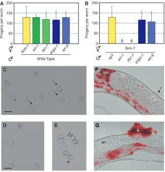
tion in the sperm-producing germline, which causes XX hermaphrodites to produce XO male offspring (Gent et al. 2009). We tested if these germline defects were shared by ergo-1(), eri-9(), rrf-3(mg373), or dcr-1(mg375Eri) animals. dcr-1(mg375Eri) and rrf-3(mg373) animals exhibited ts sterile and Him phenotypes (Fig-ure 5 andFigure S3). In contrast,ergo-1()anderi-9() animals exhibited wild-type levels of fertility and normal frequencies of male progeny (Figure 5). Thus, the class I Eri genes, but not the class II Eri genes, are required for fertility at elevated temperatures. Finally, animals har-boring both class I and class II Eri mutations are sterile at 25° (and exhibit brood sizes similar to class I Eri animals when reared at temperatures slightly below the nonpermissive temperature), indicating that class I Eri alleles are epistatic to class II alleles with regards to sterility (Figure 5C and data not shown).
Sterility in hermaphrodites can arise from defects in sperm or oocyte function. Mating with wild-type males rescued the ts sterility of class I Eri hermaphrodites, a result suggestive of a sperm-specific defect (Figure 6A). Mating assays with fem-1 hermaphrodites (which pro-duce only oocytes) crossed to class I Eri males confirmed this hypothesis; sperm fromeri-1()anddcr-1(mg375Eri) males, reared at the nonpermissive temperature, were unable to fertilizeeri-1(1)(fem-1) oocytes (Figure 6B). Sperm from class II Eri mutant males were functional in these assays (Figure 6B). Together, these data establish a role in sperm function for the class I, but not class II, Eri genes.
We examined the mature gametes of males harboring class I Eri alleles for defects in morphology or motility that might underlie sperm-specific sterility. Wild-type adult males store round, immotile spermatids in the seminal vesicle that, upon insemination, are activated by an extracellular signal to generate amoeboid crawl-ing spermatozoa with extended pseudopods (Wolf et al. 1978; Ward et al. 1981). The in vivo activation
signal can be mimicked in vitro by proteases or com-pounds that increase intracellular pH (Nelson and
Ward1980; Wardet al.1983; Shakesand Ward1989).
We assessed the ability ofdcr-1(mg375Eri)spermatids to extend pseudopods and form crawling spermatozoa by treatment with the in vitro activators monensin or triethanolamine.In vitroactivation of wild-type sperma-tids was successful 82% of the time (Figure 6C). In contrast, only 7% of dcr-1(mg375Eri) spermatids were capable of forming pseudopods, and the majority of dcr-1(mg375Eri)spermatids exhibited an aberrant morphol-ogy (Figure 6, D and E). Motile spermatozoa localize to the spermatheca, the site of fertilization, in the her-maphrodite reproductive tract.In vivoactivation of class I Eri animal spermatids was also defective, as indicated by a failure ofdcr-1(mg375Eri)spermatids to localize to the spermatheca. Vital staining of male sperm prior to mating indicated that dcr-1(mg375Eri) sperm can be transferred successfully. Temporal analysis following Figure 4.—Class I Eri-mediated RNA regulation occurs
sperm transfer argued that dcr-1(mg375Eri) sperm are rapidly displaced from the hermaphrodite reproductive tract by passing oocytes and expelled through the vulva (compare Figure 6F to 6G). Similar results were ob-served foreri-1,eri-3, andrrf-3sperm (data not shown). Thus, class I Eri males are defective in spermatid acti-vation and cell motility, which are essential for fertility (L’Hernault2006).
C. elegans hermaphrodites undergo spermatogenesis prior to oogenesis, while male animals continue to produce sperm throughout adult life (Wolfet al.1978).
Class I Eri hermaphrodites were sterile if reared at the nonpermissive temperature during spermatogenesis, but fertile if the temperature was raised during oogenesis (after spermatogenesis was completed) (Table 2). Class I Eri males were sterile if reared at the nonpermissive temperature, but eventually regained fertility following a downshift in temperature, indicating that the tempera-ture-sensitive period for fertility associated with class I Eri mutants coincides with spermatogenesis (Table 2).
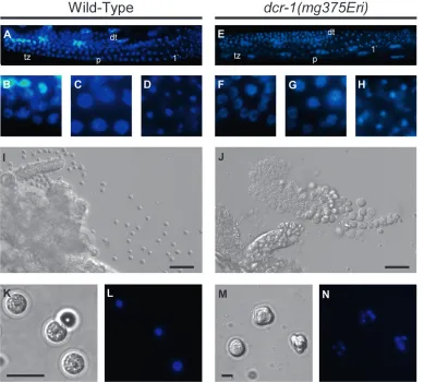
We next examined the gonads of class I Eri males for defects in sperm development. In wild-type males, a mitotically proliferating population of syncytial stem cells in the distal end of the germline gives rise to a transition zone at the onset of meiosis. Further pro-gression through meiosis I produces pachytene nuclei that condense and cellularize into primary spermato-cytes. Completion of the two meiotic divisions produces four haploid spermatids that separate from the residual body (L’Hernault 2006). Early events during
sper-matogenesis ofdcr-1(mg375Eri)mutant males appeared to be unaffected, as indicated by the normal number and morphology of germ cells in the mitotic and meiotic regions of the germline (Figure 7, A–H). The first visible defect that we observed was a change in cytology at the spermatid stage. Wild-type animals produce small spherical spermatids that each contains a single, highly condensed nucleus (Figure 7, I, K, and L). In contrast, young adult males harboring thedcr-1(mg375Eri) muta-tion accumulated sperm cells in the seminal vesicle that were large and misshapen and often contained multiple nuclei (Figure 7, J, M, and N; note the difference in scale in K and M). Similar defects have been reported for sperm mutants (e.g.,spe-26) that exhibit aberrant chro-mosome segregation (Varkeyet al.1995). The observed
defects in the dcr-1(mg375Eri) mutant may reflect delayed progression through the sperm developmental program, as older males possessed spermatids that were more similar in size and shape to wild-type spermatids (see Figure 6, C–E). Defects similar to those described above were found in sperm for all class I Eri animals, whereas sperm of class II Eri animals exhibited normal morphology and function (data not shown). Taken together, the data indicate that class I Eri mutations produce multiple aberrations in spermatogenesis, beginning with cytokinesis at the meiotic divisions and extending through terminal differentiation into Figure 5.—Class I, but not class II, Eri animals exhibit
crawling spermatozoa. These defects likely explain the ts sterility associated with class I Eri animals.
DISCUSSION
Here we report the identification and characteriza-tion of genes required for the produccharacteriza-tion of endoge-nous siRNAs, small regulatory RNAs that are thought to mediate silencing of cellular transcripts. We establish
thateri-9encodes a novel component of the endo RNAi
machinery and thateri-4 encodes a mutant variant of
DCR-1harboring a mutation within a conserved region of the DCR-1 helicase domain. We demonstrate that ERI-9 and the helicase domain ofDCR-1play a role in endo RNAi processes inC. elegans. Finally, we show that the class I Eri factors (includingdcr-1), but not the class II Eri factors (including eri-9), are required for development of, and endo RNAi processes in, sperm.
We show that animals harboring a mutation in dcr-1
exhibit defects in endo siRNA production, mRNA regulation, X-chromosome segregation, and sperm de-velopment that are indistinguishable from phenotypes Figure6.—Sterility of class I Eri animals is attributable to defects in sperm function. (A) Wild-type males were crossed with hermaph-rodites of the indicated genotype reared at the nonpermissive temperature (25°), and the number of cross-progeny were scored (n¼8,6SD). (B) The number of progeny from hermaphrodite [fem-1(hc17ts)] animals mated with males of the indicated genotype at the nonpermissive temperature (25°) (n¼ 8,6SD). (C–E) Spermatids dissected from wild-type (C) ordcr-1(mg375Eri)(D and E) adulthood males (3 days after final molt) were treated with thein vitroactivator monen-sin and visualized by DIC Nomarski. Arrows indicate pseudopods of crawling spermato-zoa. Thedcr-1(mg375Eri)spermatids are mis-shapen and fail to activate (D) or extend thin immotile projections (asterisk in E). (F and G) Wild-type ordcr-1(mg375Eri) mid-adult-hood males were stained with a vital fluores-cent dye and then mated withfem-1(hc17ts) females. Mated females were visualized by both DIC Nomarski and fluorescence mi-croscopy to assess sperm transfer and locali-zation. The fluorescent and DIC images were overlaid to generate a composite image. Arrows indicate vulva. SP, spermatheca. (F) Sperm (in red) from wild-type males migrate to the spermatheca. (G) dcr-1(mg375Eri) sperm either remain at the vulva at insemina-tion or are expelled from the uterus by pass-ing oocytes. Intestinal autofluorescence seen in these images is also observed in the ab-sence of dye staining.
TABLE 2
Temperature-sensitive period for class I Eri animals coincides with spermatogenesis
$fem-1a3#N2 $fem-1a3#eri-1 eri-1(hermaphrodite self-cross)
Embryo L4b Adultc Progenyd Embryo L4b Adultc Progenyd Embryo L4e Adultf Progenyd
15° 25° 1 15° 25° 1 15° 25° 1
25° 25° 1 25° 25° 25° 25°
25° 15° 1 25° 15° 1 25° 15°
a
Feminized animals used to prevent self-fertilization.
b
Temperature of male animals during development and early spermatogenesis.
c
Temperature of male animals during mating and later spermatogenesis.
dThe presence (1) or absence () of progeny 4 days after temperature shift. e
Temperature of hermaphrodite animals during development and spermatogenesis.
f
exhibited by eri-1(), rrf-3(), and eri-3() animals.
ERI-1, RRF-3, and ERI-3 coprecipitate with DCR-1
(Duchaineet al.2006). This latter observation has led
to the hypothesis that the ERI proteins assemble into a protein complex with DCR-1. Our results provide genetic and biochemical evidence supporting the exis-tence of a functional multi-protein complex composed ofDCR-1and the Eri factors and support the model that this complex is important for generating and stabilizing endo siRNAs. Interestingly, the N-terminal helicase domain of Dicer is well conserved throughout eukar-yotes; however, the function of this domain remains enigmatic. Our identification and characterization of
DCR-1(G492R) establishes that, inC. elegans, the heli-case domain of Dicer is required for endo siRNA expression and spermatogenesis.
We have shown thateri-9is a component of the endo RNAi machinery: ERI-9 functions in the eri-1 genetic pathway, encodes a DCR-1 coprecipitating protein (Duchaineet al.2006), and is required for endo siRNA
expression and for cognate mRNA regulation. Taken together, these data argue strongly that ERI-9 is a component of ERI/DICER complexes. Interestingly,
we have shown that components of the ERI/DCR complex are not all functionally equivalent. Our di-vision of the Eri genes into two groups on the basis of the presence [class I:eri-1,eri-3,rrf-3, anddcr-1(mg375Eri)] or absence (class II:ergo-1anderi-9) of germline pleiotro-pies and endogenous RNAi processes in the sperm-producing germline indicate that these factors can have distinct biological function(s). In the simplest model, the class I proteins form a core ERI/DICER complex that is required for endo siRNA production and endogenous RNAi in both the soma and germline, whereas the class II proteins serve as accessory factors that modify core complex activity in tissues other than the male germline. In support of this hypothesis, expression analysis of Eri genes indicates that class I Eri mRNAs are expressed at equivalent levels during sperm and oocyte production, while the class II Eri mRNAs are enriched in the oocyte-producing germline (Figure S5). Future analyses that focus on endo RNAi defects in the sperm-producing germline may identify sperm equivalents ofergo-1anderi-9.
development. The failure of class I Eri mutants to engage in sperm endo RNAi may directly cause sper-matogenesis defects. Alternatively, it is possible that the sperm defects that we have observed result from non-RNAi and siRNA related functions of the class I Eri factors. We favor the former model for the following reasons: First, we have observed a one-to-one correlation between those Eri genes required for sperm-specific endo RNAi and those Eri genes required for normal sperm development. Second, large-scale sequencing of small RNAs has shown that endo siRNAs are enriched for sequences with the ability to target mRNAs ex-pressed in sperm (Rubyet al.2006). Third, several class
I Eri factors, such as the RNA-dependent RNA poly-merase RRF-3 and the RNase III enzyme DCR-1, are proteins whose only known biochemical function re-lates to small regulatory RNA biogenesis. Finally, our genetic screens have identified missense alleles of both
dcr-1 and rrf-3 [encoding DCR-1(G492R) and
RRF-3(G187E)], which do not affect expression of their encoded proteins or the ability of these mutant pro-teins to assemble into ERI/DCR complexes (data not shown).DCR-1(G492R) andRRF-3(G187E) animals do, however, exhibit fully penetrant endo RNAi defects and ts sperm defects, strongly suggesting that it is the small RNA products ofDCR-1andRRF-3that are important for spermatogenesis. Taken together, these data argue that loss of endo siRNA expression in the male germline triggers defects in sperm development.
We have shown that mutant animals lacking Eri gene function overexpress mRNAs that exhibit sequence homology to Eri-dependent endo siRNAs. The most parsimonious explanation for these observations is that endo siRNAs negatively regulate their cognate mRNAs (and/or are the end product of this regulation). We consider two possible models for the role of endo siRNAs and endo RNAi during spermatogenesis.
1. In model 1, spermatogenesis defects are triggered by overexpression of sperm mRNAs, the negative regu-lation of which is important for sperm development. In such a case, either the target mRNA may be grossly overexpressed or the temporal expression may be precocious and/or persistent. In support of model 1, microarray analysis comparing wild-type animals to
eri-1() or rrf-3() animals indicates that sperm
RNAs are negatively regulated by ERI-1 andRRF-3
(Asikainen et al. 2007). We revisited the eri-1/rrf-3
microarray data sets and compared these data to existing microarray data sets, which identified genes preferentially expressed in the sperm (Reinke et al.
2000, 2004). This analysis showed that 96% oferi-1/
rrf-3-regulated RNAs are sperm-enriched RNAs (
Ta-ble S3). This remarkable degree of overlap between these two microarray data sets indicates that a large percentage of transcript misregulation observed in
botheri-1andrrf-3mutants at this stage of
develop-ment occurs among genes with elevated expression during spermatogenesis. At present, it is unclear which, if any, of these RNAs are responsible for the spermatogenesis defects that we have observed. We conducted RNAi-mediated knockdown of these 68 RNAs (individually) and failed to identify any sup-pressors of class I Eri sterility (data not shown). In addition, we screened several million genomes (fol-lowing EMS mutagenesis) for suppressors of Eri-mediated sterility and failed to identify a single suppressor (data not shown). Taken together, these data hint that, if model 1 is correct, the sterility of class I Eri animals is unlikely to be due to over-expression of a single sperm mRNA.
2. In model 2, spermatogenesis defects result from loss of endo siRNA-directed heterochromatin in sperm. Some Eri-dependent endo siRNAs, such as those complementary to the C. elegans X-cluster, map to regions of the genome not predicted to encode functional mRNAs. InSchizosaccharomyces pombe, the RNAi machinery plays a role in heterochromatin formation at centromeres (Reinhart and Bartel
2002; Bu¨ hleret al.2006; Colmenareset al.2007).S. pombe mutants that lack components of the RNAi machinery exhibit chromosome nondisjunction phe-notypes (Hallet al.2003). We have observed an X
chromosome nondisjunction phenotype in the class I Eri mutant animals. Thus, model 2 posits that C. elegansendo siRNAs function analogously toS. pombe siRNAs to regulate heterochromatin formation in such a way as to permit chromosome segregation and sperm function at elevated temperatures.
One puzzling aspect of our results is the temperature sensitivity of class I Eri germline phenotypes. The class I Eri mutants appear to be nonconditional for loss of endo siRNA accumulation and misregulation of target RNAs, as these defects occur at all temperatures (data not shown). Increased X chromosome nondisjunction and sperm-specific sterility of the class I Eri mutants, how-ever, are manifested more robustly at elevated tem-peratures. Most of the class I Eri mutants that we characterized carry predicted null alleles, indicating that the ts sterility phenotype associated with the class I Eri mutants is very likely not due to temperature-sensitive proteins. Rather, data from wild-type animals suggest that both chromosome segregation and sperm develop-ment are inherently sensitive to perturbation by temper-ature. For example, brood size is reduced by25% in wild-type animals reared at elevated temperature (Hirsh
and Vanderslice 1976). Since fecundity is sperm
Baillie1979). Thus, the sperm defects associated with
class I Eri animals may result from enhancement of inherently temperature-sensitive processes.
C. elegansexpresses a diverse array of small regulatory RNAs. Our genetic screens continue to identify novel factors required for the biogenesis of endo siRNAs. To date, we have identified two classes of Eri factors, one of which promotes thermotolerance in sperm. Interest-ingly, animals lacking theC. elegansPIWI homologs (and consequently lacking piRNAs) exhibit temperature-sensitive spermatogenesis defects, hinting that other small RNAs may play important roles in spermatogen-esis (Batista et al. 2008; Das et al. 2008; Wang and
Reinke 2008). Furthermore, we have identified
differ-ential germline functions among the Eri factors. Taken together, these results indicate that the mechanisms of small RNA biogenesis and function in C. elegans are complex and may vary in a tissue-dependent manner. Our genetic screens have not yet reached saturation. For example, two other Eri factors,eri-10anderi-11, identi-fied in our screens exhibit class II Eri phenotypes, are defined by single alleles, and are not allelic with any of the known endo RNAi genes, indicating that additional components of the cellular endo RNAi machinery remain to be identified. The identification and charac-terization of the full complement of endo RNAi machinery will likely facilitate the unraveling of the remarkably complex world of small regulatory RNAs.
LITERATURE CITED
Ambros, V., R. C. Lee, A. Lavanway, P. T. Williamsand D. Jewell, 2003 MicroRNAs and other tiny endogenous RNAs in C. ele-gans. Curr. Biol.13:807–818.
Asikainen, S., M. Storvik, M. Laksoand G. Wong, 2007 Whole ge-nome microarray analysis of C. elegans rrf-3 and eri-1 mutants. FEBS Lett.581:5050–5054.
Batista, P. J., J. G. Ruby, J. M. Claycomb, R. Chiang, N. Fahlgren
et al., 2008 PRG-1 and 21U-RNAs interact to form the piRNA complex required for fertility in C. elegans. Mol. Cell31:67–78. Bu¨ hler, M., A. Verdeland D. Moazed, 2006 Tethering RITS to a nascent transcript initiates RNAi- and heterochromatin-depen-dent gene silencing. Cell125:873–886.
Cam, H. P., T. Sugiyama, E. S. Chen, X. Chen, P. C. FitzGeraldet al., 2005 Comprehensive analysis of heterochromatin- and RNAi-mediated epigenetic control of the fission yeast genome. Nat. Genet.37:809–819.
Colmenares, S. U., S. M. Buker, M. Buhler, M. Dlakicand D. Moazed, 2007 Coupling of double-stranded RNA synthesis and siRNA generation in fission yeast RNAi. Mol. Cell27:449–461. Czech, B., C. D. Malone, R. Zhou, A. Stark, C. Schlingeheyde
et al., 2008 An endogenous small interfering RNA pathway in Drosophila. Nature453:798–802.
Das, P. P., M. P. Bagijn, L. D. Goldstein, J. R. Woolford, N. J. Lehrbachet al., 2008 Piwi and piRNAs act upstream of an en-dogenous siRNA pathway to suppress Tc3 transposon mobility in the Caenorhabditis elegans germline. Mol. Cell31:79–90. Duchaine, T. F., J. A. Wohlschlegel, S. Kennedy, Y. Bei, J. D. Conte
et al., 2006 Functional proteomics reveals the biochemical niche of C. elegans DCR-1 in multiple small-RNA-mediated path-ways. Cell124:343–354.
Gent, J. I., M. Schvarzstein, A. M. Villeneuve, S. G. Gu, V. Jantsch
et al., 2009 Caenorhabditis elegansRNA-directed RNA polymerase in sperm development and endogenous RNAi. Genetics 183: 1297–1314.
Ghildiyal, M., H. Seitz, M. D. Horwich, C. Li, T. Du
et al., 2008 Endogenous siRNAs derived from transposons and mRNAs in Drosophila somatic cells. Science 320: 1077– 1081.
Guang, S., A. F. Bochner, D. M. Pavelec, K. B. Burkhart, S. Harding et al., 2008 An Argonaute transports siRNAs from the cytoplasm to the nucleus. Science321:537–541.
Hall, I. M., G. D. Shankaranarayana, K.-i.Noma, N. Ayoub, A. Cohenet al., 2002 Establishment and maintenance of a hetero-chromatin domain. Science297:2232–2237.
Hall, I. M., K.-i.Nomaand S. I. S. Grewal, 2003 RNA interference machinery regulates chromosome dynamics during mitosis and meiosis in fission yeast. Proc. Natl. Acad. Sci. USA 100: 193–198.
Hamilton, A., O. Voinnet, L. Chappell and D. Baulcombe, 2002 Two classes of short interfering RNA in RNA silencing. EMBO J.21:4671–4679.
Hill, K. L., and S. W. L’Hernault, 2001 Analyses of reproductive interactions that occur after heterospecific matings within the ge-nus Caenorhabditis. Dev. Biol.232:105–114.
Hirsh, D., and R. Vanderslice, 1976 Temperature-sensitive develop-mental mutants of Caenorhabditis elegans. Dev. Biol.49:220–235. Hodgkin, J., H. R. Horvitzand S. Brenner, 1979 Nondisjunction
mu-tants of the nematodeCaenorhabditis elegans.Genetics91:67–94. Kamath, R. S., A. G. Fraser, Y. Dong, G. Poulin, R. Durbinet al.,
2003 Systematic functional analysis of the Caenorhabditis ele-gans genome using RNAi. Nature421:231–237.
Kawamura, Y., K. Saito, T. Kin, Y. Ono, K. Asai et al., 2008 Drosophila endogenous small RNAs bind to Argonaute [thinsp]2 in somatic cells. Nature453:793–797.
Kennedy, S., D. Wangand G. Ruvkun, 2004 A conserved siRNA-degrading RNase negatively regulates RNA interference in C. elegans. Nature427:645–649.
Ketting, R. F., S. E. J. Fischer, E. Bernstein, T. Sijen, G. J. Hannon
et al., 2001 Dicer functions in RNA interference and in synthe-sis of small RNA involved in developmental timing in C. elegans. Genes Dev.15:2654–2659.
Kim, V. N., 2005 Small RNAs: classification, biogenesis, and func-tion. Mol. Cells19:1–15.
Knight, S. W., and B. L. Bass, 2001 A role for the RNase III enzyme DCR-1 in RNA interference and germ line development in Cae-norhabditis elegans. Science293:2269–2271.
Lee, R. C., R. L. Feinbaumand V. Ambros, 1993 The C. elegans het-erochronic gene lin-4 encodes small RNAs with antisense com-plementarity to lin-14. Cell75:843–854.
Lee, R. C., C. M. Hammelland V. Ambros, 2006 Interacting endog-enous and exogendog-enous RNAi pathways in Caenorhabditis elegans. RNA12:589–597.
Lee, Y., K. Jeon, J. T. Lee, S. Kimand V. N. Kim, 2002 MicroRNA mat-uration: stepwise processing and subcellular localization. EMBO J.21:4663–4670.
Lee, Y. S., K. Nakahara, J. W. Pham, K. Kim, Z. He et al., 2004 Distinct roles for Drosophila Dicer-1 and Dicer-2 in the siRNA/miRNA silencing pathways. Cell117:69–81.
L’Hernault, S. W., 2006 Spermatogenesis, inWormBook, edited by The C. elegans Research Community. WormBook, http://
www.wormbook.org.
Llave, C., K. D. Kasschau, M. A. Rectorand J. C. Carrington, 2002 Endogenous and silencing-associated small RNAs in plants. Plant Cell14:1605–1619.
Lund, E., S. Guttinger, A. Calado, J. E. Dahlbergand U. Kutay, 2004 Nuclear export of microRNA precursors. Science303:95–98. Mochizuki, K., and M. A. Gorovsky, 2004 Small RNAs in genome rearrangement in Tetrahymena. Curr. Opin. Genet. Dev.14:181– 187.
Mochizuki, K., N. A. Fine, T. Fujisawa and M. A. Gorovsky, 2002 Analysis of a piwi-related gene implicates small RNAs in genome rearrangement in Tetrahymena. Cell110:689–699. Motohashi, T., H. Tabaraand Y. Kohara, 2006 Protocols for large
scale in situ hybridization on C. elegans larvae, inWormBook, edited by TheC. elegans Research Community.WormBook,
http://www.wormbook.org.
Nelson, G. A., K. K. Lewand S. Ward, 1978 Intersex, a tempera-ture-sensitive mutant of the nematode Caenorhabditis elegans. Dev. Biol.66:386–409.
Plasterk, R. H. A., 2002 RNA silencing: the genome’s immune sys-tem. Science296:1263–1265.
Reinhart, B. J., and D. P. Bartel, 2002 Small RNAs correspond to centromere heterochromatic repeats. Science297:1831. Reinke, V., H. E. Smith, J. Nance, J. Wang, C. VanDorenet al.,
2000 A global profile of germline gene expression in C. ele-gans. Mol. Cell6:605–616.
Reinke, V., I. S. Gil, S. Wardand K. Kazmer, 2004 Genome-wide germline-enriched and sex-biased expression profiles in Caeno-rhabditis elegans. Development131:311–323.
Rose, A. M., and D. L. Baillie, 1979 The effect of temperature and parental age on recombination and nondisjunction in Caenorhab-ditis elegans. Genetics92:409–418.
Ruby, J. G., C. Jan, C. Player, M. J. Axtell, W. Leeet al., 2006 Large-scale sequencing reveals 21U-RNAs and additional microRNAs and endogenous siRNAs in C. elegans. Cell127:1193–1207. Shakes, D. C., and S. Ward, 1989 Initiation of spermiogenesis in C.
elegans: a pharmacological and genetic analysis. Dev. Biol.134: 189–200.
Simmer, F., M. Tijsterman, S. Parrish, S. P. Koushika, M. L. Nonet
et al., 2002 Loss of the putative RNA-directed RNA polymerase RRF-3 makes C. elegans hypersensitive to RNAi. Curr. Biol.12: 1317–1319.
Tam, O. H., A. A. Aravin, P. Stein, A. Girard, E. P. Murchisonet al., 2008 Pseudogene-derived small interfering RNAs regulate gene expression in mouse oocytes. Nature453:534–538. Tang, G., B. J. Reinhart, D. P. Barteland P. D. Zamore, 2003 A
biochemical framework for RNA silencing in plants. Genes Dev. 17:49–63.
Timmons, L., D. L. Courtand A. Fire, 2001 Ingestion of bacterially expressed dsRNAs can produce specific and potent genetic inter-ference in Caenorhabditis elegans. Gene263:103–112. vanderKeyl, H., H. Kim, R. Espey, C. V. Okeand M. K. Edwards,
1994 Caenorhabditis elegans sqt-3mutants have mutations in the
col-1collagen gene. Dev. Dyn.201:86–94.
Varkey, J. P., P. J. Muhlrad, A. N. Minniti, B. Doand S. Ward, 1995 The Caenorhabditis elegans spe-26 gene is necessary to form spermatids and encodes a protein similar to the actin-asso-ciated proteins kelch and scruin. Genes Dev.9:1074–1086. Verdel, A., S. Jia, S. Gerber, T. Sugiyama, S. Gygiet al., 2004
RNAi-mediated targeting of heterochromatin by the RITS complex. Science303:672–676.
Wang, D., S. Kennedy, D. Conte, J. K. Kim, H. W. Gabelet al., 2005 Somatic misexpression of germline P granules and en-hanced RNA interference in retinoblastoma pathway mutants. Nature436:593–597.
Wang, G., and V. Reinke, 2008 A C. elegans Piwi, PRG-1, regulates 21U-RNAs during spermatogenesis. Curr. Biol.18:861–867. Ward, S., Y. Argonand G. A. Nelson, 1981 Sperm morphogenesis
in wild-type and fertilization-defective mutants of Caenorhabditis elegans. J. Cell Biol.91:26–44.
Ward, S., E. Hoganand G. A. Nelson, 1983 The initiation of sper-miogenesis in the nematode Caenorhabditis elegans. Dev. Biol. 98:70–79.
Watanabe, T., Y. Totoki, A. Toyoda, M. Kaneda, S. Kuramochi -Miyagawa et al., 2008 Endogenous siRNAs from naturally formed dsRNAs regulate transcripts in mouse oocytes. Nature 453:539–543.
Wianny, F., and M. Zernicka-Goetz, 2000 Specific interference with gene function by double-stranded RNA in early mouse de-velopment. Nat. Cell Biol.2:70–75.
Wolf, N., D. Hirsh and J. R. McIntosh, 1978 Spermatogenesis in males of the free-living nematode, Caenorhabditis elegans. J. Ultrastruct. Res.63:155–169.
Yigit, E., P. J. Batista, Y. Bei, K. M. Pang, C.-C. G. Chenet al., 2006 Analysis of the C. elegans Argonaute family reveals that dis-tinct Argonautes act sequentially during RNAi. Cell127:747–757.
Supporting Information
http://www.genetics.org/cgi/content/full/genetics.109.108134/DC1
Requirement for the ERI/DICER Complex in Endogenous RNA
Interference and Sperm Development in
Caenorhabditis elegans
Derek M. Pavelec, Jennifer Lachowiec, Thomas F. Duchaine, Harold E. Smith
and Scott Kennedy
D. M. Pavelec et al.
2 SI
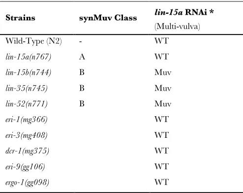
TABLE S1
Test for synMuvB phenotype
lin-15a RNAi *
Strains synMuv Class
(Multi-vulva) Wild-Type (N2) - WT
lin-15a(n767) A WT
lin-15b(n744) B Muv
lin-35(n745) B Muv
lin-52(n771) B Muv
eri-1(mg366) WT
eri-3(mg408) WT
dcr-1(mg375) WT
eri-9(gg106) WT
ergo-1(gg098) WT
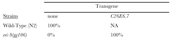
D. M. Pavelec et al. 3 SI
TABLE S2 Complementation of eri-9
Percentage of animals with viable progeny following lir-1 RNAi *
Transgene
Strains none C26E6.7
Wild-Type (N2) 100% NA
eri-9(gg106) 0% 100%
D. M. Pavelec et al.
4 SI
TABLE S3
Comparison of microarray datasets
Gene designation1 rrf-3 / WT2 eri-1 / WT2 fem-3(gf) / fem-13
D. M. Pavelec et al. 5 SI
T01C3.5 4.51 4.23 14.58 F58D2.2 2.67 2.92 14.37 W08E3.4 5.14 4.53 12.89 W03C9.1 2.92 2.51 12.42 F54A3.4 2.68 3.58 11.84 T16G12.7 5.61 5.71 11.44 C33F10.8 3.19 3.41 11.08 B0218.5 4.07 3.88 10.85 K07F5.4; kin-24 3.07 2.86 10.66 K07F5.6 3.77 3.84 10.46 F53C3.1 3.10 3.01 9.86 F59A6.2 7.49 5.57 9.63 C10C6.3 3.27 2.28 8.46 F59A3.8 2.58 2.88 8.46 F26F4.2 2.82 2.75 8.17 F56A11.6 2.16 2.06 8.02 ZK507.1 3.03 3.92 7.90 F09G8.4; ncr-2 4.11 3.95 7.38 Y6E2A.9 4.28 3.00 7.32 F25B3.4 4.54 4.52 6.27 Y38H6C.15 4.24 3.40 6.17 ZK666.8 4.54 4.02 6.10 C34F11.1 2.05 2.17 5.94 Y106G6G.3; dlc-6 9.39 8.43 5.75 ZK973.8 3.00 2.59 5.71 Y95B8A.4 2.00 2.20 5.49 R05H5.4 3.05 2.52 3.67 C25G4.4 2.24 2.05 2.12 Y39E4B.11 7.02 5.74 1.88 Y54G11A.6; ctl-1 4.47 5.01 0.88 C41H7.6 2.33 2.10 0.42 Y54G11A.13; ctl-3 6.13 6.83 N/A Y53F4B.45 3.66 3.90 N/A F58D5.7 3.38 3.22 N/A Y71F9AL.2 2.62 2.78 N/A
1Unique gene identifier from WormBase, release WS190. 2Microarray expression ratios from (ASIKAINEN et al. 2007).
3Microarray expression ratios from (REINKE et al. 2004) N/A, not available on
D. M. Pavelec et al.
6 SI
FIGURE S1.—Schematics of two genetic screens that identified eri genes. (A) Previously, we screened for mutant animals that exhibit an enhanced RNAi phenotype (1st screen) (DUCHAINE et al. 2006; KENNEDY et al. 2004). We revisited this screen and
identified additional alleles of previously known Eri factors (2nd screen). Specifically, GFP RNAi inefficiently silences unc-47::GFP
in wild-type animals (KENNEDY et al. 2004). Animals expressing unc-47::GFP were mutagenized with ethyl methanesulphonate (EMS) and F2 progeny were grown on E. coli expressing GFP dsRNA. Mutant animals able to silence unc-47::GFP in response to GFP dsRNA were kept for further analysis. These mutant animals were then subjected to dpy-13, lir-1, unc-22, and unc-73 RNAi to identify animals with generalized enhanced sensitivity to dsRNAs. eri alleles were assigned via complementation analysis for Eri phenotypes to the indicated complementation groups. It should be noted that eri-4/dcr-1 animals initially showed a strong and generalized enhanced RNAi phenotype. Following 5x outcrossings, however, eri-4/dcr-1 mutant animals exhibited a weaker but reproducible enhanced RNAi phenotype. (B) Wild type animals do not exhibit noticeable phenotypes in response to lin-15b
RNAi. eri-1, rrf-3, and eri-3 mutant animals, however, exhibit a multi-vulva (Muv) phenotype in response to lin-15b RNAi (GUANG et al. 2008). We EMS-mutagenized wild-type animals and screened for animals that exhibited a Muv phenotype in response to lin-15b RNAi. These mutant animals were then subjected to dpy-13, lir-1, unc-22, and unc-73 RNAi to identify animals with
D. M. Pavelec et al. 7 SI
D. M. Pavelec et al.
8 SI
FIGURE S3.—rrf-3(mg373) is defective for endogenous RNAi and displays a ts sterile phenotype. (A) Total RNA isolated from mixed stage animals of the indicated genotype was subjected to Northern blot analysis to detect the X-cluster small regulatory RNAs. Bottom panel, 5S RNA loading control stained with ethidium bromide. (B) cDNA generated from total RNA isolated from animals of the indicated genotype was subjected to quantitative PCR (qRT-PCR) analysis. mRNA levels of K02E2.6 and
D. M. Pavelec et al. 9 SI
FIGURE S4.—ssp-16, C25G4.6, and F18C5.4 mRNAs are enriched in the sperm-producing germline. Total RNA isolated
D. M. Pavelec et al.
10 SI
FIGURE S5.—mRNAs encoding Class II Eri factors are enriched in the female germ line. cDNA generated from total RNA isolated from age-synchronized larval stage four animals was subjected to qRT PCR analysis assessing levels of the indicated mRNAs. mRNA levels were normalized to eft-3 control, and data is expressed as ratio of mRNA abundance in animals lacking oocyte-producing germ line (fem-3(q96TSgf)) over mRNA abundance in animals lacking sperm-producing germ line (fem-1(hc17ts)).
D. M. Pavelec et al. 11 SI
FILE S1
Supporting Materials and Methods
C. elegans Strains:
The Hawaiian strain CB4856 was used for snip-SNP mapping of eri genes (WICKS et al. 2001). EG1285; lin-15(n765ts); oxls12
(unc-47::GFP ), YY009: eri-1(mg366), YY011: dcr-1(mg375), YY014: dcr-1(mg375);lin-15(n765ts); oxls12[unc-47::gfp], YY015:
eri-1(mg366);lin-15(n765ts);oxls12[unc-47::gfp], YY013: rrf-3(mg373), YY018: rrf-3(pk1426), YY019: eri-3(mg408), YY033:
eri-1(mg366);dcr-1(mg375), YY034: eri-1(mg366); eri-3(mg408), DP38: unc-119(ed3), YY166: ergo-1(gg098),YY173: eri-1(mg366);
ergo-1(gg098), YY206: eri-9(tm2346), YY211: eri-9(gg101), YY216: eri-9(gg106), YY221: eri-1(mg366);eri-9(gg106), YY223;
unc-119(ed3);eri-9(gg106), YY242: ergo-1(gg098);eri-9(gg106), YY174: nrde-3(gg066); ggls01[nrde-3::gfp], YY175: eri-1(mg366);
nrde-3(gg066); ggls01[nrde-3::gfp], YY219: eri-9(gg106); ggls01[nrde-3::gfp], YY237: dcr-1(mg375); ggls01[nrde-3::gfp], JK1973: fem-3(q96),
YY273: eri-1(mg366);fem-3(q96), BA17: fem-1(hc17), YY299: eri-1(mg366); fem-1(hc17), YY369: unc-119(ed3);
eri-9(gg106);ggEx001[eri-9::3xFlag::gfp], WM27: rde-1(ne219).
RNAi experiments:
RNAi experiments were conducted as described previously (TIMMONS et al. 2001). Bacteria expressing dsRNA, including sqt-3,
lir-1, unc-73, cel-1, tra-2, dpy-13, lin-1, unc-22, and lin-15a, were obtained from the Ahringer RNAi library (KAMATH et al. 2003) and
sequenced to verify their identities. lin-15b RNAi was performed as described previously (GUANG et al. 2008). Phenotypes elicited
by each RNAi clone were scored as either the percentage of animals exhibiting the appropriate phenotype or on a scale of -
(unaffected) to ++++ [same severity as eri-1(mg366) animals].
Transgenic rescue:
eri-9(gg106); unc-119(ed3) hermaphrodites were injected with a plasmid containing the wild-type unc-119 and C26E6.7 genes.
Transgenic lines were identified by rescue of the Unc motility defect, then screened for lethality of lir-1 RNAi. The HA-tagged
dcr-1 rescuing transgene has been described previously (Duchaine et al. 2006). Rescue of dcr-1(mg375Eri) was assessed by the
restoration of fertility at 25˚C.
Phenotypic analyses:
Genetic screens for enhanced sensitivity to RNAi are described in Supporting Materials. To determine brood size, individual
adult hermaphrodites were propagated at the indicated temperature and transferred to new plates every 24 hours. Total numbers
D. M. Pavelec et al.
12 SI
determined by counting no less than 1154 progeny from hermaphrodite animals reared at 23˚C. Mating experiments to assess
fertility of Eri hermphrodites were performed with wild-type males harboring an unc-47::GFP transgene marker to allow
identification of cross-progeny. Mating experiments to assess fertility of Eri males were performed with fem-1(hc17ts)
hermaphrodites reared at 25˚C.
qRT-PCR primers (all shown 5’->3’):
eft-3 -(acttgatctacaagtgcggagga) and (aaagatcccttacccatctcctg)
act-1 - (ccgtgacatcaaggagaagc) and (cctgtccgtcaggaagttcg)
E01G4.5 - (gccaaacagcttctagaagccgc) and (cgggttgacgtccattacaagtcc)
W04B5.1 - (gctacacgttttcaaaatgtgtggct) and (ccgccaagtggaattttcttctccg)
C40A11.10 - (tgtggatttcaacgtggcgg) and (gatgctatcgcttagcggtgg)
K02E2.6 - (ccagtggtacaagtgggagtaaacg) and (cgctgcgtgagctgtagttgatagg)
msp-3 - (cggcgagcagatgaattgatcacc) and (ccaaacccagccgggtacg)
ssp-16 - (tgatcactcgcactgctg) and (gccgacattggaattgtcac)
C25G4.6 - (cgtcccgattcgattgatg) and (ggcttcaatcctggaagagc)
F18C5.4 - (ccgtcttacattggaaagatcc) and (tttggaggaggaagcatcac)
T14G10.8 - (caagtagttccggcaactcatcgg) and (gaatactcgcgcgaaacgatgattaaac)
C55C3.5 - (gcgatctcggcggtggc) and (cttcgagtcttgtagctcgcgg).
Northern blot probe sequences:
ssp-16 mRNA- (5’-tggtgcgaaatgaacgacaagtttgtcctcctttg/3 StarFire/-3’)
act-1 mRNA- (5’-ggtggttcctccggaaagaacagtgttggcgtaca/3 StarFire/-3’)
E01G4.5 siRNA- (5’-gaccaaaccgcgcgcttcagaggtcattggcttcatacactcaaaagc/3 StarFire/-3’)
X-cluster siRNA- (5’-acctcataccgcgtatctattc/3 StarFire/-3’)
let-7 miRNA- (5’-aactatacaacctactacctcaccggatcc/3 StarFire/-3’)
21U-1 piRNA- (5’-gcacggttaacgtacgtacca/3 StarFire/-3’)
ssp-16 siRNA- atacgaaacaacaaccaaagctttaccatgtcgctcactgctgatccaccagcctgcactgtgcc/3 StarFire/-3’),
(5’-ggacaaacttgtcgttcatttcgcaccagccccagctgatgctactgatgctcaggccgc/3 StarFire/-3’), (5’-
D. M. Pavelec et al. 13 SI
Supporting References
ASIKAINEN,S., M.STORVIK, M.LAKSO and G.WONG, 2007 Whole genome microarray analysis of C. elegans rrf-3 and eri-1
mutants. FEBS Letters 581: 5050-5054.
DUCHAINE,T.F., J.A.WOHLSCHLEGEL, S.KENNEDY, Y.BEI, J.D.CONTE et al., 2006 Functional Proteomics Reveals the
Biochemical Niche of C. elegans DCR-1 in Multiple Small-RNA-Mediated Pathways. Cell 124: 343-354.
GUANG,S., A.F.BOCHNER, D.M.PAVELEC, K.B.BURKHART, S.HARDING et al., 2008 An Argonaute Transports siRNAs from the Cytoplasm to the Nucleus. Science 321: 537-541.
KAMATH,R.S., A.G.FRASER, Y.DONG, G.POULIN, R.DURBIN et al., 2003 Systematic functional analysis of the Caenorhabditis elegans genome using RNAi. Nature 421: 231-237.
KENNEDY,S., D.WANG and G.RUVKUN, 2004 A conserved siRNA-degrading RNase negatively regulates RNA interference in
C. elegans. Nature 427: 645-649.
REINKE,V., I.S.GIL, S.WARD and K.KAZMER, 2004 Genome-wide germline-enriched and sex-biased expression profiles in
Caenorhabditis elegans. Development 131: 311-323.
TIMMONS,L., D.L.COURT and A.FIRE, 2001 Ingestion of bacterially expressed dsRNAs can produce specific and potent genetic
interference in Caenorhabditis elegans. Gene 263: 103-112.
WICKS,S.R., R.T.YEH, W.R.GISH, R.H.WATERSTON and R.H.A.PLASTERK, 2001 Rapid gene mapping in Caenorhabditis