ABSTRACT
Drake, Stephenie Lynn. Characterization of the response of Vibrio vulnificus to sublethal stresses during oyster handling and processing (Under the direction of Dr. Lee-Ann Jaykus)
Vibrio vulnificus, a naturally occurring marine bacterium, causes severe disease in at-risk individuals consuming contaminated raw shellfish. The organism can be difficult to discriminate from natural microflora present in the product, complicating the
evaluation of process control efficacy. The purpose of this study was to construct a strain of V. vulnificus expressing green fluorescent protein (Vv-GFP-K) which could be readily distinguished from background flora. Once constructed, the objectives were to compare the physiological characteristics of Vv-GFP-K to the wild-type parent (Vv-WT); to use Vv-GFP-K to evaluate survival of the bacterium under various environmental stresses relevant to food processing; and to assess the effect of sodium pyruvate media
supplementation on recovery efficiency, with particular reference to sublethally injured cells.
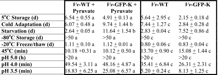
V. vulnificus strain ATCC 27562 was engineered to express GFP and kanamycin resistance using methods of conjugation. Comparisons were made between Vv-GFP-K and Vv-WT with respect to growth characteristics, heat tolerance (45oC), freeze/thaw tolerance (-20o and -80oC), acid tolerance (pH 5.0, 4.0, and 3.5), cold storage (5oC), cold adaptation (15oC) and starvation. Recoveries were evaluated using non-selective [tryptic soy agar-2%NaCl, (TSAN2)] medium with and without sodium pyruvate
designated (i) rapid cooling (iced); (ii) conventional cooling (5oC); and (iii) mild abusive cooling (temperature dropped to 5oC over 8 hr) were evaluated. Acetic and citric acids at pH values ranging between 3.5 to 5.0 were evaluated in the acid studies.
In most cases, Vv-GFP-K was comparable to Vv-WT with respect to growth, survival, thermal inactivation, and freeze thaw survival. There were differences between Vv-WT and Vv-GFP-K with respect to acid tolerance, although these differences
disappeared with sodium pyruvate supplementation of media. In broth studies dealing with organic acids, Vv-GFP-K was rapidly inactivated with acetic acid. Similar, but not as dramatic results were seen for citric acid. As pH values declined, the positive impact of pyruvate supplementation on cell recovery disappeared.
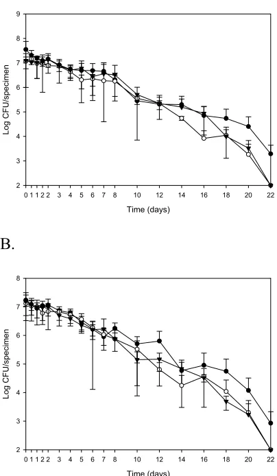
In the refrigeration studies done in the matrix, there were no apparent differences in Vv-GFP-K levels for all three treatments within the first few days of storage. In all cases, levels dropped 1 log10 after 8 days refrigerated storage. By the end of the study
(21 d), Vv-GFP-K levels were nondetectable for both iced and conventionally cooled product, however mild abusively cooled oysters still had levels approximating 103 CFU/oyster. Vv-GFP-K levels remained stable for up to 24 hrs within the oyster meat under acidic conditions at various pH values. The oyster meat provided a protective environment that prevented inactivation of Vv-GFP-K.
CHARACTERIZATION OF THE RESPONSE OF VIBRIO VULNIFICUS TO SUBLETHAL STRESSES DURING OYSTER HANDLING AND PROCESSING
By
STEPHENIE LYNN DRAKE
A thesis submitted to the Graduate Faculty of North Carolina State University
in partial fulfillment of the requirements for the Degree of
Master of Science
FOOD SCIENCE
Raleigh 2004
APPROVED BY:
________________________ ______________________
Dr. Lee-Ann Jaykus Dr. David Green
Chair of Advisory Committee
ii
BIOGRAPHY
Stephenie Lynn Drake was born February 6, 1979 in Sunnyside, Washington. She grew up in the city of Wenatchee in the foothills of the Cascade Mountains. She graduated from Wenatchee High School in 1998, where she was very active in FFA and 4-H. She then moved to Starkville, Mississippi to escape shoveling snow and to start her undergraduate college education at Mississippi State University. While at MSU, she was very active in the Food Science club especially as president for 3 years. She completed an underdergraduate research project under the direction of Dr. Marshall. She received second place in the undergraduate research competition at the Institute of Food
Technologists/ Meeting in June, 2002. She obtained her Bachelor of Science degree in Food Science in 2002.
The confidence gained during her undergraduate research project was invaluable when it came time for her to begin her own graduate research under the direction of Dr. Jaykus. She received second place for her oral presentation in the Food Microbiology Division at the Institute of Food Technologists’ Metting in July, 2003.
iii
ACKNOWLEDGEMENTS
Thank you to the North Carolina Sea Grant Program for supporting this project. I would like to express a sincere gratitude to major professor, Dr. Lee-Ann Jaykus. Thank you for your patience and guidance throughout my time here.
I would also like to thank my committee members; Dr. David Green and Dr. Donn Ward for their invaluable assistance during this project.
I would like to thank my husband, Robert May, who did not know what he was getting into when he married me. I could not have done this without your support.
I would like to thank my sister, MaryAnne, who was always there at the building on the weekends and was also there to listen to me.
I would like to thank my loving parents, Stephen and Dorothy. Thank you for your encouraging words and never ending support and love.
iv
TABLE OF CONTENTS
Page
LIST OF TABLES... vi
LIST OF FIGURES... vii
CHAPTER 1. Comprehensive Review of the Literature... 1
1.1 Introduction...1
1.1.1 Classification...1
1.1.2 Habitat...2
1.1.3 Distribution ...3
1.2 Pathogenicity...4
1.3 Epidemiology...6
1.3.1 General...6
1.3.2 Wound infection...7
1.3.3 Primary septicemia...8
1.3.4 Gastroenteritis ...8
1.4 Enumeration and Detection Methods...8
1.5 Environmental Factors ...12
1.5.1 Temperature ...12
1.5.2 Salinity ...13
1.5.3 pH...14
1.5.4 Cold stress...15
1.5.5 Starvation ...16
1.5.6 VBNC ...18
1.6 Techniques to eliminate V. vulnificus from oysters ...22
1.6.1 Commercial heat shock...23
1.6.2 Depuration and relaying...24
1.6.3 GRAS compounds ...26
1.6.4 Ionizing irradiation...27
1.6.5 Thermoradiation...28
1.6.6 Refrigeration ...28
1.6.7 Freezing and vacuum packaging...29
1.6.8 Hydrostatic pressure...30
1.6.9 Heat treatment...31
1.7 Summary...32
1.8 Reference Cited...36
v
2.1 Abstract ...46
2.2 Introduction...48
2.3 Materials and Methods...51
2.3.1 Bacteriological media ...51
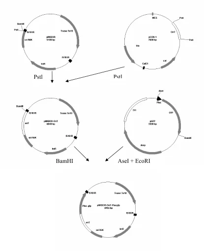
2.3.2 Development of mutant (Vv-GFP-K) of V. vulnificus...52
2.3.3 Comparison of growth and survival characteristics of Vv-GFP-K to wildtype Vv-WT...53
2.3.4 Recovery of Vv-GFP-K in oyster matrix ...53
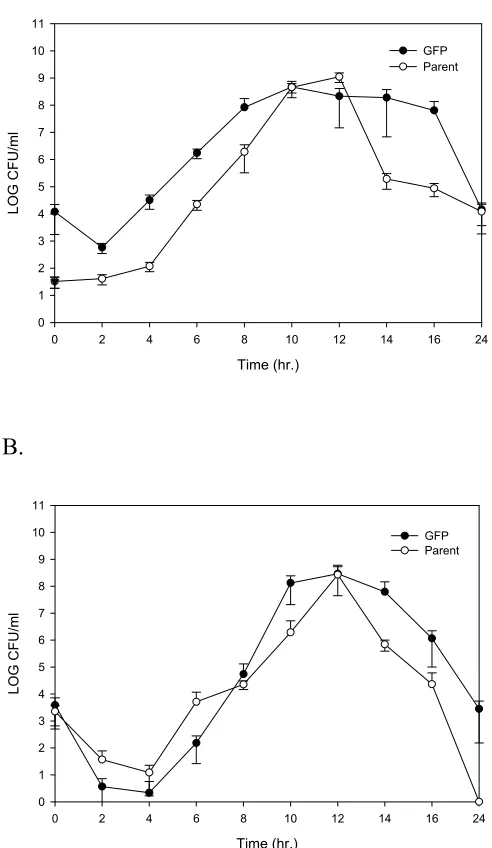
2.3.5 Growth curves...54
2.3.6 Refrigerated storage and starvation ...54
2.3.7 Frozen storage...55
2.3.8 Thermal inactivation ...55
2.3.9 Acid tolerance ...56
2.4 Statistical Analysis...56
2.5 Results...56
2.6 Discussion...60
2.7 References Cited ...74
CHAPTER 3. Using a green fluorescent protein strain of Vibrio vulnificus to evaluate microbial stress in the oyster matrix...78
3.1 Abstract ...78
3.2 Introduction...80
3.3 Materials and Methods...82
3.3.1 Strain...82
3.3.2 Bacteriological media ...82
3.3.3 Refrigerated storage ...83
3.3.4 Acid exposure ...84
3.3.5 Joint effect of refrigeration and acidic storage ...85
3.3.6 Statistical Analysis...85
3.4 Results...86
3.5 Discussion...88
3.6 Conclusions...93
3.7 References Cited ...101
APPENDICES...103
Appendix A. Appearance of Vv-GFP-K expressing fluorescencing under ultraviolet light on TSAN2 compared to Vv-WT. ...104
Appendix B. Morphological and biochemical comparisons between Vv-WT and Vv-GFP-K...105
Appendix C. Recovery of injured cells due to starvation at 5oC using different concentrations of sodium pyruvate (0-320mg). ...106
vi
LIST OF TABLES
CHAPTER 1.
1.1 Preliminary biochemical tests ... 35 CHAPTER 2.
2.1 Comparison of Vv-WT to Vv-GFP-K on pyruvate supplemented and
non-supplement media ...73 2.2 Comparison of pyruvate supplemented and non-supplement media with
vii
LIST OF FIGURES
CHAPTER 1.
1.1 Schematic of oyster...34 CHAPTER 2.
2.1 Schematic representation of the procedure for construction of plasmid ...67 2.2 Growth Curves at 37oC of GFP-K and parent strains of V. vulnificus...68 2.3 Survival of GFP-K and parent strain of V. vulnificus at various conditions.69 2.4 Survival of GFP-K and parent strain of V. vulnificus during different
freezing temperatures...70 2.5 Survival of GFP-K and parent strain of V. vulnificus during thermal
inactivation at 45oC ... 71 2.6 Survival of GFP-K and parent strain of V. vulnificus under various acidic
conditions...72 CHAPER 3.
3.1 Survival of Vv-GFP-K in shellstock oysters plated with and without
sodium pyruvate supplementation ...96 3.2. Survival of Vv-GFP-K in acidified TSBN2 with acetic acid plated with
and without sodium pyruvate supplementation ...97 3.3. Survival of Vv-GFP-K in acidified TSBN2 with citric acid plated with
and without sodium pyruvate supplementation ...98 3.4. Survival of Vv-GFP-K in shucked oysters and acidified TSBN2 held at
23oC and plated with and without sodium pyruvate supplementation ...99 3.5. Survival of Vv-GFP-K in acidified TSBN2 with citric acid plated with
CHAPTER 1 Literature Review Introduction
In the U.S., contaminated seafood is responsible for 26.5% of all foodborne disease outbreaks (Mead et al., 1999). The majority of these seafood-related human illnesses are associated with the consumption of raw bivalve molluscan shellfish such as oysters, clams, and mussels (Cook, 1991). Bivalves are filter feeding organisms that pump seawater through their digestive systems to obtain oxygen and food. In this process they accumulate and concentrate microorganisms in their digestive tracts. These include harmless environmental organisms as well as pathogens, the most significant of which are the human enteric viruses and the pathogenic Vibrio species. Since shellfish are frequently consumed whole and raw, they can serve as passive carriers of foodborne disease agents.
Classification
The bacterial genus Vibrio is in the family Vibrionaceae, which also includes the genera Aeromonas, Plesiomonas, and Photobacterium (Atlas, 1997). All Vibrios are environmentally ubiquitous in the marine environment. V. cholerae O1 and V. mimicus are the only Vibrio species that do not grow in media with added sodium chloride. There are 30 species in the genus Vibrio. Thirteen of these species are known to be pathogenic to human and fish, including V. cholerae O1, V. cholerae non-O1, V. mimicus, V.
Vibrios have been reported to cause foodborne disease; however, V. cholerae O1, V. parahaemolyticus, and V. vulnifiucus are the most significant agents. Members of the Vibrio genus are straight or curved Gram negative, non-spore forming rods, 0.5 to 0.8 µm in width and 1.4 to 2.6 µm in length (McLaughlin, 1995). However, when they are grown in the laboratory, they frequently revert to a straight rod morphology (Atlas, 1997). They are motile by a single polar flagellum and are either aerobic or facultatively anaerobic. Most species produce oxidase and catalase and ferment glucose without producing gas (McLaughlin, 1995). V. vulnificus is similar phenotypically to V.
parahaemolyticus (Oliver, 1989). The two most distinctive reactions of V. vulnificus are fermentation of lactose and production of B-D-galactosidase and these tests can be used to distinguish it from the related Vibrio parahaemolyticus (Hollis et al., 1976).
Habitat
Vibrio species are environmentally ubiquitous to estuarine waters and can frequently be isolated in high numbers from bivalves, sediment, and plankton. V. vulnificus can be isolated directly from seawater and a variety of other marine foods as well (Kelly, 1982: Oliver et al., 1983; Tamplin et al., 1982; Oliver et al., 1983; Oliver, 1989; O’Neil et al., 1992; Ruple and Cook, 1992; Tamplin and Capers, 1992; Cook, 1994; DePaola et al., 1994; Wright et al. 1996). The numbers of V. vulnificus in Gulf Coast oysters peak during the summer followed by a gradual reduction in the winter (Motes et al., 1998). Higher densities of V. vulnificus are found in oyster digestive tissue (DePaola et al., 1997) as compared to muscle tissue.
Although V. vulnificus levels are higher in the estuarine environment during the warm summer months as compared to the winter, the organism remains persistent
have found that V. vulnificus survives in plankton and the organism has in fact been found in plankton, suggesting that this bacterium inhabits similar niches to V. cholera and V. parahaemolyticus (Wright et al, 1996 and Vanoy et al.,1992). Others believe that V. vulnificus lives in sediment. For instance, Vanoy et al. (1992) found V. vulnificus during the months of January to March in marine sediment, suggesting winter survival in the floc zone at the sediment interface. It may be possible that V. vulnificus survives in the sediment interface during the winter and when conditions are more conducive for growth (summer months), V. vulnificus will go on to colonize plankton.
Distribution
shellfish samples collected from Mid-Atlantic waters. V. vulnificus accounted for 8-10% of the total aerobic bacteria isolated from Chesapeake Bay oysters, suggesting that levels of V. vulnificus in Chesapeake waters may be comparable to those in Gulf of Mexico waters (Wright et al., 1996).
Pathogenicity
There are virulent and avirulent strains of V. vulnificus. Many virulence factors have been reported for V. vulnificus and include (i) the presence of a polysaccharide capsule; (ii) various extracellular enzymes; (iii) the ability to obtain iron from transferrin; and (iv) the absence of estrogen in the host (Linkous and Oliver, 1999).
The presence of a capsule, which is also related to colony opacity, is probably the best known virulence factor. The transformation of encapsulated isolates to the non-encapsulated form is dependent on growth phase and other variables in the environment, which in turn affect bacterial cell morphology. Encapsulated isolates have an opaque colony morphology but can undergo a reversible phase variation to a translucent colony phenotype that is correlated with reduced capsular polysaccharide (CPS). Translucent strains are less virulent than opaque strains. For instance, Moreno and Landgraf (1998) found that translucent colonies of V. vulnificus were avirulent in mice and Wright et al. (1990) reported that nonencapulated strains produced by transposon mutagenesis had a lethal dose over four times higher than that of the encapsulated strains (Wright et al., 1990).
protease of V. vulnificus may be of extreme importance to the pathogenicity of this bacterium (Linkous and Oliver 1999). A metalloprotease containing a zinc atom, this enzyme degrades a number of biologically important proteins including elastin, fibrinogen, and plasma protease inhibitors (Miyoshi et al., 1995). The most dramatic pathological action of the metalloprotease is its vascular permeability-enhancing action (Shinoda and Miyoshi, 2000). V. vulnificus also produces both hydroxymate and
phenolate (catechol) siderophores which have been shown through production of isogenic mutants to be associated with enhanced virulence (Strom and Paranjpye, 2000).
Extracellular proteins produced by V. vulnificus are also important in the organism’s ability to survive in the estuarine environment and perhaps cause disease in infected hosts. For example, V. vulnificus exports a chitinase that may be used by the bacterium to colonize and adhere to the chitin exoskeletons of zooplankton. The production of the metalloprotease and a hemolysin may allow the organism to colonize and multiply in molluscan shellfish by breaking down tissue at the site of colonization, promoting release of necessary nutrients (Strom and Paranjpye, 2000).
The amount of iron available in the host is an important factor influencing the lethality of both virulent and avirulent strains of V. vulnificus. Stelma et al. (1992) found that an iron overloaded mouse was killed at doses of <100 CFU for a virulent strain of V. vulnificus and at a dose >4000 CFU for an avirulent strain. On the other hand, Reyes et al. (1987) showed a lethal oral infective dose of V. vulnificus to be <105 CFU/ml for virulent strains and >109 CFU/ml for avirulent strains when mice were not iron
in the amount of transferrin, may be associated with the pathogenesis of V. vulnificus infection (Morris et al., 1987 and Brennt et al., 1991). Transferrin is an iron transport protein, and because free iron is virtually absent in the human body, pathogenic bacteria like V. vulnificus may have evolved a mechanisms to scavenge iron from the iron transport proteins (Strom and Paranjpye, 2000).
Recent studies suggest that men are more susceptible to V. vulnificus infection than are women, and it appears that estrogen may be protective against infection.
According to a study conducted by Shapiro et al. (1998), 86% of the reported cases of V. vulnificus infection occurred in men. Although this may be due to the fact that men are more likely to consume raw oysters, or that men are more likely to have underlying liver disease, a recent study by Linkous and Oliver (1999) offers an alternative explanation. This investigation showed that male rats injected with V. vulnificus LPS had an 82% fatality rate, whereas, normal female rats treated identically had a fatality rate of only 21%. When these female rats that were ovariectomized, which lowered their estrogen levels, fatality rates increased to 75%. Estrogen may protect women by reducing iron availability. Women have a tendency to be anemic, which reduces overall iron availability in this gender.
Epidemiology General
vulnificus infection, with 80% of those who die from the infection having underlying liver dysfunction (Strom and Paranjpye, 2000).
In one of the first of its kind, Desenclos et al. (1991) used the case control study design to show an estimated annual incidence of Vibrio infectious at 95.4 per million for raw oyster consumers with liver disease, 9.2 per million for raw oyster consumers without liver disease, and 2.2 per million for those who do not consume raw oysters. Another case control study conducted by Hlady and Klontz (1996) reported disease manifestation proportions of 51%, 24%, and 17% for gastroenteritis, wound infection, and septicemia, respectively. Fatality rates were only 1% for gastroenteritis, 5% for wound infection, and 44% for septic disease. Sixty-eight percent of gastroenteritis and 83% of primary septicemia cases were associated with raw oyster consumption. Ninety-one percent of the primary septicemia cases and 86% of the wound infections occurred in the months of April through October, with 48% of those with primary septicemia
reporting pre-existing liver disease. Wound infection
Klontz (1996), it was reported that other Vibrio species can also be responsible for wound infections.
Primary septicemia
Primary septicemia is defined as a systemic illness characterized by fever and shock and for which V. vulnificus is isolated from blood or an otherwise sterile sites (Strom and Paranjpye, 2000). The consumption of contaminated raw oysters is the cause of this disease in almost all cases. Desenclos et al. (1991) showed that raw oyster
consumers were 10 times more likely to develop primary septicemia than non-consumers. Hlady and Klontz (1996) showed that V. vulnificus, V. cholera non O1, and V.
parahaemolyticus can all cause septic disease. Liver disease due to cirrhosis, alcoholism, or hepatitis is the most important risk factor for fatality from V. vulnificus infections, especially primary septicemia (Shapiro et al., 1998).
Gastroenteritis
Enumeration and Detection Methods
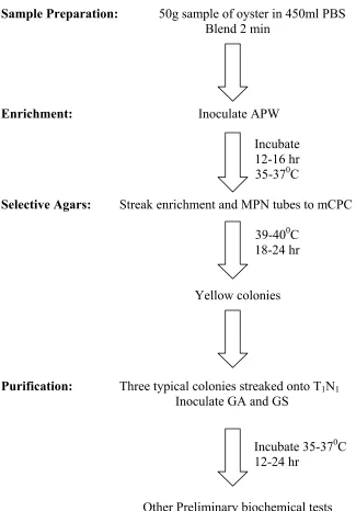
There are several different methods recommended for the detection and/or enumeration of V. vulnificus. The FDA Bacteriological Analytical Manual (1998) cites the standard procedures for recovery of V. vulnificus (Figure 1). In this method,
phosphate buffered saline (PBS) dilutions are followed by alkaline peptone water (APW) enrichments in a MPN format. PBS enrichment broth can be used in parallel, and in both cases, enrichments are incubated at 35-370C for 16-18 hrs. MPN tubes having turbidity are streaked onto modified Cellobiose Polymysin B Colstrin (mCPC), with the PBS enrichment broth also streaked onto mCPC agar plates as a control. The mCPC plates streaked from the PBS enrichment broth serves as a control in case APW tubes lack turbidity. Modified CPC plates are incubated for 18-24hr at 39-400C after which they are examined for typical V. vulnificus colonies which are flat and yellow with opaque centers and translucent peripheries, about 2mm in diameter. These presumptive colonies are then transferred to APW for the monoclonal antibody-Enzyme Immunoassay (EIA) test for rapid identification of V. vulnificus. For biochemical identification, three or more typical colonies are selected from mCPC, streaked onto 1% trypton, 1% NaCl (T1N1) agar, and
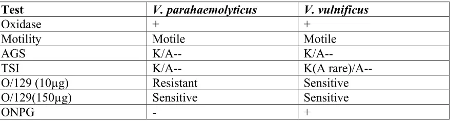
also inoculated into gelatin agar (GA) and gelatin salt (GS) agar, followed by incubation for 18-24 hr at 35-370C. Other preliminary biochemical tests for V. vulnificus include oxidase, motility, arginine-glucose slant (AGS), triple sugar iron (TSI) agar, O/129 Vibriostat sensitivity, and the ONPG test. The different reactions for V. vulnificus and V. parahaemolyticus to the preliminary biochemical tests are outlined in Table 1.
For instance, Azanza et al. (1996) found that peptone (0.1%) solution containing 3% NaCl was a better diluent than was phosphate buffered saline. Hagen et al. (1994) found that alkaline peptone water (APW) was better at recovering V. parahaemolyticus and V. vulnificus than was salt-polymyxin B broth (SPB). Alam et al. (2001) suggested using nutrient agar with 2% NaCl as the primary culture medium followed by transfer to a selective medium such as Thiosulfate-citrate-bile salt sucrose (TCBS), since this two-step method recovered significantly higher numbers of V. parahaemolyticus and other Vibrios from seawater samples. Hoi et al. (1998) found that pre-enrichments followed by
selective plating on cellobiose-colistin (CC) agar were more effective in increasing and enumerating V. vulnificus than were cellobiose-polymyxin B-colistin (CPC), modified cellobiose-polymyxin B-colistin (mCPC), and thiosulfate-citrate-bile salt sucrose (TCBS) agars. However, TCBS was the only agar for which these recovery differences were significant. It was observed in this research that levels of colistin and polymyxin B retard the growth of V. vulnificus. TCBS agar was generally a good medium for cultivating V. vulnificus; however, it was not good at differentiating V. vulnificus from other sucrose negative Vibrios and CPC agar outperformed in this respect (Oliver et al., 1992).
traditional 5 to 7 days. Alam et al. (2001) found that replicate plating produced better cell recovery and higher counts than did the MPN method.
It is known that low temperature induces membrane damage that tends to make cells more sensitive to salt, in which case salt levels generally found in marine and estuarine media can be lethal to stressed cells. Both V. vulnificus and V.
parahaemolyticus require a minimum of 0.5% NaCl, but large amounts of salt can negatively affect the recovery of these organisms. Oliver (1981) found that TCBS, estuarine salts (ES), and brain heart infusion (BHI) agars all produced comparable results for V. vulnificus and V. parahaemolyticus as plating media on survival assays, especially after cold shock. However, the addition of 3% salt to BHI greatly affected cell viability, with V. vulnificus killed after 30 min and V. parahaemolyticus after 24 hours.
The development of DNA based methods for the detection of V. vulnificus has aided in both the identification and discrimination of Vibrio species from one another. Wright et al. (1993) developed an oligonucleotide DNA probe (VVAP) to identify V. vulnificus, which could effectively differentiate V. vulnificus from other Vibrio species. Gooch et al. (2001) found that direct plating colony lifts followed by hybridization using probes specific for V. vulnificus and V. parahaemolyticus produced results similar to MPN methods. In their study, the differences between direct plating followed by colony lift hybridization and MPN methods occurred only at lower densities which may imply different detection sensitivities; MPN methods were more sensitive (3 MPN/g for a 0.1g) than direct plating methods (10 CFU/g for 0.1g sample). The hydrophobic grid
placed on tryptic soy magnesium sulfate-sodium chloride (TSAMS) agar for 3 hours at 350C and then transferred to Vibrio parahaemolyticus sucrose agar (VPS) and incubated at 420C overnight. Blue to blue-green colonies are counted and differentiation is done using a TDH-specific gene probe for V. parahaemolyticus and a gene probe specific for the cytotoxin-hemolysin gene of V. vulnificus (Kaysner, 1994).
PCR has also been used to identify V. vulnificus. Brauns et al. (1991) detected both culturable and non-culturable V. vulnificus by PCR amplification using primers flanking a 340-bp fragment of the cytotoxin-hemolysin gene. As little as 72 pg of DNA from culturable cells and 31 ng of DNA from nonculturable cells could be detected. Brasher et al. (1998) designed a multiplex PCR assay to simultaneously detect several foodborne pathogens, including V. vulnificus, V. parahaemolyticus, and V. cholerae. When applied to artificially inoculated oyster homogenate, they were able to detect <101 -102 cells/g. Amplification targets were invA, ctx, and tl genes for Vibrio vulnificus, V. cholerae, and V. parahaemolyticus, respectively. In addition, Wang et al. (1997)
developed a different PCR method to detect 13 species of foodborne pathogens, including Vibrio cholerae, V. parahaemolyticus, and V. vulnificus. Detection limits per PCR
reactions for Vibrio cholerae, V. parahaemolyticus, and V. vulnificus are 40, 4, and 100 cells, respectively, and no interference was observed using the PCR assay when food samples were artificially inoculated with other competing bacterial species.
Environmental Factors Temperature
optimum growth temperature of 370C (Kelly, 1982). V. vulnificus was rarely isolated from surface water samples collected from the Gulf of Mexico in January through March, when water temperatures were below 200C. Peak recovery occurred in September
indicating seasonal variation in organism prevalence (Kelly, 1982). The inability to culture V. vulnificus from cold waters is probably due to the entry of cells into a “viable but not culturable (VBNC)” state (Wolf and Oliver, 1992; Oliver, 1993; 1995; Oliver et al., 1995). In a study conducted by Kaspar and Tamplin (1993), V. vulnificus grew quite well in the temperature range of 130C to 220C, but did not grow well below 8.50C. Wright et al. (1996) were able to retrieve V. vulnificus from estuarine waters of the Chesapeake Bay at temperatures as low as 80C, however, V. vulnificus could not be recovered at temperatures lower than 12.50C from the Gulf of Mexico waters (Simonson and Siebeling, 1986).
Salinity
Salinity also seems to play an important role on the prevalence of V. vulnificus. Tamplin et al. (1982) found that V. vulnificus could not be isolated during winter months but could be isolated at high densities during summer months. These researchers
concluded that both temperature and salinity play important and interrelated roles, as V. vulnificus was not isolated until temperatures of 170C and salt content of 17 ppt. were reached, and higher levels of V. vulnificus were found when the water temperature
exceeded >290C and salt content was greater than 23 ppt. Further research by Kaspar and Tamplin (1993) demonstrated that at salinities between 5 and 25 ppt., V. vulnificus levels increased; however, when salinities were 30, 35, and 38 ppt, V. vulnificus levels
(1998) who observed lower numbers of V. vulnificus cells at salinities above 28 ppt, which is typical of Atlantic coastal sites. This high salinity may explain in part why V. vulnificus could not be routinely isolated from oysters harvested from waters off of North or South Carolina. High V. vulnificus levels, however, were found in oysters harvested from intermediate salinities between 5 and 25 ppt (Motes et al., 1998).
pH
There are two strategies for bacteria to survive low pH conditions. One
mechanism is the rpoS gene that encodes for the alternative sigma factor. A sigma factor is a subunit of bacterial RNA. The sigma factor attaches to the bacterial RNA
polymerase which in turn attaches to a DNA promoter, signaling the transcription of a particular gene (Lewin, 1997). The RpoS alternative sigma factor regulates at least 30 proteins, including the expression of genes that control thermotolerrance, starvation survival, osmotic stress response, and acid response (Jay, 2000).
The acid tolerance response is another mechanism for survival of V. vulnificus. In many instances, when bacteria are exposed to a pH <5.9, the acid tolerance response is induced and this enables cells to survive at pH values as low as 3.3 (Waterman and Small, 1996). It has been suggested that this response may also reduce the infectious dose of certain bacteria (Waldor and Mekalanos, 1996), theoretically because the cells may be “conditioned” to survive at low pH, promoting their survival through the GI tract.
cells from a pH as low as 3.5. Just like V. vulnificus, when A. hydrophila in broth was shifted from pH 7.2 to 5.0 then to 3.5, the cells were able to survive in culture (Karem et al., 1994). This study showed that proteins synthesized during adaptation to pH 5.0 were involved in the subsequent survival during exposure to pH 3.5. Koo et al. (2000)
reported strain to strain differences in acid tolerance, although at pH 2.0 there was no variability in reduction between strains suggesting that after a certain pH all strains were killed. It appears that low temperature offers protection against the anitibacterial effects of both pH and salinity. Hijarrubia et al. (1996) observed a more rapid inactivation of V. vulnificus cells kept at low pH and salinity at 200C as compared to storage under the same conditions at 50C.
Cold Stress
Some organisms such as V. vulnificus are able to adapt and endure at very low temperatures, when previously conditioned by exposure to less cold temperature. V. vulnificus is more susceptible to low temperatures when in its logarithmic growth phase as compared to its stationary phase (Oliver et al., 1991). Kim and Dunn (1997) found that lactic acid bacteria freeze-thaw survivability improved when cells were cold shocked at 100C for 2 hrs prior to freezing at -200C, and that a gene homologous to the major cold shock protein of E. coli and B. subtilus appeared responsible. Bryan et al. (1999)
suggested that cold adaptive “protective” proteins in V. vulnificus may enhance survival and tolerance to cold temperatures and that iron plays a role in adaptation at cold
that of E. coli. Nonetheless, even though overall protein synthesis for V. vulnificus
showed a sharp decline, forty unique proteins were synthesized at higher levels during the cold stress response (McGovern and Oliver, 1995). When V. vulnificus cells are cold shocked, their numbers initially decline but after a brief lag, cell growth resumes at a slower rate.
V. vulnificus’s entry into the VBNC state can be slowed by exposure to a slightly colder temperature prior to the final cold temperature, this slower entry into the VBNC state may be attributed to an adaptative response. In a study conducted by Bryan et al. (1999), V. vulnificus entered the so-called VBNC state when the temperature shifted from 350C to 60C; however, when the culture was subjected to 150C prior to further
temperature downshift, the cells remained culturable, showing an adaptive response to cold temperature. The culture was also able to survive better during freezing at -780C when freezing was preceded by cold shock. In a study conducted by Bang and Drake (2002), cold stress had no effect on V. vulnificus heat resistance or freeze/thaw resistance, but cold adaptation improved cold temperature tolerance.
Starvation
and decreased rate of recovery. This leads to a rapid loss of available
poly-B-hydroybutyrate that in turn results in the formation of outer membrane vesicles that are related to the continuous size reduction that occurs during starvation survival.
In a study conducted by Nystrom et al. (1990) using Vibrio spp. strain S14, which resembles Vibrio proteolyticus, the investigators described long-term multiple nutrient starvation as a three-phase process. The first step was the accumulation of guanosine5’-diphosphate (ppGpp) and a decrease in RNA and protein synthesis. The second phase was characterized by a temporary increase in the rates of RNA and protein synthesis, lasting for 1-3 hr and paralleling a decrease in the ppGpp pool. Finally, a gradual decline in macromolecular synthesis occurred. A total of 66 proteins were identified as
starvation inducible (Sti), and these were temporally expressed throughout the three phases of starvation. Chronologically, the early class of starvation proteins was the most essential for long-term survival. When strain S14 was prestarved (1 hr.) for glucose, amino acids, ammonium, or phosphate and then briefly (1hr.) exposed to CdCl2, the strain
showed an enhanced survival during a subsequent multiple-nutrient starvation. Exposure to CdCl2 during single nutrient starvation allowed cross protection to occur in the cell
during multiple starvation. In general, the total number of proteins induced by single nutrient starvation less than that of a multiple starvation.
addition, 14 polypeptides were found to be exclusively expressed during multiple nutrient starvation, which indicates that other signals may be involved when cells are starved of several different nutrients at the same time.
VBNC
The term viable but nonculturable (VBNC) was introduced in 1985 to describe bacterial cells that do not form colonies on high nutrient solid media, but are considered alive because metabolic activity can still be detected (Oliver, 2000; Gauthier, 2000). The VBNC state can be contrasted to cell injury in that injured cells lose their ability to grow on selective media, but can still be cultured on nutrient-rich media; VBNC cells cannot be cultured at all. The VBNC state is a mechanism for bacteria to survive adverse
conditions. There appear to be several environmental factors that induce the VBNC state, including cold temperature, starvation, and suboptimal pH (Gauthier, 2000).
There are many changes observed in bacteria upon entry into the VBNC state. Morphologically, V. vulnificus cells in the VBNC state are small cocci (0.3µm), whereas after resuscitation the cells become rod shaped (3um length and 0.7 width) (Linder and Oliver, 1989, and Nisson et al., 1991). In another study, Holmquist and Kjelleberg (1993) found that carbon and multiple-nutrient starvation resulted in small cells, whereas all starved for nitrogen and phosphorous became filamentous or swollen large rods. VBNC cells also clump, suggesting the production of exopolysaccharides resulting in an outer membrane that is “blebbed” (Johnston and Brown, 2002). Membrane blebbing is a modification to the outer membrane that is frequently associated with bacterial resistance mechanisms (Jones et al., 1989). Light microscopy shows that V. vulnificus cells
again. Oliver and Colwell (1973) summarized that as temperature decreased, there was a switch to more unsaturated fatty acids in the cell membrane. Indeed, the fatty acid content of palmitic (C16) and palmitoleic (C16:1) was decreased by 57%, whereas, short
chain fatty acid content increased from 5.4% to 29.0% while cells were entering the VBNC state (Linder and Oliver, 1989).
It appears that starvation combined with cold stress play a role in the induction of the VBNC state. For instance, Linder and Oliver (1989) found that V. vulnificus become nonculturable after 23 days in artificial seawater (ASW) at 50C, while Nilsson et al. (1991) found V. vulnificus became nonculturable after 27 days at 50C in nutrient limited ASW. Regardless, V. vulnificus was able to be revived from the VBNC state within 3 days after a temperature shift to 210C. Nilsson found that chloramphenicol and
ampicillin inhibited VBNC cell resuscitation for 24 hrs., however after 72 hrs. the effects were minimal, suggesting that active protein and peptidoglycan synthesis are ongoing during all stages of resuscitation.. According to Oliver et al. (1991), V. vulnificus
prestarved for 24 hr. at room temperature before exposure to 50C did not enter the VBNC state, whereas cells starved for the same amount of time while at 50C entered the VBNC
could be resuscitated in artificial seawater (ASW) with an increase in temperature to 220C. Yet, VBNC cells incubated in BHI for 12 hrs., followed by incubation in ASW for 12 hrs, could be readily resuscitated. The resuscitation of cells from the VBNC state appear not to be possible when cells are not pre-exposed to nutrients. Oliver and Wanucha (1989) showed that V. vulnificus cells readily took up and metabolized amino acids at 130C, with cells held at 20C and 90C showing a reduced rate of uptake. This study suggesting that V. vulnificus survives at reduced temperatures, however with reduced rates of activity.
The relationship between VBNC state and virulence is of great interest. In a study done by Colwell et al. (1985), VBNC cells of V. cholerae, at a concentration of 5 x 10⁶ cells/ml by direct viable count, were fed to human volunteers and caused fecal shedding at levels of 2.9 x 10³ cfu/g. Another study by Linkous and Oliver (1989) suggested that VBNC cells of V. vulnificus lost virulence in mice, however a low level of inoculum (5 x 10⁴ cells) was used in these experiments. Most recently, Oliver and Bockian (1995) showed that injection of a population as low as < 0.1 cfu/ml of VBNC cells of V. vulnificus into mice resulted in lethality.
Historically, media supplemented with sodium pyruvate or catalase improves the recovery of injured cells. It is believed that calatase is produced by most aerobic
microorganisms for the degradation of toxic hydrogen peroxide. Standard recovery procedures are based on aerobic incubation of selective media, which requires the cell to function under a respiratory metabolism involving the reduction of oygen to water. This process dictates the need for detoxifying enzymes which degrade hydrogen peroxide (Calabrese et al., 1990). Unlike catalase, which most likely acts to degrade peroxides in and around the cell surface, pyruvate may be absorbed into the cell and degrade
endogenous sources of H2O2 (Rayman et al., 1978). When bacteria are transferred from
one environment to another, the process may challenge the adaptive resources of the bacteria. Because the bacteria do not have time to adapt, a resulting metabolic imbalance occurs causing rapid production of superoxide and free radicals which cells cannot detoxify, resulting in cell death (Bloomfield et al., 1998).
The beneficial effect of pyruvate was first described by Baird-Parker and
Bogosian et al. (2000) were the first to use catalase or pyruvate supplemented media with V. vulnificus. These investigators forced V. vulnificus cells into the VBNC state by starving them in sterile seawater stored at 5oC. While they noted a logarithmic decline in cell numbers over time when plating on nutrient-rich medium. Higher culturable cell counts, were observed using plates containing catalase or sodium pyruvate. They proposed that at least a proportion of VBNC cells may be hydrogen peroxide-sensitive culturable cells, and that the use of sodium pyruvate in the nutrient rich media eliminated hydrogen peroxides produced during autoclaving. Bogosian et al. (2000) also showed that warming samples of hydrogen peroxide-sensitive cells to room temperature led to the growth of these residual culturable cells, using nutrients provided by the nonculturable cells. However, when the hydrogen peroxide-sensitive culturable cells had declined to undetectable levels and only nonculturable cells were present, warming had no effect. Evaluation of different concentrations of sodium pyruvate (20, 40, 80, 160, and 320mg) showed no higher cell recovery than with 40mg of sodium pyruvate (Bogosian et al., 2000). Throughout these studies different levels of catalase and sodium pyruvate have been used; however, the optimal level of pyruvate may vary based on the type of injury to which the microorganisms have been exposed (Lee and Hartman, 1989). The use of hydrogen peroxide degrading compounds may help to bridge the gap between the contrasting theories about the nature of the VBNC state.
Techniques to eliminate V. vulnificus from oysters
for control of this organism (Tamplin et al., 1982). Hood et al. (1983) confirmed that fecal coliform levels do not correlate with the levels of V. cholerae and V.
parahaemolyticus in fresh oysters, although total bacterial levels correlated with levels of total Vibrio spp. after 7 days. This correlation did not, however, hold up after extended (14 days) refrigerated storage of oysters. O’Neil et al. (1992) also found no correlation between fecal coliforms levels and V. vulnificus levels. Most recently, Ruple and Cook (1992) suggested a correlation between the fecal coliform level and V. vulnificus during the warm weather months (May-Sept) but not in the cooler months of the year.
Commercial Heat Shock
Early work by Hood et al. (1983, 1984) demonstrated conflicting data regarding the persistence of Vibrio species in shucked versus shellstock oysters. In general, these researchers found that processing reduced the overall microbial load in oysters, but the individual Vibrio species examined remained statistically the same throughout
processing. Later work demonstrated that in artificially contaminated shellstock and shucked oysters, V. vulnificus, survived for 14 days at 20C and could be isolated from the drip exuded from the shellstock oyster (Kaysner et al., 1989). Research by Ruple and Cook (1992) showed that commercial heat shock processing of oysters does not reduce the levels of V. vulnificus per se, but immediate storage on ice did reduce the level of V. vulnificus by a 1 to 2 logs. Murphy and Oliver (1992) found that the levels of V.
vulnificus in shellstock oysters stored at temperatures ranging from 0.50C to 220C declined, suggesting that temperature abuse may not be as important a factor in increasing the public health risk of V. vulnificus as previously believed. Overall, the commercial heat shock process cannot be relied upon to eliminate V. vulnificus from oysters.
Depuration and Relaying
not reduced by depuration. It may be that V. vulnificus becomes part of the normal microflora or that it attaches to the oyster gut tissue quite tenaciously. Eyles and Davey (1984) also confirmed that depuration did not reduce V. parahaemolyticus nor V. cholera levels in shellfish, concluding that depuration is not effective in eliminating naturally occurring Vibrio species in oysters. Tamplin and Capers (1992) found that recirculation through 60 W of UV light did not effectively decontaminate depuration waters held at elevated temperatures (>210C). It was hypothesized that V. vulnificus multipled in the oyster tissue rapidly at this elevated temperature, being excreted into the waters at higher rates than the bactericidal activity of UV light. At lower temperatures (150C), V.
vulnificus levels in depuration waters were, however, effectively reduced by this same treatment (Tamplin and Capers, 1992).
Relaying is another controlled purification method that involves moving shellfish from a restricted harvesting area to an open area where natural cleansing can occur. Cook and Ellender (1986) found that the temperature of the relaying water and its microbiological quality had an impact on the length of time needed to hold oysters in relay locations. Optimal relaying time appears to be 16 to 17 days. Oysters that were physiologically stressed took longer to cleanse than did unstressed oysters, presumably due to slower metabolic activity. While pathogens such as Salmonella can be eliminated within 5 days by relaying (Cook and Ellender, 1986), Motes and DePaola (1996)
GRAS compounds
As a possible aid in controlling V. vulnificus contamination in shellfish,
investigators have also examined FDA approved food preservatives. Several Generally Recognized as Safe (GRAS) compounds have been found to have antimicrobial
properties, and some have been applied to control Vibrio contamination in shellfish. Ascorbic acid has been shown to have antibacterial effects on some bacteria such as E. coli, presumably due to the formation of diketones during autooxidation (Myrvik et al., 1954). The antimicrobial effect of diacetyl has been shown to be most effective on Gram-negative bacteria, with Pseudomonas species being the most sensitive (100ug/ml) (Jay, 1982). The greater sensitivity of Gram-negative bacteria to α-dicarbonyl inhibitors appears to be due to their capacity to inactivate amino acid binding proteins in the periplasm of the cell, especially the arginine binding proteins. Sun et al. (1994) found that diacetyl at a concentration of 0.05% was lethal to V. vulnificus strains occurring naturally in oysters, while in comparative experiments, lactic acid and BHA compounds did not appear to decrease the level of V. vulnificus. Interestingly, the antimicrobial activity of diacetyl did not have the same effect on the overall microflora in the oysters, suggesting that diacetyl has an enhanced effect on V. vulnificus. Diacetyl appears to affects the permeability of cell membranes and accumulates in the membrane lipid bilayer (Johnson and Steele, 2001).
hemocytes did not have a significant lethal effect on either opaque or translucent strains of V. vulnificus, although the opaque strains of V. parahaemolyticus were more resistant to the hemocytes than were the translucent strains. Unfortunately, most oysters (C. virginica) found in mid-Atlantic and Gulf Coast waters are heavily infected with Perkinsus marinus, an oyster pathogen responsible for severe oyster population losses throughout this region. P. marinus produces a serine protease which is capable of digesting oyster connective tissues by degrading extracellular matrix proteins. Tall et al. (1999) found oyster hemocytes treated with serine protease produced by Perkinsus marinus were not as effective at lowering the level of naturally occurring V. vulnificus compared to untreated oysters hemocytes. The presence of the protease secreting oyster pathogen P. marinus appears to suppress the natural ability of oyster hemocytes to eliminate V. vulnificus.
Ionizing Irradiation
Gamma irradiation can eliminate V. vulnificus from both shellstock and shucked oysters. Novak et al. (1966) found that a 0.2 Mrad dose of gamma radiation could be applied for pasteurization of oyster meat without causing changes in organoleptic quality. After this treatment, total bacterial counts decreased by 99%. After storage on ice for 7, 14, and 21 days, these oysters had better flavor, odor, and appearance compared to untreated oysters. In fact, irradiated oysters were found to be acceptable after 21 days of refrigerated storage whereas untreated oysters had spoiled by that time. Matches and Liston (1971) found that in most cases, V. parahaemolyticus was reduced 4-6 log10 using
eliminated with doses less than 0.1 kGy of 60Co (Mallett et al., 1991). Interestingly, little additional work on irradiation control strategies has been completed in recent years. Thermoradiation
The combination of heat and radiation is known as thermoradiation and this technique appears to have excellent potential to eliminate V. vulnificus. V. vulnificus cells are more sensitive to thermoradiation than to heating or radiation alone (Ama et al., 1994), having been eliminated from oysters using treatments of 400C plus 0.480 or 0.875 kGy. Low initial numbers of V. vulnificus cells (104 CFU/ml) in pH 7.0 buffer were more rapidly inactivated by thermoradiation (400C with 0.048 kGy) compared to higher cell numbers (107 cfu/ml). It is possible that higher cell numbers allow the cells to clump together which in turn protected the interior cells, resulting in the need for a higher dose in order to achieve an equivalent degree of inactivation.
Refrigeration
Refrigeration is an excellent method to control the multiplication of V. vulnificus in oysters. Cook (1994) observed that V. vulnificus did not multiply in oysters stored a <130C; however, significant growth did occur in oysters stored at 180C or above. Cook
(1997) later observed that the largest increase in numbers of V. vulnificus in freshly harvested shellstock oysters occurred during the first 3.5 to 5 hours after harvest. The growth of V. vulnificus is rapid in unrefrigerated oysters and peaks after only 12 hours. Although refrigeration controls growth, Cook and Ruple (1992) observed it took 14 to 21 days of refrigerated storage to reduce V. vulnificus in shellfish to undetectable levels.
In 1993, the National Shellfish Sanitation Program stipulated the first
or depuration must be placed under temperature control within 20 hours of harvest (April-November) and within 36 hours for product harvested from December through March. Once placed under temperature control, shellstock must be iced or the storage area or conveyance otherwise continuously maintained at 7.20C or below until final sale to the consumer. In 1995, time controls were changed so that after harvesting, shellstock were required to be placed under temperature control within 12-14 hours depending on the average monthly maximum water temperature (Cook, 1997). Also in 1995, the Interstate Shellfish Sanitation Conference (ISSC) adopted an additional control plan for states that had been confirmed as the originating site of shellstock products associated with two or more V. vulnificus illnesses. In this case, if water temperature was between 180C and 230C, shellstock was required to be placed under temperature control within 14 hrs; if greater than 230C and less than 28 0C, the time limit was less than 12 hrs; and if the water temperature was greater than 280C, the time limit was less than 6 hrs (Associated Press, 1996). However, in more recently approved shellfish harvesting and handling guidelines, shellfish may remain unrefrigerated for as long as 10 hours after harvest, even when water temperatures exceed 270C (U.S. Department of Health and Human Services,
1999).
Freezing and Vacuum packaging
Cook and Ruple (1992) reported that freezing reduces the levels of V. vulnificus although it does not eliminate the organism, even after frozen storage for up to 12 weeks. Temperatures of -200C were more effective in killing V. vulnificus than were
(Boutin et al., 1985). Dombroski et al. (1999) showed that freezing inactivated 95 to 99% of V. vulnificus in oyster homogenates irregardless of the type of freezing conditions (conventional freezing (-20oC), cold blast freezing (-85oC), and cryogenic freezing (liquid nitrogen). .Johnston et al. (2002) showed that the total cell numbers were the same for freshly cultured V. vulnificus, V. cholerae, and V. parahaemolyticus both before and after freezing (-200C), with thawing providing a maximum reduction of 1.5 logs. Similar results were obtained from VBNC cells. In a study conducted by Parker et al. (1994), the combination of vacuum packaging and freezing decreased V. vulnificus levels in oysters by 3 to 4 logs within 7 days post-freezing, and levels continued to drop throughout frozen storage up to day 70 although, complete elimination was never achieved. The
combination of vacuum packaging and freezing controlled V. vulnificus levels more effectively than did freezing with conventional packaging (Parker et al., 1994). Hydrostatic pressure
Most microorganisms are baroduric, meaning they can survive high pressures but normally grow best at atmospheric pressures. The use of high pressure has been a promising emerging technology to control pathogens in certain foods. When using pressure to inactivate microorganisms, treatment will depend on both the intensity of the pressure and the length of exposure (Hoover et al. 1989). It appears that high hydrostatic pressure causes a change in cell morphology (Hoover et al., 1989). For example,
(1991) demonstrated that V. parahaemolyticus is rapidly reduced to non-detectable levels at pressures higher than 1,700 atm. when suspendended in clam juice. Later research uses the MPa in place of the atm, the relationship between the two being 10:1 (atm:Mpa). Research shows that treatment with hydrostatic pressure of 250 MPa for 10 min at 250C provided a 5 log reduction of V. vulnificus in culture to undetectable levels, again in clam juice (Styles et al. 1991). However, cells of V. vulnificus in the viable but noncultural (VBNC) state appear to be more resistant to the lethal effects of high hydrostatic pressure (Berlin et al. 1999).
Heat Treatment
Heat is a very effective means to eliminate V. vulnificus. While not inactivated rapidly at 400C, V. vulnificus cells are rapidly and exponentially inactivated at 500C or higher (Ama et al., 1994). Cook and Ruple (1992) found that naturally occurring numbers of V. vulnificus could be reduced to non-detectable levels at a temperature of 500C for 10 min in oysters. Cultures of V. vulnificus, V. cholera and V. parahaemolyticus showed D-values 12 sec, 22.5 sec, and 1.75 min respectively at 550C, and all three
organisms, were suspended in broth, were reduced by >7 log10 when treated at 700C for 2
min (Johnston & Brown, 2002). In broth, V. parahaemolyticus was found to be more resistant to heat inactivation at 470C when it was previously heat shocked at 420C for 30 min, yet unconditioned V. parahaemolyticus cells were readily inactivated at 470C (Wong et al., 2002). The combination of a low pH (5.5) and heating (500C) effectively
inactivated V. vulnificus by 7 log10 in a broth model (Wong et al., 2002). V. vulnificus
According to Kim et al. (1997), the V. vulnificus morphotype influences thermal-death times such that opaque strains have higher D and Z values than translucent strains, indicating that they are more resistant. The range of D-values for opaque colonies was 3.44-3.66 min and for translucent colonies was 3.18-3.38 min at 470C; the range of Z-values for opaque colonies was 2.45-2.51 0C while that for translucent colonies was 1.89-2.070C.
Summary
V. vulnificus infections, with fatality rates in septic patients approaching 60%, are associated with the consumption of raw seafood, particularly oysters, where this
bacterium becomes concentrated during the natural process of filter feeding. The occurrence of this bacterium in oysters is a significant concern to both the shellfish industry and public health agencies. Yet, conflicting evidence exists regarding the efficacy of proposed processing strategies to control V. vulnificus. The methods of heat, ionizing radiation and the combination of these methods known as thermoradiation appears to work well to reduce levels or even eliminate V. vulnificus from shellfish; however, the organoleptic quality of the product may be negatively impacted by these processes. While refrigeration may prevent the growth of V. vulnificus in oysters, it does not eliminate this bacterium. Likewise, relaying and depuration do not eliminate V. vulnificus cells that have become part of the digestive tract of the oyster and these methods can be time consuming and expensive. Furthermore, there is conflicting
these processes due to the viable but non- culturable (VBNC) state. Furthermore, cross-protection provided by a previous exposure to processing stresses such as reduced pH or low temperatures, may result in V. vulnificus cells that are more resistant to these
processing technologies.
Figure 1.1: Schematic of oyster
Sample Preparation: 50g sample of oyster in 450ml PBS Blend 2 min
Enrichment: Inoculate APW
Incubate
12-16 hr
35-370C
Selective Agars: Streak enrichment and MPN tubes to mCPC
39-400C
18-24 hr
Yellow colonies
Purification: Three typical colonies streaked onto T1N1
Inoculate GA and GS
Incubate 35-370C
12-24 hr
Table 1.1: Preliminary Biochemical Tests
Test V. parahaemolyticus V. vulnificus
Oxidase + +
Motility Motile Motile AGS K/A-- K/A--
TSI K/A-- K(A rare)/A--
O/129 (10µg) Resistant Sensitive
O/129(150µg) Sensitive Sensitive
Reference
Alam, M.J., K.I. Tomochika, S.I. Miyoshi, and S. Shinoda. 2001. Analysis of seawaters for the recovery of culturable Vibrio parahaemolyticus and some other vibrios. Microbiol Immunol. 45:393-397.
Ama, A.A., M.K. Hamdy, and R.T. Toledo. 1994. Effects of heating, pH and thermoradiation on inactivation of Vibrio vulnificus. Food Microbiol. 11:215-227. Associated Press. 1996. Process scalds oyster danger. Herald-Sun, Durham, NC 3/8/96. Atlas, R.M. 1997. Principles of Microbiology. Bacterial Diversity. Ch. 17. pp. 980. J.E. Fishback, (Ed.), 2nd ed. Wm. C. Brown Publishers, Boston, MA.
Azanza, P.V., K.A. Buckle, and G.H. Fleet. 1996. Effect of diluents on the enumeration of Vibrio vulnificus. Int. J. Food Microbiol. 30:385-390.
Bacteriological Analytical Manual, 8th ed. 1998. U.S. Food and Drug Administration. Association of Official Analytical Chemists, Arlington, VA.
Baird-Parker, A.C., and E. Davenport. 1965. The effect of recovery medium on isolation of Staphylococcus aureus after heat treatment and after storage of frozen dried cells. J. Appl. Bacteriol. 28:390-402.
Bang, W. and M.A. Drake. 2002. Resistance of cold- and starvation-stressed Vibrio vulnificus to heat and freeze-thaw exposure. J. Food. Prot. 65:975-980.
Berlin, D.L., D.S. Herson, D.T. Hicks, and H.G. Hoover. 1999. Response of pathogenic Vibrio species to high hydrostatic pressure. Appl. Environ. Microbiol. 65:2276-2780. Bloomfield, S.F., G.S. Stewart, C.E. Dodd, I.R. Booth, and E.G. Power. 1998. The viable but nonculturable phenomenon explained? Microbiol. 144:1-3.
Bogosian, G., N.D. Aardema, E.V. Bourneuf, P.J. Morris, and J.P. O’Neil. 2000. Recovery of hydrogen peroside-sensitive culturable cells of Vibrio vulnificus gives the appearance of resuscitation from a viable but nonculturable state. J. Bacteriol. 182:5070-5075.
Boutin, B.K., A.L. Reyes, J.T. Peeler, and R.M. Twedt. 1985. Effect of temperature and suspending vehicle on survival of Vibrio parahaemolyticus and Vibrio vulnificus. J. Food Prot. 48:875-878.
Brauns, L.A., M.C. Hudson, and J.D. Oliver. 1991. Use of the polymerase chain reaction in detection of culturable and nonculturable Vibrio vulnificus cells. Appl. Environ. Microbiol. 57:2651-2655.
Brennt, C.E., A.C. Wright, S.K. Dutta, and J.G. Morris, Jr. 1991. Growth of Vibrio vulnificus in serum from alcoholics: association with high transferring iron saturation [letter]. J. Infect Dis. 164:1030-1032.
Brenton, C.E., G.J. Flick, M.D. Pierson, R.E. Croonenberghs, and M. Pierson. 2001. Microbiological quality and safety of quahog clams, Mercenaria mercenaria, during refrigeration and at elevated storage temperatures. J. Food Prot. 64:343-347.
Bryan, P.J., R.J. Steffan, A. DePaola, J.W. Foster, and A.K. Beji. 1999. Adaptive response to cold temperatures in Vibrio vulnificus. Current Microbiol. 38:168-175. Calabrese, J.P., and G.K. Bissonnette. 1990. Improved detection of acid mine water stressed coliform bnacteria on media containing catalase and sodium pyruvate. Can. J. Microbiol. 36:544-550.
Cook, D.W. 1991. Microbiology of bivalve molluscan shellfish. In Microbiology of Marine Food Products. Ch. 2. pp. 19-39. Donn R. Ward and Cameron Hackney (Ed.). Van Nostrand Reinhold, New York, NY.
Cook, D.W. 1994. Effect of time and temperature on multiplication of Vibrio vulnificus in postharvest Gulf coast shellstock oysters. Appl. Environ. Microbiol. 60:3483-3484. Cook, D.W. 1997. Refrigeration of oyster shellstock: Conditions which minimize the outgrowth of Vibrio vulnificus. J. Food Prot. 60:349-352.
Cook, D.W. and R.D. Ellender. 1986. Relaying to decrease the concentration of oyster-associated pathogens. J. Food Prot. 49:196-202.
Cook, D.W. and A.D. Ruple. 1992. Cold storage and mild heat treatment as processing aids to reduce the numbers of Vibrio vulnificus in raw oysters. J. Food Prot. 55:985-989. Cook, D.W., P. O’Leary, J.C. Hunsucher, E.M. Sloan, J.C. Bowers, R.J. Blodgett, and A. DePaula. 2002. Vibrio vulnificus and Vibrio parahaemolyticus in U.S. retail shell oysters: a national survey from June 1998 to July 1999. J. Food Prot. 65:79-87.
Colwell, R.R., P.R. Brayton, D.J. Grimes, D.B. Roszak, S.A. Huq, and L.M. Palmer. 1985. Viable but non-culturable Vibrio cholerae and related pathogens in the
environment: implications for the release of genetically engineered microorganisms. Bio/Technology 3:817-820.
DePaola, A., M.L. Motes, D.W. Cook, J. Veazey, W.E. Garthright, and R. Blodgett. 1997. Evaluation of an alkaline phosphatase-labeled DNA probe for enumeration of Vibrio vulnificus in Gulf Coast oysters. J. Microbiol. Meth. 29:115-120.
Desenclos, J.C.A., K.C. Klontz, L.E. Wolfe, and S. Hoecherl. 1991. The risk of Vibrio illness in the Florida raw oyster eating population, 1981-1988. J. Epidemiol. 134:290-297.
Dombroski, C.S., L.A. Jaykus, D.P. Green, and B.E. Farkas. 1999. Use of a mutant strain for evaluating processing strategies to inactivate Vibrio vulnificus in oysters. J. Food Prot. 62:592-600.
Eyles, M.J. and G.R.Davey. 1984. Microbiology of commercial depuration of the Sydeny rock oyster, Crassostrea commercialis. J. Food. Prot. 47:703-706.
Gauthier, M.J. 2000. Nonculturable Microorganisms in the Environment. Ch. 6. pp. 87. R.R. Colwell and D.J. Grimes, (Ed). ASM Press, Washington, D.C.
Genthner, F.J., A.K. Volety, L.M. Oliver, and W.S. Fisher. 1999. Factors influencing in vitro killing bacteria by hemocytes of the easter oyster (Crassostrea virginica). Appl. Environ. Microbiol. 65:3015-3020.
Gooch, J.A., A. DePaola, C.A. Kaysner, and D.L. Marshall. 2001. Evaluation of two direct plating methods using nonradioactive probes for enumeration of Vibrio
parahaemolyticus in oysters. Appl. Environ. Microbiol. 67:721-724.
Groubert, T.N. and J.D. Oliver. 1994. Interaction of Vibrio vulnificus and the eastern oysters, Crassostrea virginica. J. Food Prot. 57:224-228.
Hagan, C.J., E.M. Sloan, G.A. Lancette, J.T. Peeler, and J.N. Sofos. 1994. Enumeration of Vibrio parahaemolyticus and Vibrio vulnificus in various seafoods with two
enrichment broths. J. Food Prot. 57:403-409.
Hesselman, D.M., M.L. Motes, and J.P. Lewis. 1999. Effects of a commercial heat-shock process on Vibrio vulnificus in the American oyster, Croassostrea virginica, harvested from the Gulf Coast. J. Food Prot. 62:1266-1269.
Hijarrubia, M.J., B. Lazaro, E. Sunen, and A. Fernandez-Astorga. 1996. Survival of Vibrio vulnificus under pH, salinity and temperature combined stress. Food Microbiol. 13:193-199.
Hoi, L., I. Dalsgaard, and A. Dalsgaard. 1998. Improved isolation of Vibrio vulnificus from seawater and sediment with cellobiose-colistin agar. Appl. Environ. Microbiol. 64:1721-1724.
Hollis, D.G., R.E. Weaver, C.N. Baker, and C. Thornsberry.1976. Halophilic Vibrio species isolated from blood cultures. J. Clin. Microbiol. 3:425-431.
Holmquist, L. and S. Kjelleberg. 1993. The carbon starvation stimulon in the marine Vibrio sp. S14 (CCUG15956) includes three periplasmic space protein responders. J. Gen. Microbiol. 139:209-215.
Hood, M.A., G.E. Ness, G.E. Rodrick, and N.J. Blake. 1983. Effects of storage on microbial loads of two commercially important shellfish species, Crassostrea virginica and Mercenaria campechiensis. Appl. Environ. Microbiol. 45:1221-1228.
Hoover, D.G., C. Metrick, A.M. Papineau, D.F. Farkas, and D. Knorr. 1989. Biological effects of high hydrostatic pressure on food microorganisms. J. Food Technol. 43:99-107. Jay, J.M. 1982. Antimicrobial properties of diacetyl. Appl. Environ. Microbiol. 44:525-532.
Jay, J.M. 2000. Modern Food Microbiology. Ch. 22. pp. 425., 6th ed. Aspen Publishers, Inc., Gaithersburg, Maryland.
Johnson, M.E. and J.L. Steele. 2001. Food Microbiology: Fundamentals and Frontiers. Ch. 3. pp.656. M.P. Doyle, L.R. Beuchat, and T.J. Montville, (Ed.), 2nd ed.ASM Press, Washington, D.C.
Johnston, M.D. and M.H. Brown. 2002. An investigation into the changed physiological state of Vibrio bacteria as a survival mechanism in response to cold temperatures and studies on their sensitivity to heating and freezing. J. Appl. Microbiol. 92:1066-1077. Jones, M.V., T.M., Herd, and H.J. Christie. 1989. Resistance of Pseudomonas aeruginosa to amphoteric and quaternary ammonium biocides. Microbios 58: 49-61.
Karem, K.L., J.W. Foster, and A.K. Bej. 1994. Adaptive acid tolerance response (ATR) in Aeromonas hydrophila. Microbiol. 140:1731-1736.
Kaspar, C.W. and M.L. Tamplin. 1993. Effects of temperature and salinity on the survival of Vibrio vulnificus in seawater and shellfish. Appl. Environ. Microbiol. 59:2425-2429.
Kaysner, C.A., M.L. Tamplin, M.M. Wkell, R.F. Stott, and K.G. Colburn. 1989. Survival of Vibrio vulnificus in shellstock and shucked oysters (Crassostrea gigas and Crassostrea virginica) and effects of isolation medium on recovery. Appl. Environ. Microbiol.
55:3072-3079.
Kaysner, C.A., C. Abeyta, K.C. Jinneman, and W.E. Hill. 1994. Enumeration and differentiation of Vibrio parahaemolyticus and Vibrio vulnificus by SNA-DNA colony hybridization using hydrophobic grid membrane filtration technique for isolation. J. Food Prot. 57:163-165.
Kelly, M.T. 1982. Effect of temperature and salinity on Vibrio (beneckea) vulnificus occurrence in a gulf coast environment. Appl. Environ. Microbiol. 44:820-824.
Kim, C.M., K.C. Jeong, J.H. Rhee, and S.H. Choi. 1997. Thermal-death times of opaque and translucent morphotypes of Vibrios vulnificus. Appl. Environ Microbiol. 63:3308-3310.
Kim, W.S. and N.W. Dunn. 1997. Identificantion of cold shocked gene in lactic acid bacteria and the effect of cold shock on cryotolerance. Current Microbiol. 35:59-63. Koo, J., A. DePaola, and D.L. Marshall. 2000. Impact of acid on survival of Vibrio vulnificus and Vibrio vulnificus phage. J. Food Prot. 63:1049-1052.
Koo, J., A. DePaola, and D.L. Marshall. 2000. Effect of simulated gastric fluid and bile on survival of Vibrio vulnificus and Vibrio vulnificus phage. J. Food Prot. 63:1665-1669. Lee, R.M. and P.A. Hartman. 1989. Optimal pyruvate concentration for the recovery of coliforms from food and water. J. Food. Prot. 52:119-121.
Lewin, B. 1997. Genes. Ch.11. pp.297. Oxford University Press, New York.
Linder, K. and J.D. Oliver. 1989. Membrane fatty acid and virulence changes in the viable but nonculturable state of Vibrio vulnificus. Appl. Environ. Microbiol. 55:2837-2842.
Linkous, D.A. and J.D. Oliver. 1999. Pathogenesis of Vibrio vulnificus. FEMS Microbiol. Lett. 174:207-214.
Lorca, D.A., M.D. Pierson, G.J. Flick, and C.R. Hackney. 2001. Levels of Vibrio vulnificus and organoleptic quality of raw shellstock oysters (Crassostrea virginica) maintained at different storage temperatures. J. Food Prot. 64:1716-1721.
Marden, P. A. Tunlid, K. Malmcrona-Friberg, G. Odham, and S. Kjelleberg. 1985.
Physiological and morphological changes during short term starvation of marine bacterial isolates. Arch. Microbiol. 142:326-332.
Matches, J.R. and J. Liston. 1971. Radiation Destruction of Vibrio parahaemolyticus. J. Food Sci. 36:339-340.
McGovern, V.P. and J.D. Oliver. 1995. Induction of cold-responsive proteins in Vibrio vulnificus. J. Bacteriol. 177:4131-4133.
McLaughlin, J.C. 1995. Vibrio. In Manual of Clinical Microbiology. Ch. 35. pp. 465-476. E. JoBaron, M.a. Pfaller, F.C. Tenover, R.H. Yolken, P.R. Murray (Ed.), 6th ed. American Society for Microbiology (ASM) Press, Washington, D.C.
Mead, P.S., L. Slutsker, V. Dietz, L.F. McCaig, J.S. Bresee, C. Shapiro, P.M. Griffin, and R.V. Tauze. 1999. Food-related illness and death in the United States. Emerg. Infect. Dis. 5:607-625.
Micelli, G.A., W.D. Watkins, and S.R. Rippey. 1993. Direct plating procedure for enumerating Vibrio vulnificus in oysters (Crassostrea virginica). Appl. Environ. Microbiol.59:3519-3524.
Miyoshi, S., H. Narukawa, K. Tomochika, and S. Shinoda. 1995. Actions of Vibrio vulnificus metalloprotease on human plasma proteinase-proteinase inhibitor systems: A comparative study of native protease with its derivative modified by polyethylene glycol. Microbio. Immunol. 39:235-288.
Mizunoe, Y., S.N. Wai, A. Takade, S. Yoshida. 1999. Restoration of culturability of starvation-stressed and low-temperature-stressed Escherichia coli O157 cells bu using H2O2-degrading compound. Arch. Microbiol. 172:63-67.
Moreno, M.L. and M. Landgraf. 1998. Virulence factors and pathogenicity of Vibrio vulnificus strains isolated from seafood. J. Appl. Microbiol. 84:747-751.
Morris, J.G., A.C. Wright, L.M. Simpson, P.K. Wood, DE. Johnson, and J.D. Oliver. 1987. Virulence of Vibrio vulnificus: association with utilization of transferring-bound iron, and lack of correlation with levels of cytotoxin or protease production. FEMS Microbiol Lett. 40:55-90.
Motes, M.L. and A. DePaola. 1996. Offshore suspension relaying to reduce levels of Vibrio vulnificus in oysters (Crassostrea virginica). Appl. Environ. Microbiol. 62:3875-3877.