ABSTRACT
DEAN, TIMOTHY RICHARD. Characterization of
In Vitro Selected RNA Aptamers for the RB69 RegA Translational Repressor Protein and Optimization of in vitro Selection Utilizing Complex Targets. (Under the direction of Eric S. Miller.)The purpose of the research has been to characterize the nucleic acid sequence requirements necessary for recognition by the translational repressor protein RegA. RegA, from bacteriophage RB69 and T4 related bacteriophages, is a translational repressor protein that is highly conserved and capable of repressing translation of numerous transcripts. The mRNA binding sites for RegA are devoid of structure and are AU rich. in vitro selection was carried out for 5 rounds generating specific sequences capable of binding to RB69 RegA. Characterization of those sequences included sequencing, dissociation constant (Kdapp)
determination, and use of the selected sequences to search the RB69 genome for potential binding sites.
The second aspect of the research focused on optimization of conditions for completing successful in vitro selection experiments against complex biological targets. Bacteriophage R17 is an F+ coliphage infecting E. coli found in the intestinal tracts of swine.
Characterization of
In Vitro
Selected RNA Aptamers for the RB69 RegA
Translational Repressor Protein and Optimization of
in vitro
Selection Utilizing
Complex Targets
by
Timothy R. Dean
A dissertation submitted to the Graduate Faculty of North Carolina State University
in partial fulfillment of the requirements for the degree of
Doctor of Philosophy
Microbiology
Raleigh 2002
Approved By:
__________________________
__________________________
DEDICATION
To my wife Cindy and my two lovely children Kaylie and Teun. Without your support I would never have been able to complete my degree. I thank you from the bottom of my heart and hope you all know that this work is as much yours as it is mine. Your loving husband and father,
PERSONAL BIOGRAPHY
TIMOTHY RICHARD DEAN
ACKNOWLEDGEMENTS
First and foremost I would like to thank my family for all their love and support. I wish to express my sincere gratitude to my parents for their financial support as well as for their words of encouragement. I would like to thank Dr. Miller for allowing me the opportunity to study and work in his laboratory, and to my committee members Dr. Dennis Brown, Dr. Mike Hyman, and Dr. James Brown for all their support and suggestions. My committee has enabled me to reach the position I am at today. I would like to thank all the staff and students in the Microbiology department. It has been with everyone's input that my work has progressed. In particular I want to thank Sherrice Allen who showed me the ropes and got me off to a great start in my research. Tom Hall, with all his helpful discussions and calm demeanor kept me on the strait and narrow. Andy Andrews for numerous suggestions and for his ability to listen to my problems when I'm sure he would rather have been elsewhere. A special thanks goes out to the best technician that I will ever work with, Bridget Szczypinski. Her friendship and support will never be forgotten. Thanks to Frances Trouth for being such a wonderful labmate and to Jong Lee for being a lab lightening rod and a good friend. Thanks to Dawn, Annette, and Yvonne for keeping me in line, and last but not least, my thanks goes out to Jimmy Cooper for keeping the department and our lab in tip top shape. It is with everyone's support that I am where I am today!! Thank you all.
TABLE OF CONTENTS
Page
LIST OF TABLES……….………...………viii
LIST OF FIGURES ……….……….….ix
IN VITRO SELECTION, FROM BIOLOGY TO BIOTECHNOLOGY... 1
I. INTRODUCTION ... 2
II. HISTORY ... 3
III. IN VITRO SELECTION - THE PROCESS ... 5
A. Nucleic acid library...6
B. Incubation with the target...8
Binding considerations...9
C. Partitioning bound nucleic acid from unbound...10
D. RT-PCR of binding molecules...12
E. Periodic assay for binding during the course of SELEX ...13
F. Post SELEX activities...14
IV. INCREASING NUCLEIC ACID STABILITY IN IN VITRO SELECTION ... 15
Incorporating modified nucleotides during RNA SELEX...16
V. SELEX: EXAMPLE EXPERIMENTS ... 19
Small molecule targets and the RNA World...19
in vitro selection of catalytic nucleotides ...24
VI. COMPARISON TO ANTIBODY TECHNOLOGY ... 26
VII. CONCLUSION: THE FUTURE OF SELEX. ... 28
REFERENCES:... 29
CHARACTERIZATION OF IN VITRO SELECTED RB69 REGA RNA BINDING SITES.. 38
I. BACTERIOPHAGE T4 TRANSLATIONAL REPRESSORS ... 39
T4 translational repressor Gp32 ...39
T4 translational repressor Gp43 ...40
T4 translational repressor RegA ...40
II. USING SELEX TO EXPLORE REGA-RNA INTERACTIONS... 42
T4 RegA:RNA analysis using SELEX ...42
RB69 RegA:RNA analysis using SELEX ...45
REFERENCES:... 46
IN VITRO SELECTED PHAGE RB69 REGA RNA BINDING SITES YIELDS UAA TRIPLETS AND A POTENTIAL ROLE IN TRANSLATIONAL REINITIATION ... 48
ABSTRACT ... 49
INTRODUCTION... 50
MATERIALS AND METHODS... 53
CONCLUSIONS ... 72
REFERENCES... 74
FIGURE LEGENDS ... 88
OPTIMIZATION OF SELEX USING BEAD IMMOBILIZED BACTERIOPHAGE R17 ... 90
INTRODUCTION... 91
MATERIALS AND METHODS... 95
RESULTS ...104
DISCUSSION...116
CONCLUSIONS ...118
LIST OF TABLES
Table 1.1 Successful DNA SELEX experiments………..15
Table 1.2 Selections carried out using modified ribonucleotides………..……….16
Table 1.3 Nucleic acid aptamers for small molecules………...21
Table 1.4 Protein targets utilized in SELEX experiments………..……….……24
Table 2.1 RegA sensitive mRNAs……….……….…41
Table 2.2 Hierarchy of biological RNA affinities for T4 and RB69 Reg A proteins….42 Table 2.3 Binding affinity of selected RNA sequences for T4 RegA………...……..44
Table 2.4 Biological T4 mRNAs that are regulated by RegA……….……….44
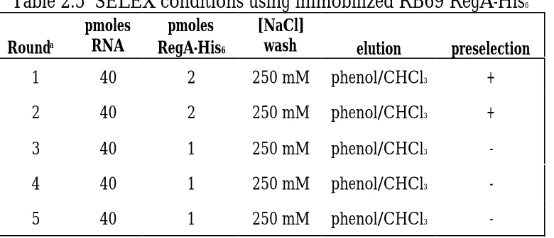
Table 2.5 SELEX conditions using immobilized RB69 RegA-His6……….….…………80
Table 2.6 Cloned sequences after five rounds of SELEX………..…………81
Table 2.7 Kdapp values for RB69 RegA on defined RNAs……….………..82
Table 2.8 Potential RB69 RegA binding sites in RB69 genome……….…………..83
Table 3.1 Optimized reaction conditions………..………..…………99
Table 3.2 Selection of RNAs that bind to Phage R17 using N40 Nitrocellulose Partitioning……….……….….…………107
LIST OF FIGURES
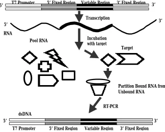
Figure 1.1 Typical selection scheme for RNA SELEX……….……….…...…5
Figure 1.2 Template: Primer arrangement with subsequent RNA transcript….………….7
Figure 2.1 Sequence of SELEX N14 DNA pools……….……….……….84
Figure 2.2 Gel shift analysis of RB69 RegA binding to selected and variant RNA ….85
Figure 2.3 Kdapp analysis of RB69 RegA-RNA interactions………..………….……...86
Figure 2.4 SELEX derived sequence logo……….…….….………87
Figure 3.1 Alignment and comparison of R17 and Qβ coat proteins………….…....……94
Figure 3.2 N40 template design……….…….….96
Figure 3.3 Biotinylated phage bound to nitrocellulose membranes…..……..….……….101
Figure 3.5 SDS-PAGE of purified phage……….……….………104
Figure 3.6 PCR and in vitro transcription optimization………..……….…..106
Figure 3.7 Modified western blot……….……….……111
Chapter 1
Literature Review
I. Introduction
In vitro selection, in vitro evolution, or SELEX (Systematic Evolution of Ligands by Exponential enrichment)is a combinatorial technique wherein random pools of nucleic acids (RNA or DNA) are used as the starting materials to isolate rare functional molecules (1, 2, 3). Originally, in vitro selection was used to generate functional molecules that could bind to large
or small ligands. Over the years numerous improvements to the procedure have greatly increased its potential while at the same time increasing the potential functionality of the nucleic acid aptamers. Functionality is determined by the conditions of the selection and include, but are not limited to, protein binding, small molecule binding, enzyme inhibition, and catalysis (4,5,6,7). In vitro selection straddles the biology/biotechnology interface being used to examine the interactions between proteins and their nucleic acid targets, as well as provide reagents for environmental, therapeutic, and diagnostic applications (8,9,10). The results of in vitro selection experimentation have caused a rethinking of the capabilities of
nucleic acids, broadening their already diverse functionality. Selection results have also given plausibility to the “RNA World” hypothesis, where RNA stored information within its code while also carrying-out the reactions essential for “life” at the time.
modified nucleotides, which can greatly increase the stability and half-life of potential therapeutic aptamers (12,13). Additional modification, by appending polyethylene glycol to an aptamer, have greatly increased the time it takes a therapeutic nucleic acid to be cleared from the human body (14). To fully understand the current research being performed using in
vitro selection a review of its history is useful.
II. History
It is generally assumed that SELEX was initiated around 1990. However, there was quite a bit of groundwork laid out before the modern methods of selection came into being. When it was learned that nucleic acids could carry-out both informational and functional roles it became evident to many researchers that evolution could be carried out at the molecular level. The first to experiment with these characteristics was Sol Spiegelman who worked with bacteriophage Qβ (15). Due to the lack of pure enzymes and amplification techniques such
as the polymerase chain reaction Spiegelman showed that Darwinian selection could operate in a cell free system (Reviewed in 16). The bacteriophage Qβ genome is replicated by the Qβ
replicase protein. Using serial dilutions numerous generations of replication could be carried out quickly. The phenotype under the selective pressure was the replication speed itself. Due to the high inherent error rate of the replicase the Qβ genome deleted segments that were not
size decreased. Even though these experiments were carried-out years ago they formed the framework for much of the current work using in vitro selection.
With the development of automated methods for chemically synthesizing highly diverse nucleic acid pools, and amplification methods such as the polymerase chain reaction came the ability to create increasingly complex libraries of nucleic acids. These developments also allowed for selection experiments to be carried out in the absence of bacteriophage Qβ.
These advances as well as the previous work by Spiegelman show that SELEX was not an idea generated by a single mind at a specific time, but instead a growing body of knowledge being put to use in numerous laboratories.
Loeb and Struhl were some of the first pioneers to take advantage of these new advancements as they probed for oligonucleotide function. They selected functional promoter sequences both in vitro and in vivo (17,18). The use of random sequences continued to grow with many researchers identifying binding sites for regulatory proteins (19,20).
(3). Both of these papers showed that nucleic acid aptamers could be developed to bind to numerous targets including basically any conceivable molecule. Since the early days of in vitro
selection, experimentation has moved to include applications that have medical diagnostic, environmental diagnostic, and therapeutic capabilities.
III.
in vitro
Selection - The Process
RNA
T7 Promoter 5’ Fixed Region Variable Region 3’ FixedRegion
5’ 3’
dsDNA
T7 Promoter 5’ Fixed Region Variable Region 3’ FixedRegion RT-PCR
Partition Bound RNA from Unbound RNA
Transcription
5’ 3’
PoolRNA Incubation
with target
Target
5’ 3’
A complete review of the methods of in vitro selection will allow for the comparison to other combinatorial methods, highlighting the capabilities that are possible with the procedure. The following description will outline a scheme geared toward generating nucleic acids that bind a target molecule (protein). However the same considerations would apply when performing a selection with the goal of generating a catalytic nucleic acid. The ligands that are generated using in vitro selection are typically called aptamers. This word is derived from the Greek word aptus, which means, "to fit" and describes early selection results with a
lock and key mode of interaction between the nucleic acid and the target (21).
A. Nucleic acid library
T7 fixed Primer T7 promoter
5’-CCGAAGCTTAATACGACTCACTATAGGAGTCAAAGCCGTAGCGA 3’
CCTCAGTTTCGGCATCGCT N14 CCAGTAGACGCGTCACCTAGGACCTAG 5’
SLXN14 CGTCACCTAGGACCTAG 5’
3’ Fixed Primer dsDNA 86 bp
mRNA 61 bases 5’ GGAGUCAAAGCCGUAGCGA N14 GGUCAUCUGCGCAGUGGAUCCUGGAUC3’
(T7 Transcript)
The synthetic oligonucleotide is converted into double stranded DNA using the Polymerase Chain Reaction (PCR) with 5' T7 fixed and 3' fixed primers (24). At this point the strands of DNA can be separated and single stranded DNA (ssDNA) can be used in the selection experiment or the DNA can be transcribed into RNA which can then be used in the selection experiment. The use of DNA vs. RNA is a case of fitting the right nucleic acid with the desired terminal function. Due to the 2’ OH group present in RNA it is inherently less stable than ssDNA of the same basic sequence (25). Therefore, in applications, which may require a stable aptamer it may be preferable use ssDNA.
Some selection experiments wish to better define a known binding site while some wish to identify a novel binding site. Each of the aforementioned experiments requires a random region with different characteristics. When doing an experiment to better identify a novel binding site it is appropriate for each base position of the random region to be synthesized with an equal probability of an A, C, G, or T. These types of selections can be done with different length randomized regions. A random region containing 40 nucleotides has the potential to have 440 or ~1024 possible sequences. Selections have been carried out
using as few as 8 random nucleotides to as many as 220 nucleotides (26). Typically when doing a binding experiment, randomized regions are kept below 70 nucleotides (23).
When doing a selection to better define a known binding site, the degree of randomization can be adjusted. For example, there may only be 10% "doping" at each base position using the known binding site as the original sequence. In this case if the wild type base is a C it would be present in the pool 70% of the time, while A, G, and T would all be at the same position 10% of the time. An excellent example of this type of experiment using a partially randomized pool was a selection to define the important residues of the Rev-response element (RRE) of HIV-I Rev (27). In these experiments the known binding site is the basis for the SELEX generated binding site.
B. Incubation with the target
Due to the unique chemical characteristics of each base the folding properties of the different members in the nucleic acid pool are different, allowing them to adopt different secondary structures (28). Each of these secondary structures makes up part of a “shape” library, with each molecule having different characteristics. The different properties of each molecule allow the pool to be placed under a specific pressure, usually a unique or novel function (binding to a target). Prior to incubation with the target the nucleic acid is heated sufficiently (approximately 70° C) to allow for it to be denatured completely. Following
pool to adopt their proper “native” conformation. Once the pool of nucleic acids is prepared it is then incubated with the target protein. Incubation time should be long enough for the reaction to reach a binding equilibrium, usually between 30-60 minutes.
Binding considerations
Important considerations for a successful binding reaction include the content of the binding buffer and the temperature of the reaction. Many binding buffers have been used successfully in SELEX experiments (29,30). A major consideration when selecting a binding buffer should be the concentration of divalent metal cations, specifically MgCl2. Divalent
cations allow for the stable formation of RNA or ssDNA secondary structure by shielding the negative charges on the phosphate backbone of nucleic acids (31). The more stable the nucleic acid secondary structure the greater the percentage of nucleic acid molecules within the pool that will be in the correct conformation. The temperature of the binding reaction is also an important consideration. Many successful selections have been carried-out with binding taking place at room temperature (23° C) (32). Temperatures that are too high
C. Partitioning bound nucleic acid from unbound
Negative selection entails passing the pool of nucleic acids through the membrane, while collecting the flow through. The end result is that any RNA or DNA that does not bind to the nitrocellulose membrane will be caught in the flow-through and subsequently incubated with the protein target. Individual nucleic acid species that have an affinity for nitrocellulose will be removed from the pool allowing the selection to continue with little or no background binding. It is usually sufficient to perform negative selections when background binding reaches levels where it is difficult to discern a protein-dependent binding pattern.
Following binding of the target:nucleic acid to the membrane the bound nucleic acid must be removed from the protein. This elution step is carried out using a strong denaturant. 7 M urea and phenol/chloroform extraction have all been used successfully to separate bound nucleic acid molecules from the target (37,38).
target molecule over the column transferring the binding species to the free target (39). The main consideration using this method is that the concentration of free target must be significantly higher than the concentration of the immobilized target. One other method of partitioning involves separation of nucleic acids that bind to the target from molecules that do not bind via gel shift mobility.
The use of gel shift mobility has proven to be a successful method of separating bound nucleic acids from unbound molecules (40). In this case the target is incubated with the pool of RNA or ssDNA in solution. The binding reaction is then run on a non-denaturing polyacrylamide gel. Separation is based upon the mobility difference between “free” unbound nucleic acid vs. bound molecules. Bound molecules will be much greater in size and their mobility will be greatly retarded with respect to non-binding species. This difference in mobility allows for the extraction and purification of binding molecules over non-binding molecules. Partitioning and separating the aptamers from the target allows for continued enzymatic manipulation.
D. RT-PCR of binding molecules
experiment is a DNA SELEX, the purified bound DNAs are simply subjected to PCR. Both cases result in the generation of a dsDNA molecule which is used as the input for the next round of selection. These final enzymatic steps can be used to monitor the progress of a SELEX experiment.
The use of the final enzymatic steps (RT-PCR) in each round of selection to monitor the progress of the experiment can prove very beneficial. By tracking the number of cycles of PCR needed to generate sufficient double stranded product from round to round the significance of binding molecules from round to round can be analyzed. Analysis should focus on the difference in the number of cycles of PCR needed to generate dsDNA in the actual round of selection versus the number of cycles needed to amplify dsDNA using a pool of nucleic acids that is not selected with target. As the selection progresses it should take fewer cycles of PCR to amplify dsDNA than the number of cycles needed to amplify dsDNA in the "negative" control. Hence, as a greater percentage of the pool is enriched for binding species the number of PCR cycles will decrease.
E. Periodic assay for binding during the course of SELEX
nucleic acid is kept constant while the concentration of target protein is increased in each assay. As the pool of nucleic acids is enriched for binding species there should be a dose-dependant curve generated in the binding assay (43). This dose- dose-dependant curve is evidence that binding species are being selected. Rounds of selection are carried-out iterativly until the nucleic acid pool shows no improvement in binding over successive rounds. An excellent example using the binding assay to monitor the progress of a selection can be seen in a selection using the Ricin A-chain as the nucleic acid target (4). The importance of monitoring the progress of the selection is clearly seen by round 7 where ~ 20% of the input pool bound to the target. However, there was 7.8% background binding. In the following 2 rounds there was heavy negative selection incorporated into the procedure and at round 9 target binding was increased to 51% while background binding was reduced to ~2.5% (4). This example clearly demonstrates the usefulness of monitoring the selection by assaying for target binding, while at the same time using the assay results to incorporate negative selection into the scheme to reduce potential non-specific binding nucleic acid molecules.
F. Post SELEX activities
based on the sequence data. Using lowest energy of formation algorithims, programs such as mfold (47) are capable of predicting secondary structure. In addition, individual molecules can also be assayed for their ability to bind the target and this affinity is usually expressed as a dissociation constant (Kd). Individual Kd values can range from µM to low nM values
depending on the target and the experiment.
IV.
Increasing nucleic acid stability in
in vitro
selection
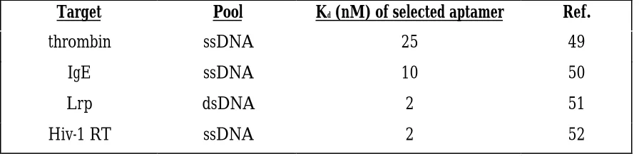
Nucleic acid selections are highly successful inside the laboratory, however, this success quickly coorelates into failure if the efficacy of the selected molecules is poor due to quick degradation, in real world applications. This degradation is more acute when using RNA over ssDNA because of the ribonucleases present in cells and blood serum. The half life of many RNAs are in the range of minutes or less (48). This quick degradation does not allow for successful target identification and binding. One method of circumventing the relative instability of RNA is to use ssDNA. The half life of many ssDNA molecules is increased to 1-2 hours (48). Conducting selection with ssDNA may allow for affective therapeutic or diagnostic use (Table 1).
Table 1.1 Successful DNA SELEX experiments.
Target Pool Kd (nM) of selected aptamer Ref.
thrombin ssDNA 25 49
IgE ssDNA 10 50
A highly successful method of increasing the stability of RNA aptamers is the use of modified nucleotides. Increased stability of ribonucleotides can be accomplished by the chemical modification at specific positions (Table 2). Modifications to ribonucleotides are at the 2’ position of the ribonucleotide or at the phosphodiester backbone because modifications at these positions greatly interfere with nucleases that degrade RNA (48). The two main strategies that have been developed for using modified nucleotides are use of modified nucleotides during the selection, incorporating them in each round (48). The second method involves completing the SELEX experiment using unmodified nucleotides and then substituting modified nucleotides at the conclusion of the experiment (48). Each of these strategies will be reviewed.
Table 1.2 Selections carried out using modified ribonucleotides.
Target Kd (nM) Ref.
thrombin 400 53
HIV-1 Rev 1 55
elastase 15 56
bFGF 0.3 57
Incorporating modified nucleotides during RNA SELEX
polymerases to incorporate the modified nucleotide into the synthesized RNA or DNA molecule (53). There have now been many experiments that have used incorporation of modified nucleotide incorporation by polymerases. The first successful experiment to use presubstitution was a selection using thrombin as a target. The anti-thrombin aptamer contained 5-(1-pentynyl)-2’-deoxyuridine in the molecules in the DNA pool (53). Interestingly, the modified thrombin aptamers showed decreased affinity for thrombin when compared to a selection carried out using natural unmodified nucleotides. Numerous
modified nucleotides have been used successfully in selections. These include 2’-aminouridine, and 2’-aminocytidine (54). These two modified nucleotides have been used because they inhibit ribonuclease activity, while at the same time are still recognized by T7 RNA polymerase and AMV reverse transcriptase, allowing for their incorporation into successive rounds of selection.
Aptamers that bind to bFGF and VerF were selected using presubstitution with modified pyrimidines (57). Using these modified bases allowed for a successful selection with molecules having Kds in the picomolar range. In addition to having such a strong affinity for
during the process of SELEX. Equally as successful has been the incorporation of modified nucleotides into aptamers at the post-SELEX stage.
Post-SELEX incorporation of modified ribonucleotides
Addition of modified nucleotides into selected aptamers has been shown to increase both affinity for the target as well as increase the stability of the aptamer. A key example of such a success was seen in a selection using Rous sarcoma virus (RSV) and a random pool of unmodified RNA molecules (60). Following selection, binding aptamers were completely substituted with 2’-fluoropyrimidines. With complete substitution there was a total loss of affinity by the aptamers for RSV. Three more rounds of selection were carried out using modified nucleotides, resulting in modified aptamers that could bind RSV and interfere with viral replication as well as the unmodified pool. The result of the final three rounds of selection was to remove all unsubstituted pyrimidines that were not critical for binding, while increasing the stability of the aptamer.
These examples clearly show that using modified nucleotides incorporated into the selection experiment, or incorporated following the generation of binding aptamers, leads not only to more stable molecules, but also molecules with increased binding affinity. Future advancements such as interference analysis (61) should lead to the generation of aptamers that are highly nuclease resistant as well as maintaining high binding affinity.
V.
SELEX: Example Experiments
Small molecule targets and the RNA World
SELEX has been used extensively to develop aptamers to many small molecules. Aptamers directed at small molecules have helped delve into the "RNA World" hypothesis (62). In the RNA World RNA, is proposed to have stored the genetic code, as well as performed the role of chemical catalyst by functioning as an enzyme. For this to happen, RNA must have been able to recognize small molecules and substrates with high affinity and in a highly specific manner. In vitro selection is an excellent experimental procedure capable
of showing the capabilities RNA has for recognizing small organic molecules.
length of 100 nucleotides were used to select for molecules that could bind to the dyes. These experiments were very successful, and specific aptamers were derived for each of the six organic dyes used. The surprising result of these experiments was that the chemical makeup of the four bases was sufficient to enable stable interactions between the RNA and the organic dyes. Other small molecules such as the carbohydrate cellobiose (34) have generated successful aptamers, clearly showing that RNA has the capability to recognize and bind to molecules that have little diversity in chemical makeup.
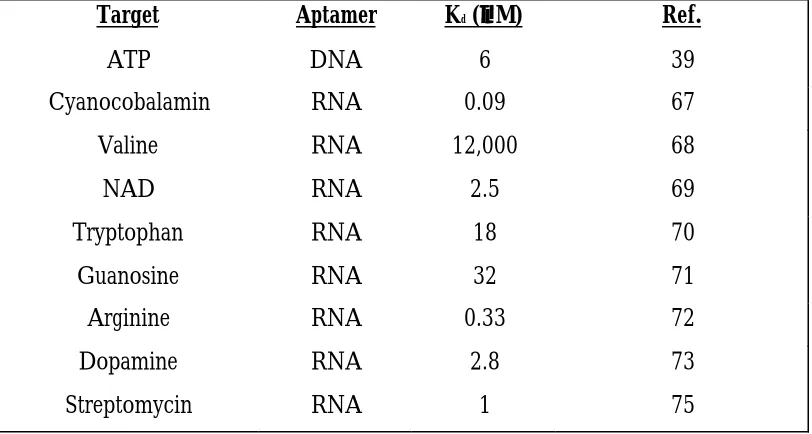
In addition to cellobiose, other biological molecules have been used as targets for SELEX experiments. The first to be used was ATP (39). ATP is directly involved in the of energy of living systems (63). This selection was carried out by using ATP immobilized on a column matrix. RNA was passed over the column and binding species eluted by passing an excess of “free” ATP over the column. Following the selection, analysis of the cloned sequences showed that while highly divergent, all contained an 11 nucleotide stretch that folded into the same secondary structure. This structure was deduced to be a hairpin loop with an internal, asymmetric purine rich bulge. Once again SELEX was used to show that the four bases present in RNA were capable of recognizing small organic molecules, relatively simple chemical structures in biologically relevant molecules (Table 1.3).
do not function when they are converted into RNA, which is not surprising. The 2’-OH group on RNA not only makes it more nuclease sensitive but also gives it added chemical makeup causing it to fold into secondary structures differing from ssDNA which lacks the 2’-OH.
Table 1.3 Nucleic acid aptamers for small molecules.
Target Aptamer Kd (µµM) Ref.
ATP DNA 6 39
Cyanocobalamin RNA 0.09 67
Valine RNA 12,000 68
NAD RNA 2.5 69
Tryptophan RNA 18 70
Guanosine RNA 32 71
Arginine RNA 0.33 72
Dopamine RNA 2.8 73
Streptomycin RNA 1 75
Protein targets
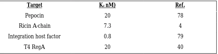
In addition to small organic targets the next logical approach for SELEX is the generation of aptamers against proteins. It would make sense that RNA or ssDNA would be able to form tight interactions with protein targets, which possess twenty amino acids with differing reactive groups. Proteins fold into complex three-dimensional structures enabling nucleic acid aptamers to bind in clefts or other surface structures. Numerous selections have been carried out using proteins that bind nucleic acids as their normal biological activity, as well as selections against proteins that do not function as nucleic acid binding proteins.
The first protein SELEX experiment was for bacteriophage T4 DNA polymerase (Gp43) (3). The binding of Gp43 to a hairpin structure located in the translation initiation region of its own mRNA allows for autogenous regulation. In this experiment the SELEX procedure was utilized to elucidate what is responsible for the loop nucleotide bias in this protein:RNA interaction. The RNA pool consisted of a random region of 8 nucleotides. Following 5 rounds of selection on nitrocellulose the pool of RNA exhibited the same affinity for Gp43 as the wild type binding sequence (Kd approximately 5 nM) (3). The end results of
the experiment showed that there were two different loop sequences with very similar binding constants. In addition, the results showed that there were very few loop nucleotides that could tolerate substitution without loss of binding affinity.
SELEX protocol could be used to predict a high affinity RNA binding site. The R17 coat protein is well characterized and naturally binds an RNA hairpin repressing expression of the replicase gene whose translation initiation region is contained in the hairpin (74). An RNA pool with a random region 32 nucleotides long was subjected to 11 rounds of selection. 47 individual sequences were cloned and sequenced, 38 were unique while 36 contained the hairpin structure necessary for high affinity to the R17 coat protein. These results showed that the SELEX procedure can accurately predict an RNA binding site for the R17 coat protein, and that this success could be expanded to other targets in the same class.
Table 1.4 Protein targets utilized in SELEX experiments.
Target Kd nM) Ref.
Pepocin 20 78
Ricin A-chain 7.3 4
Integration host factor 0.8 79
T4 RegA 20 40
in vitro
selection of catalytic nucleotides
Discussion thus far has focused on the ability of RNA or ssDNA to recognize and bind small organic molecules and proteins, both with high specificity and high affinity. The main goal of many SELEX experiments is to force polynucleotides to carry out some unique function. In addition to binding to targets, SELEX experiments have also been carried-out with the goal of finding RNA or ssDNA that can display enzyme-like functions. This section will deal with some of the experiments and advances that have been made on the road to polynucleotide catalysis.
molecule. The experiment required 10 rounds to isolate ribozymes capable of catalyzing the reaction at a rate of 0.06 min-1
Researchers also quickly realized that RNAs of known function could be used as the starting point in SELEX experiments designed to create molecules capable of completing different tasks (80). The first successful attempt at re-designing a known functional RNA was undertaken to create an RNA capable of catalyzing self phosphorylation (81). An initial pool of ~ 1015 different molecules that were already biased for binding to adenosine 5’-triphosphate
was subjected to 13 rounds of selection for RNAs that acquire a thiophosphate group when incubated with γ-thioATP. The end result was a pool consisting of RNAs that were capable of
self-phosphorylating at a rate of ~0.05 min-1.
isomerization of the biphenyl substrate. The observed Kcat was 2.8 X 10⁻⁵ min-1 and the
observed KM was 542 µM.
The above examples are by no means exhaustive; catalytic RNA and ssDNA have been developed to perform numerous chemical reactions including RNA alkylation (86), transacylation and amide bond formation (87), porphyrin metalation (88), and DNA ligation (89). The results of these experiments show the diverse functions that RNA and ssDNA can possess, lending credence to their potential diagnostic and therapeutic abilities as well as strengthening the RNA World hypothesis.
VI.
Comparison to antibody technology
Antibodies have been used for decades and are the most widely used class of molecules providing molecular recognition. The use of antibodies in detecting molecules predates the 1950’s (90). During the 1970’s, monoclonal antibody technology became available allowing for the production of large amounts of specific antibodies to meet the increasing demands in diagnostics (91). The use of antibodies has continued to grow over the years, however, there are certain limitations to antibodies (92), which bear mention.
issue. A second major issue with antibody generation is the potential time and cost expenditure. There may be a considerable time and money expenditure searching for a rare antibody, screening it, and purifying it for laboratory use. The performance of multiple antibody preparations is frequently different from preparation to preparation, most likely due to the differing immune systems of different animals. Finally, antibody preparations are subject to irreversible denaturation due to temperature sensitivity and have a definite “shelf” life (92). Many of these issues can be overcome by the use of oligonucleotides generated by the SELEX procedure.
VII. Conclusion: The future of SELEX.
Significant advancements have been made over the last decade in the generation of aptamers, and in the variety of aptamers that have been developed. Their use in molecular recognition has made their use in therapeutics and diagnostics commonplace. Only eight years after the inception of SELEX, aptamers were already in clinical trials (92). Other areas that are beginning to use nucleic acid aptamers are in vivo diagnostics, and in vivo imaging
REFERENCES:
1. Ellington, A.D. and Szostak, J.W. (1990) Nature 346: 818-822.
2. Joyce, G.F. (1989) Gene 82: 83-87.
3. Tuerk, C. and Gold, L. (1990) Science 249: 505-510.
4. Hesselberth, J.R., Miller, D., Robertus, J., and Ellington, A.D. (2000) J Biol Chem 275: 4937-4942.
5. Huizenga, D.E. and Szostak, J.W. (1995) Biochemistry 34: 656-665.
6. Rusconi, C.P., Yeh, A., Lyerly, H.K., Lawson, J.H. and Sullenger, B.A. (2000) Thromb Haemost 84: 841-848.
7. Joyce, G.F. (2001) Methods Enzymol 341: 503-517.
8. Bruno, J.G. and Kiel, J.L. (1999) Biosens Bioelectron 14: 457-464.
9. Brody, E.N. and Gold, L. (2000) J Biotechnol 74: 5-13.
10. Davis, K.A., Abrams, B., Lin, Y. and Jayasena, S.D. (1996) Nucleic Acids Res 24: 702-706.
12. Verma, S., and Eckstein, F. (1998) Annual Rev. Biochem. 67: 99-134.
13. Andreola, M., Calmels, C., Michel, J., Toulme, J., and Litvak, S. (2000) Eur. J. Biochem. 267: 5032-5040.
14. White, R., Sullenger, B., and Rusconi, C. (2000) Journal of Clinical Inv. 106: 929-934.
15. Mills, D., Peterson, R., and Spiegelman, S. (1967) Proc. Natl. Acad. Sci. 58: 217.
16. Spiegelman, S. (1971) Q. Rev. Biophys. 4: 213-53.
17. Oliphant, A., and Struhl, K. (1987) Methods Enzymologyl. 155: 558-568.
18. Horwitz, M., and Loeb, L. (1986) Proc. Natl. Acad. Sci. 83: 7405-7409.
19. Thiesen, H., and Bach, C. (1990) Nucleic Acids Res. 18: 3203-3209.
20. Sun, X., and Baltimore, D. (1991) Cell 64: 459-470.
21. Klug, S., and Famulok, M. (1994) Mol. Bio. Rep. 20: 97-107.
22. Pfleiderer, W., Matysiak, S., Bergmann, F., and Schnell, R. (1996) Acta Biochimica Polonica 43: 37-44.
24. Erlich, H.A. (1989) in PCR Technology: Principles and Applications for DNA Amplification, pp. 1-7, Stockton Press, New York, NY.
25. Moran, L., Scrimgeour, K., Horton, R., Ochs, R., and Rawn, J. (1994) in Biochemistry, pp. 24.1-24.33, Neil Patterson Publishers/Prentice-Hall, Inc., Englewood Cliffs, NJ.
26. Bartel, D., and Szostak, J. (1993) Science 261: 1411-1418.
27. Bartel, D., Zapp, M., Green, M., and Szostak, J. (1991) Cell 67: 529-539.
28. Woodson, S. (2000) Cell Mol. Life Sci. 57: 796-808.
29. Seiwert, S., Nahreini, T., Aigner, S., Ahn, N., and Uhlenbeck, O. (2000) Chemistry and Biology 7: 833-843.
30. Geiger, A., Burgstaller, P., Eltz, H., Roeder, A., and Famulok, M. (1996) Nucleic Acids Research 24: 1029-1036.
31. Saenger, W. (1984) in Principles of Nucleic Acid Structure (Cantor, C. Ed.) pp. 201-219, Springer - Verlag, New York, NY.
32. Rusconi, C., Yeh, A., Lyerly, H., Lawson, J., and Sullenger, B. (2000) Thromb Haemost. 84: 841-848.
34. Yang, Q., Goldstein, I., Mei, H., and Engelke, D. (1998) Proc Natl Acad Sci USA. 95: 5462-5467.
35. Conrad, R., Giver, L., Tian, Y., and Ellington, A. (1996) Methods Enzymology 267: 336-367.
36. Jhaveri, S., and Ellington, A. (1990) In Vitro Selection of RNA Aptamers to a Protein Target by Filter Immobilization (personal communication).
37. Baskerville, S., Zapp, M., and Ellington, A. (1995) Journal of Virology 69: 7559-7569.
38. Allen, S. (1999) Thesis pp. 94-100.
39. Sassanfar, M., and Szostak, J. (1993) Nature 364: 550-553.
40. Brown, D., Brown, J., Kang, C., Gold, L., and Allen, P. (1997) Journal Biological Chemistry 272: 14969-14974.
41. Tuerk, C. (1995) in Methods in Molecular Biology PCR Cloning Protocols: From Molecular Cloning to Genetic Engineering (White, B. Ed.) pp. 219-230, Humana Press Inc., Totowa, NJ.
42. Stone, S., Hughes, M., and Jost, J. (1991) in A Laboratory Guide to In Vitro Studies of Protein-DNA Interactions (Saluz, H., and Becker, M. Eds.) pp. 163-176, Birkhauser-Verlag, Boston, MA.
44. Validis, B. (1998) in Basic Cloning Procedures (Berzins, V. Ed.) pp. 1-169, Springer, New York, NY.
45. Graham, C., and Hill, A. (2001) in Methods in Molecular Biology: DNA Sequencing Protocols (Vol 167) pp. 1-244, Humana Press Inc., Totowa, NJ.
46. Patel, D., and Suri, A. (2000) Journal of Biotechnology 74: 39-60.
47. Zuker, M., Mathews, D., and Turner, D. (1999) RNA Biochemistry and Biotechnology
288: 911-940.
48. Osborne, S., and Ellington, A. (1997) Chemical Rev. 97: 349-370.
49. Bock, L., Griffin, L., Latham, J., Vermaas, E., and Toole, J. (1992) Nature 355: 564-566.
50. Wiegand, T., Williams, P., Dreskin, S., Jouvin, M., Kinet, J., and Tasset, D. (1996) Journal Immunology 157: 221-230.
51. Cui, Y., Wang, Q., Stormo, G., and Calvo, J. (1995) Journal Bacteriology 177: 4872-4880.
52. Schneider, D., Feigon, J., Hostomsky, Z., and Gold, L. (1995) Biochemistry 34: 9599-9610.
55. Jensen, K., Atkinson, B., Willis, M., Koch, T., and Gold, L. (1995) Proc Natl Acad Sci USA 92: 12220-12224.
56. Lin, Y., Padmapriya, A., Morden, K., and Jayasena, S. (1995) Proc Natl Acad Sci USA 92: 11044-11048.
57. Jellinek, D., Green, L., Bell, C., Lynott, C., Gill, N., Vargeese, C., Kirschenheuter, G., McGee, D., Abesinghe, P., Pieken, W., Shapiro, R., Rifkin, D., Moscatelli, D., and Janjic, N. (1995) Biochemistry 34: 14413-14424.
58. Green, L., Jellinek, D., Bell, C., Beebe, L., Feistner, B., Gill, S., Jucker, F., and Janjic, N. (1995) Chemical Biology 10: 683-695.
59. Pieken, W., Olsen, D., Benseler, F., Aurup, H., and Eckstein, F. (1991) Science
253: 314-317.
60. Pan, W., Craven, R., Qiu, Q., Wilson, C., Wills, J., Golovine, S., and Wang, J. (1995) Proc Natl Acad Sci USA 92: 11509-11513.
61. Green, L., Waugh, S., Binkley, J., Hostomska, Z., Hostomsky, Z., and Tuerk, C. 1995) Journal of Molecular Biology 247: 60-68.
62. Atkins, J., and Gesteland, R. (1993) in The RNA world : the nature of modern
RNA suggests a prebiotic RNA world, pp. 1-630, Cold Spring Harbor Laboratory Press, Cold Spring Harbor, NY.
63. Moran, L., Scrimgeour, K., Horton, R., Ochs, R., and Rawn, J. (1994) in
64. Harada, K., and Frankel, A. (1995) EMBO 14: 5798-5811.
65. Conn, M., Prudent, J., and Schultz, P. (1996) Journal American Chemical Society
118: 7012-7013.
66. Li, Y., and Sen, D. (1996) Nat Struct. Biology 3: 743-747.
67. Lorsch, J., and Szostak, J. (1994) Biochemistry 33: 973-982.
68. Majerfeld, I., and Yarus, M. (1994) Nat Struct. Biology 1: 287-292.
69. Lauhon, C., and Szostak, J. (1995) Journal American Chemical Society 117: 246-1257.
70. Famulok, M., and Szostak, J. (1992) Journal American Chemical Society 114: 3990-3991.
71. Connell, G., and Yarus, M. (1994) Science 264: 1137-1141.
72. Geiger, A., Burgstaller, P., Eltz, H., Roeder, A., and Famulok, M. (1996) Nucleic Acids Research 24: 1029-1036.
73. Mannironi, C., Di Nardo, A., Fruscoloni, P., and Tocchini-Valentini, G. (1997)
75. Wallace, S., and Schroeder, R. (1998) RNA 4: 112-123.
76. Griffin, L., Tidmarsh, G., Bock, L., Toole, J., and Leung, L. (1993) Blood 81: 3271-3276.
77. Kelly, J., Feigon, J., and Yeates, T. (1996) Journal of Molecular Biology 256: 417-422.
78. Hirao, I., Madin, K., Endo, Y., Yokoyama, S., and Ellington, A. (2000) Journal of Biological Chemistry 275: 4943-4948.
79. Goodman, S., Velten, N., Gao, Q., Robinson, S., Segall, A. (1999) Journal of Bacteriology 181: 3246-3255.
80. Connell, G., and Christian, E. (1993) Origins of Life 23: 291-297.
81. Lorsch, J., and Szostak, J. (1994) Nature 371: 31-36.
82. Schultz, P., and Lerner, R. (1995) Science 269: 185-1842.
83. Abelson, J. (1990) Science 249: 488-489.
84. Ellington, A., and Szostak, J. (1992) Nature 355: 850-852.
85. Prudent, J., Uno, T., and Schultz, P. (1994) Science 264: 1924-1927.
87. Illangasekare, M., Sanchez, G., Nickles, T., and Yarus, M. (1995) Science 267: 643-647.
88. Li, Y., Geyer, R., and Sen, D. (1996) Biochemistry 35: 6911-6922.
89. Cuenoud, B., and Szostak, J. (1995) Nature 375: 611-614.
90. Yallow, R., and Berson, S. (1959) Nature 185: 1648-1649.
91. Kohler, G., and Milstein, C. (1975) Nature 256: 496-497.
92. Jayasena, S. (1999) Clinical Chemistry 45: 1628-1650.
93. Charlton, J., Sennello, J., and Smith, D. (1997) Chemical Biology 4: 809-816.
Chapter 2
I. Bacteriophage T4 Translational Repressors
Bacteriophage T4 utilizes translational repression as a method of gene regulation (1). The T4 proteins Gp43, Gp32, and RegA have all been studied extensively and much is known about their roles in gene regulation. While all three proteins repress translation, Gp32 and Gp43 carry out their regulation in a manner differing from that of RegA. Both Gp32 and Gp43 regulate their own synthesis by preventing translation of their cognate mRNAs, while RegA has been shown to regulate numerous genes, in addition to its own transcript (1).
T4 translational repressor Gp32
Gp32 is a single stranded DNA binding protein. Its primary function is to bind and stabilize single stranded DNA allowing DNA polymerase to replicate the opposite strand (2). While Gp32 is primarily a ssDNA binding protein, it will bind to RNA when the concentration of protein reaches an elevated level. When the ssDNA is saturated with Gp32 the excess protein then binds to its own mRNA repressing further translation (1). Gp32 recognizes an RNA pseudoknot structure at its own translation initiation region (TIR), then fills an adjacent unstructured region, thereby reducing binding and translating the mRNA (3). Gp32 is a Zn2+ protein, incorporating 1 mole of Zn2+ per mole of protein (4). In addition,
T4 translational repressor Gp43
Gp43 is a DNA replication accessory protein with 5'→3' polymerase activity, as well as having 3'→5' exonuclease activity (1). Gp43 operates somewhat analogous to Gp32
function in translational repression. Gp43 binds to an RNA hairpin in the translation initiation region of the gene 43 mRNA. The minimum operator sequence to which Gp43 binds consists of 36 nucleotides that include a hairpin (containing a 5 base-pair helix and an 8 nucleotide loop) and a single-stranded segment that contains the Shine-Dalgarno sequence of the ribosome binding site (5). Binding by Gp43 at this site occludes the TIR preventing translation of the gene (5,6). Gp32 and Gp43 function in translational repression by recognizing RNA secondary structure and binding at that site. The third T4 translational repressor, RegA, functions much differently in translational repression than the other two repressors.
T4 translational repressor RegA
The T4 translational repressor RegA protein folds into two structural domains (8). Domain I of the RegA protein contains a four-stranded beta-sheet and two alpha-helices. Domain II contains a four-stranded beta-sheet and an unusual 3/10 helix (8). Mutagenesis study has shown the RNA binding site to be a surface pocket formed by residues on two loops and an alpha-helix (8).
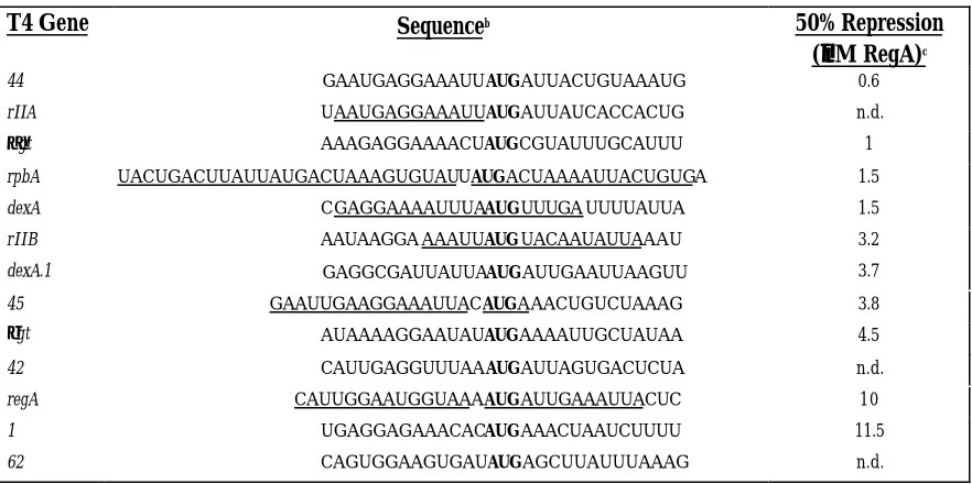
Table 2.1 RegA sensitive mRNAsa
T4 Gene Sequenceb 50% Repression
(µµM RegA)c
44 GAAUGAGGAAAUUAUGAUUACUGUAAAUG 0.6
rIIA UAAUGAGGAAAUUAUGAUUAUCACCACUG n.d.
αgt AAAGAGGAAAACUAUGCGUAUUUGCAUUU 1
rpbA UACUGACUUAUUAUGACUAAAGUGUAUUAUGACUAAAAUUACUGUGA 1.5
dexA CGAGGAAAAUUUAAUGUUUGA UUUUAUUA 1.5
rIIB AAUAAGGA AAAUUAUGUACAAUAUUAAAU 3.2
dexA.1 GAGGCGAUUAUUAAUGAUUGAAUUAAGUU 3.7
45 GAAUUGAAGGAAAUUACAUGAAACUGUCUAAAG 3.8
βgt AUAAAAGGAAUAUAUGAAAAUUGCUAUAA 4.5
42 CAUUGAGGUUUAAAUGAUUAGUGACUCUA n.d.
regA CAUUGGAAUGGUAAAAUGAUUGAAAUUACUC 10
1 UGAGGAGAAACACAUGAAACUAAUCUUUU 11.5
62 CAGUGGAAGUGAUAUGAGCUUAUUUAAAG n.d.
aAdapted from Miller et al., 1994.
bRegA sensitive TIRs aligned at the AUG initiation codon. Underlined nucleotides are protected from RNase
digestion by bound RegA protein.
c The concentration of RegA causing 50% reduction in the yield of in vitro translated product from each message.
Table 2.2 Hierarchy of biological RNA affinities for T4 and RB69 Reg A proteinsa T4 RegA Protein Sequenceb Kapp(107 M-1)
RB G45 AAGGAAAUAAA 19.9
T4 G44 GAGGAAAUUAU 4.9
T4 G45 AAGGAAAUUAC 3.2
RB G44 GAGGAAAAUUA 2.5
RB regA AUGGUAAAAAU 1.3
T4 regA AUGGUAAAAUG 0.8
RB69 RegA Protein Kapp(107 M-1)
T4 G44 GAGGAAAUUAU 34.0
RB G44 GAGGAAAAUUA 9.5
T4 G45 AAGGAAAUUAC 2.0
RB G45 AAGGAAAUAAA 1.8
RB regA AUGGUAAAAAU 0.6
T4 regA AUGGUAAAAUG 0.16
a Adapted from Sengupta et al., 2001.
b Underlining indicates RNase protected nucleotides.
II. Using SELEX to explore RegA-RNA interactions
T4 RegA:RNA analysis using SELEX
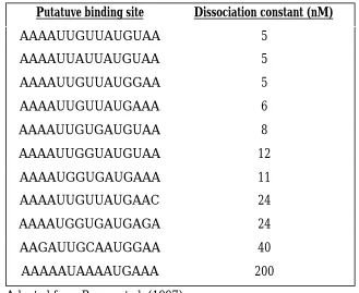
Table 2.3 Binding affinity of selected RNA sequences for T4 RegAa Putatuve binding site Dissociation constant (nM)
AAAAUUGUUAUGUAA 5 AAAAUUAUUAUGUAA 5 AAAAUUGUUAUGGAA 5 AAAAUUGUUAUGAAA 6 AAAAUUGUGAUGUAA 8 AAAAUUGGUAUGUAA 12 AAAAUGGUGAUGAAA 11 AAAAUUGUUAUGAAC 24 AAAAUGGUGAUGAGA 24 AAGAUUGCAAUGGAA 40 AAAAAUAAAAUGAAA 200
a Adapted from Brown et al. (1997)
Table 2.4 Biological T4 mRNAs that are regulated by RegAa T4 Gene RegA binding site
rIIB AAAAUUAUGUAC
44 AAAUUAUGAUU
DexA AAAAUUUAAUGUUU
RpbA AAAGGUGUAUUAUGACU
45 AAAUUACAUGAAA
αgt AAAACUAUGCGU
βgt AAAAGGAAUAUAUGAAA
1 AAACACAUGAAA
regA AAUGGUAAAAUGAUU
RB69 RegA:RNA analysis using SELEX
RB69 RegA translational repressor protein is 78% identical in amino acid sequence to the well-characterized T4 protein (15), binds both RB69 and T4 mRNAs, and displays a hierarchy of binding to RNAs that do not have a conserved sequence or structure (17). These
differences in the sequences of T4 RegA and RB69 RegA, and their different affinities for the same mRNAs suggested RB69 RegA as a useful homolog of the T4 protein for studying RNA-binding and translational regulation.
RNAs randomized at 14 positions (the SLXN14 template) were carried-through rounds of SELEX in binding reactions with immobilized His-tagged RB69 RegA (RegA-His6).
After only five rounds of selection, RNAs that preferentially bound RB69 RegA-His6 showed
extensive sequence conservation among the 18 individual RNAs that were cloned and sequenced (18).
REFERENCES:
1. Miller, E., Karam, J., and Spicer, E. (1994) in Molecular Biology of Bacteriophage T4
(Karam, J., and et al., Eds.) pp. 193-205, ASM Press, Washington, D.C.
2. Villemain, J., and Giedroc, D. (1996) Biochemistry 35: 14395-14404.
3. McPheeters, D., Stormo, G., and Gold, L. (1988) Journal of Molecular Biology201: 517-535.
4. Giedroc, D., Keating, K., Williams, K., Konigsberg, W.,and Coleman, J.(1986) Proc Natl Acad Sci U S A 83: 8452-6
5. Tuerk, C., Eddy, S., Parma, D., and Gold, L. (1990) Journal of Molecular Biology 213: 749-761.
6. Andrake, M., Guild, N., Hsu, T., Gold, L., Tuerk, C., and Karam, J. (1988) Proc Natl Acad Sci U S A 85: 7942-7946.
7. Szewczak, A., Webster, K., Spicer, E., and Moore, P., (1991) Journal of Biological Chemistry 266: 17832-17837.
8. Kang, C., Chan, R., Berger, I., Lockshin, C., Green, L., Gold, L., and Rich, A. (1995) Science 268: 1170-1173.
9. Winter, R., Morrissey, L., Gauss, P., Gold, L., Hsu, T., and Karam, J. (1987) Proc Natl Acad Sci U S A 84: 7822-7826.
10. Miller, E., Karam, J., Dawson, M., Trojanowska, M., Gauss, P., and Gold, L. (1985) )
11. Webster, K., Adari, H., and Spicer, E. (1989) Nucleic Acids Research 17: 10047-10068.
12. Daegelen, P., and Brody, E. (1990) Genetics 125: 237-248.
13. Karam, J., Gold, L., Singer, B., and Dawson, M. (1981) Proc Natl Acad Sci U S A78: 4669-4673.
14. Unnithan, S., Green, L., Morrissey, L., Binkley, J., Singer, B., Karam, J., and Gold, L. (1990) Nucleic Acids Research18: 7083-7092.
15. Jozwik, C., and Miller, S. (1992) Proc Natl Acad Sci U S A 89: 5053-5057.
16. Brown, D., Brown, J., Kang, C., Gold, L., and Allen, P. (1997) Journal of Biological Chemistry272: 14969-14974.
17. Sengupta, T., Gordon, J., and Spicer, E. (2001) Nucleic Acids Research 29: 1175-1184.
In vitro
Selected Phage RB69 RegA RNA Binding Sites Yields UAA Triplets and a
Potential Role in Translational Reinitiation
Timothy R. Dean, Sherrice V. Allen
+and Eric S. Miller
*Department of Microbiology
North Carolina State University
Raleigh, NC 27695-7615
*Corresponding author:
Phone: 919-515-7922 Fax: 919-515-7867
email: [email protected]
+Current address:
Department of Natural Sciences Fayetteville State University 1200 Murchison Road Fayetteville, NC 28301
Abstract
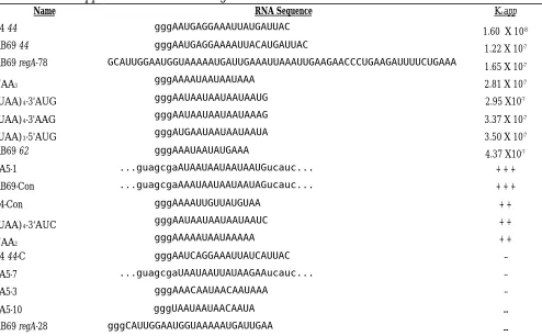
The SELEX method of in vitro selection was used to isolate RNAs that bind the RB69 RegA translational repressor protein immobilized to Ni-NTA agarose. After five rounds of SELEX, the pool of selected RNA displayed striking sequence uniformity: UAAUAAUAAUAAUA was clearly enriched in the 14 nucleotides that underwent selection.
Individual, cloned molecules displayed the (UAA)n sequence, with two RNAs having a 3'
AUG. Removing the 3' AUG reduced binding in gel shift assays, moving the AUG 5'
proximal of the (UAA)n improved binding, but (UAA)4 alone still bound the purified protein.
Dissociation constants showed that RNA shortened to (UAA)3 and (UAA)2 also retained
Introduction
Protein-mediated translational repression is an important regulatory mechanism in prokaryotes and eukaryotes (1,2). Bacteriophage T4 encodes three translational repressors: the single stranded DNA binding protein (gp32), the DNA polymerase (gp43), and RegA (3). Gp32 and gp43 regulate their own translation by binding to a pseudoknot structure or a simple hairpin, respectively, contained in the translation initiation region (TIR) of their mRNA (4-6). As for most translational repressor proteins, RNA structure is an important component of the target site for unambiguous binding and repression of their target mRNAs.
T4 RegA is an unusual translational repressor. It represses translation of several different phage mRNAs, including its own (7,8). Several studies have focused on the specificity determinants in the RegA:RNA interaction. A striking feature of RegA binding sites on RNA is the unusual absence of secondary structure. The TIRs at RegA binding sites are AU-rich. An RNA structure study revealed that one of the binding sites (T4 44 RNA) is
single-stranded, with a 2'-endo conformation of the ribose that is more characteristic of B-form DNA than of A-form RNA (9). Comparing the TIR regions recognized by RegA with all T4 transcripts fails to show a marked difference between the two sets of RNA.
SELEX (Systematic Evolution of Ligands by Exponential Enrichment) is an in vitro
ribosome binding (12). The SELEX experiments confirmed that T4 RegA recognizes RNA in a sequence-dependent manner and does not rely on RNA structure. The reported "consensus" sequence, 5'-AAAAUUGUUAUGUAA-3'' (12), again showed that T4 RegA binds AUG flanked by AU-rich bases. The crystal structure of the T4 protein has been solved (13), and mutation analyses (14) reveal that specific RNA site recognition is mediated through a unique RNA-binding domain defined by an N-terminal helix region (15,16) in a structural binding "cleft" (15).
Bacteriophage RB69 is a T4-related phage whose genes complement defects in some T4 genes (17). RB69 encodes a RegA protein that is composed of 122 amino acids and is 78% identical to T4 RegA (16). RB69 RegA can bind and translationally repress T4 mRNAs, including gene 44, 45, and rpbA, but does so with slightly different affinities for the respective RNAs (16,18,19). Thus, RB69 RegA provides a useful homolog of the T4 protein for studying RNA-binding and translational regulation.
RB69 RegA SELEX experiments were carried-out using immobilized RB69 RegA protein and a pool of RNA randomized at 14 positions. After 5 rounds of selection, all of the selected sequences were AU-rich. The preponderance of (UAA)n in the selected RNAs is
consistent with naturally occurring sites, and distinguishes the SELEX binding sites of T4 and
RB69 RegA. The results show that RB69 RegA binds (UAA)n RNA that lacks an AUG, and
Materials and Methods
Strains, plasmids, and media. E. coli JM109 and pGEM-T vector were from Promega Corp. (Madison, WI). pSA1 contains RB69 regA cloned into the polyhistidine fusion vector
pET21 for expression of RegA69-His6 (20). regA on pSA1 was sequenced throughout to
confirm the absence of mutations in the gene. Procedures for growth of bacteria in LB media have been described (21). Ni-NTA agarose, Ni-NTA spin columns, and Qiaprep Miniprep were obtained from Qiagen (Valencia, CA). Ribomax T7 in vitro transcription system, T4 DNA ligase, Taq DNA polymerase, and restriction enzymes were obtained from Promega Corp. and used as recommended. Retrotherm RT was from Epicentre Technologies (Madison, WI). Ribogreen RNA Quantitation kit was from Molecular Probes (Eugene, OR).
RegA proteins. RegA69-His6 C-terminal fusion protein was synthesized in vitro from
pSA1 and immobilized on agarose Ni-NTA Spin Columns (Qiagen) as described (20). RB69 RegA was induced in E. coli B containing a heat-inducible λ expression vector (pEC69) with
the wildtype gene, and the protein purified as described (18).
SELEX template. A fourteen nucleotide (nt) variable region DNA library, containing 3 X 10⁸ unique sequences, was constructed by the polymerase chain reaction (PCR) using a 5’
equal amounts of each base at the 14 nt positions (IDT, Coralville, IA). The 61 nt SLXN14 oligo was PAGE purified.
SELEX parameters with RegA69-His6. Four initial 50 µl PCR reactions were
performed to generate the initial dsDNA pool (22). Each reaction contained: 0.5 mM each dNTP, 7.5 mM MgCl2, 1.5 U Taq DNA polymerase, Buffer (50 mM KCl, 10 mM Tris-HCl,
pH 9.0 at 25°C, 0.1% Triton X-100), 6 µM each T7 Fixed and 3’ Fixed primers, 0.3 µM SLXN14 template. PCR was performed for 10 cycles at 93°C 30 sec; 55°C 10 sec; and 72°C
1 min. PCR products were separated by nondenaturing 8% PAGE, visualized by ethidium bromide - UV illumination, eluted in 10 mM Tris-HCl, pH 8.0, 1 mM EDTA, 0.2% SDS, and ethanol precipitated.
SLXN14 RNA used for selection was generated by in vitro transcription with T7 RNA polymerase (Ribomax, Promega Corp.). Each 25 µl reaction contained 12 pmoles of the
initial PCR product or 1/2 of the subsequent RT-PCR product (6 - 40 pmoles) in 80 mM HEPES-KOH, pH 7.5, 24 mM MgCl2, 2 mM spermidine, 40 mM DTT, 7.5 mM each rNTP
and T7 enzyme mix (T7 RNA polymerase, RNase inhibitor and yeast inorganic pyrophosphatase). Reactions were incubated at 37°C for 2 hrs. Following DNase treatment, RNA was phenol-extracted, precipitated and suspended in 30 µl RNase-free dH2O.
In vitro translation mixtures containing 1 or 2 pmoles of RegA69-His6 in 200 µl
Ni-NTA buffer I (50 mM NaH2PO4, pH 8.0; 300 mM NaCl; 10 mM imidazole) were applied to
40 µl of Ni-NTA slurry, incubated at 4°C for 30 min, followed by centrifugation at 14,000 rpm for 1 min. To remove other extract proteins, the slurry was washed twice with 200 µl
Ni-NTA buffer II (50 mM NaH2PO4, pH 8.0; 300 mM NaCl; 20 mM imidazole). Immobilized
protein was then equilibrated 3 times with 200 µl of RNA binding buffer (20 mM Tris, pH
7.5; 250 mM NaCl; 5 mM MgCl2; 1 mM β mercaptoethanol). The SLXN14 random RNA
pool (100 pmoles) in 200 µl RNA binding buffer was denatured at 65°C for 5 min and cooled on ice. In negative pre-selections, the RNA was applied to 40 µl of Ni-NTA without
RegA69-His6, incubated for 10 min at 25°C, centrifuged at 7,000 rpm for 2 min, and the supernatant
collected. RNA (40 pmoles) was then applied to 40 µl RegA69-His6 Ni-NTA, incubated and
centrifuged as above. The slurry was washed 3 times with 200 µl of RNA binding buffer to
remove unbound RNAs. The flow-through and washes for each round were used in the Ribogreen RNA assay to measure the amount of random RNA that did not bind to the
immobilized RegA69-His6 protein. Selected RNAs were extracted from the slurry with 400 µl
1:1 10 mM Tris-HCl, pH 8.0, 1 mM EDTA, 0.2% SDS and phenol/CHCl3 followed by
centrifugation at 14,000 rpm for 1 min. Extracted RNA was ethanol precipitated and
Each 50 µl RT-PCR reaction contained 20 µl of RNA (10 to 20 pmoles) from the
previous round of selection, 10 mM Tris-HCl, pH 8.3, 50 mM KCl, 1.5 mM MgCl2, 0.75 mM
MnSO4, 0.2 mM each dNTP, 1 µM 3’ Fixed and T7 Fixed primers, and 5 U Retrotherm RT
(EpicentreTechnologies). Primer annealing was done at 50°C for 5 min, first strand synthesis
at 70°C for 10 min, and PCR for 25 cycles at 93°C 30 sec, 55°C 10 sec and 72°C 1 min.
Amplified DNA was visualized by 8% non-denaturing PAGE to ensure correct product size. After rounds 1, 2, and 5, RT-PCR products were gel-purified. DNA was transcribed as above to create the RNA pool for the next round of selection. Round 5 RT-PCR product was cloned into pGEM-T. The ligation mixtures were transformed into JM109 as described (21) and insert-containing clones sequenced.
RNA binding assays and Kdapp determinations. RNA was transcribed from dsDNA
made using a sequence-specific oligo template, 5' T7 promoter primer, and a 3' primer. PCR conditions to create dsDNA for transcription were identical to those used in SELEX (see
above). RNA was labeled with [α32P-UTP] via a T7 in vitro RNA transcription system
(Ribomax, Promega Corp). Each 25 µl reaction contained 10% of PCR, 80 mM
HEPES-KOH, pH 7.5, 24 mM MgCl2, 2 mM spermidine, 40 mM DTT, 2.5 mM each rATP, rGTP, and
rCTP, 1 mM rUTP, 50 µCi α32P-UTP, and T7 enzyme mix (T7 RNA polymerase, RNase
inhibitor and yeast inorganic pyrophosphasase). Reactions were incubated at 37°C for 2 hr.
excised, and RNA extracted overnight into ribonuclease-free water. Following ethanol precipitation, purified RNA was suspended in 15 µl ribonuclease free water. RNA (1.5 µl) was mixed into 13 µl of RNA binding buffer (150 mM NaCl), heated to 70°C for 3 min and then cooled to room temperature over 10 min. Single-point gel shift assays used 1.5 µM
purified RB69 RegA protein incubated with 115 nM of α32P-UTP-labeled RNAfor 20 min at
room temperature. For Kdapp determinations, RB69 RegA from 18 nM to 739 nM was used
in a final binding volume of 15 µl. Binding reactions were placed at -20°C for 5 min and then
5 µl of loading dye added. Samples were run on 12% non-denaturing PAGE in 0.5X TBE for 3 hr at 4°C. Gels were placed on a phosphor screen, exposed for 30 min, and viewed using a
Storm 840 phosphorimager (Molecular Dynamics). ImageQuant software was used to obtain
the volume report of labeled bands and to derive the fraction of RNA shifted. Kdapp values
were obtained from Bjerrum plots of the fraction of free RNA vs. the log concentration of RB69 RegA (23). Kdapp was calculated where the fraction of free RNA = 0.5. Values are the
average of two or more separate experiments, with R ranging from 0.95 to 0.99.
Logo and walker analysis. RB69 RegA-selected RNA sequences (eleven UAA containing sequences) were aligned and used to generate a sequence logo (24). The total
information content of the selected sites (Rseq) was calculated, as was RFreq of the RB69
Results
Conditions for selecting RB69 RegA RNA binding sites. In vitro selection (SELEX) was undertaken to identify high-affinity RNA binding sites of the phage RB69 RegA protein. Because many of the shorter, biologically relevant RNA sequences (e.g., the gene 44 RNA) bound by RegA are 9 – 12 nucleotides, a randomized region of 14 consecutive bases was used.
At 414, approximately 3 X 10⁸ different RNAs are theoretically possible. Since 15 pmoles of
the initial SLXN14 template were converted to dsDNA for transcription, at least 104
over-representation of each sequence was achieved in the dsDNA. Using 40 pmoles of RNA
transcript, 103–fold excess of the different RNAs was present during the first binding reaction
with RegA. The relatively short randomized region limits the structural diversity in the pool. However, structure has not appeared relevant for RegA (9,12) and the shorter random region should reduce the number of rounds needed to select high affinity RNAs.
To partition complexes, RegA-His6, previously shown to bind T4 44 RNA (20), was
immobilized on agarose. In the first two rounds, negative pre-selection was performed with Ni-NTA agarose alone to remove potential matrix-binding RNAs. Following round 2, the
amount of immobilized RB69 RegA-His6 was reduced to one pmole (RNA:RegA ratio of 40:1)
SELEX parameters (Table 1) were used to enhance selection efficiency, while reducing the number of cycles needed to obtain high-affinity RB69 RegA binding sites.
The selected bulk RNA pool displays a repeating UAA sequence. The progress of selection, and the diversity of the RNA pool, were evaluated by sequencing a portion of the dsDNA (RT-PCR product) following the fifth round of SELEX. Figure 1 shows the sequence of the template pool prior to selection (which showed a slight over-representation of Cs), and that of the pool selected for binding to RegA-His6 after five rounds. A reduction in the
randomization of the N14 region was seen, with an apparent selection for alternating Us and
As. A clear pattern of (UAA)n is evident in the selected RNA pool. Therefore, the dsDNA at
the end of round five was cloned and the nucleotide sequence of independent inserts determined.