ISSN(Online): 2319-8753 ISSN (Print) : 2347-6710
I
nternational
J
ournal of
I
nnovative
R
esearch in
S
cience,
E
ngineering and
T
echnology
(A High Impact Factor, Monthly, Peer Reviewed Journal)
Visit: www.ijirset.com
Vol. 8, Issue 5, May 2019
Brain Tumor Detection and Prediction Using
Different Clustering Algorithms
Rahul Sanjay Yerme1, Rushikesh Joshi, Hemant Anil Yeole3, Vishal Prafulla Narkhede4,
Prof.Rahul C. Salunkhe5
Dept. of Information Technology, SITS, Pune Maharashtra, India Dept. of Information Technology, SITS, Pune Maharashtra, India Dept. of Information Technology, SITS, Pune Maharashtra, India
Dept. of Information Technology, SITS, Pune Maharashtra, India Dept. of Information Technology, SITS, Pune Maharashtra, India
ABSTRACT: Current system presents the implementation of different algorithms for detection of range and shape of tumor in brain MR images and identifies stage of tumor from the given area of tumor. Tumor is an uncontrolled growth of tissues in any part of the body. After researching a lot statistical analysis which is based on those people whose are affected in brain tumor some general Risk factors and Symptoms have been discovered. The development of technology in science day night tries to develop new methods of treatment. This image is visually examined by the physician for detection & diagnosis of brain tumor. However this method accurate determines the accurate of stage & size of tumor and identifies stage of tumor from the area of tumor. This work uses segmentation of brain tumor based on the k-means and fuzzy c-means algorithms. This method allows the segmentation of tumor tissue with accuracy and reproducibility comparable to manual segmentation. In addition, it also reduces the time for analysis and identifies stage of tumor from the given area of tumor.
KEYWORDS: Magnetic Resonance Imaging (MRI), Brain tumor, Pre-processing, K-means, fuzzy c-means,
Thresholding.
I. INTRODUCTION
Normally the anatomy of the Brain can be viewed by the MRI scan or CT scan. In this paper the MRI scanned image is taken for the entire process. The MRI scan is more comfortable than CT scan for diagnosis. It is not affect the human body. It is based on the magnetic field and radio waves. There are different types of algorithm were developed for brain tumor detection. But they may have some drawback in detection and extraction.
ISSN(Online): 2319-8753 ISSN (Print) : 2347-6710
I
nternational
J
ournal of
I
nnovative
R
esearch in
S
cience,
E
ngineering and
T
echnology
(A High Impact Factor, Monthly, Peer Reviewed Journal)
Visit: www.ijirset.com
Vol. 8, Issue 5, May 2019
A person who was affected by any kind of tumor has an increased risk of developing another brain tumor of any type. A person who has two or more close relatives (mother, father, sister, brother, or child) who are responsible for developing brain tumor has a risk factor of developing brain tumor for his own. Rarely, members of a family will have an inherited disorder that makes the brain more sensitive and increases the risk of brain tumor. About 5% of brain tumors may be linked to hereditary (genetic) factors or conditions. The objective of this work is to contract such a tool which can tell people about his/her approximate condition about brain tumor ,that is he or she in risk or not and how much?. The developing platform for the detection is java. At the end, we are providing systems that detect the tumor and its shape and identify stage of tumor from the given area of tumor.
II. RELATED WORK
Presented system of image registration and data fusion theory adapted for the segmentation of MR images. Propose a system of image registration and data fusion theory adapted for the segmentation of MR images. This system provides an efficient and fast way for diagnosis of the brain tumor.This system provides an efficient and fast way for diagnosis of the brain tumor called K-means algorithm [1].
Meena and Raja presented an approach of Spatial Fuzzy C means (PET-SFCM) clustering algorithm on Positron Emission Tomography (PET) scan image datasets. Proposed algorithm is incorporated the spatial neighborhood information with traditional FCM and updating the objective function of each cluster. This algorithm is implemented and tested on huge data collection of patients with brain neuro degenerative disorder such as Alzheimer’s disease. It has demonstrated its effectiveness by testing it for real world patient data sets. [2].
Presented system look at three algorithms namely K Means clustering, Expectation Maximization and the Normalized cuts and compare them for image segmentation. This project addresses the problem of segmenting an image into different regions. We analyze two unsupervised learning algorithms namely the K-means and EM and compare it with a graph based algorithm, the Normalized Cut algorithm. The K-means and EM are clustering algorithms, which partition a data set into clusters according to some defines distance measure [3].
Funmilola et al presented the Fuzzy K-C-means method, which carries more of Fuzzy C-means properties than that of K-means. This work has mainly focused attention on Clustering methods, specifically k-means and fuzzy c-means clustering algorithms. These algorithms were combined together to come up with another method called fuzzy k-c-means clustering algorithm, which has a better result in terms of time utilization. The algorithms have been implemented and tested with Magnetic Resonance Image (MRI) images of Human brain. Results have been analyzed and recorded [4].
Wilson and Dhas used K-means and Fuzzy C-means respectively to detect the iron in brain using SWI technique. An accurate assessment of iron accumulation is required for diagnosis and therapy of iron overload in various neurodegenerative diseases. Susceptibility Weighted Imaging (SWI) offers information about any tissue that has a different susceptibility than its surrounding structures. [5].
Presented deep study of brain tumor describes different type of diagnosis approaches. This paper presents a systematic Type-II fuzzy expert system for diagnosing the human brain tumors (Astrocytoma tumors) using T1-weighted Magnetic Resonance Images with contrast. The proposed Type-II fuzzy image processing method has four distinct modules: Pre-processing, Segmentation, Feature Extraction, and Approximate Reasoning. [6].
In the field of pattern recognition due to the fundamental involvement of human perception and inadequacy of standard Mathematics to deal with its complex and ambiguously defined system, different fuzzy techniques have been applied as an appropriate alternative [7].
Presented work has suggested a synergistic and an effective algorithm for the detection of brain tumors based on Median filtering, K Means Segmentation, FCM Segmentation, and finally, threshold segmentation. In this proposed approach we enhance the quality of the tumor images acquired by the aid of MRI and then to detect the size of the tumors, approximate, reasoning are applied. [8].
ISSN(Online): 2319-8753 ISSN (Print) : 2347-6710
I
nternational
J
ournal of
I
nnovative
R
esearch in
S
cience,
E
ngineering and
T
echnology
(A High Impact Factor, Monthly, Peer Reviewed Journal)
Visit: www.ijirset.com
Vol. 8, Issue 5, May 2019
set of contours extracted from the image. Each of the pixels in a region is similar with respect to some characteristic or computed property, such as color, intensity, or texture [9].
In this work presented k-means and C-mean clustering techniques to extract the features from the brain images and detect the tumor [10].
III. PROPOSED SYSTEM
The proposed work is a combination of two clustering algorithms. The presented system has mainly four modules: preprocessing, segmentation, feature extraction, approximate reasoning. Pre-processing step is done by image filtering using median filter. After that Image Segmentation is carried out by K-means and Fuzzy C-means algorithms. Feature extraction is by image thresholding and finally, approximate reasoning step to recognize the tumor area and position in MRI image and identify stage of tumor from result area of brain tumor. I.e. finally implement a system to identify stage of tumor which is easier, cost reducible and time savable.
Figure: Proposed System Architecture Pre-processing
In this step, firstly we removed the noise of an image using median filter also other artifacts in the image and sharpening the edges in the image. RGB to gray conversion and Reshaping also takes place here. It includes median filter for noise removal. The possibilities of arrival of noise in modern MRI scan are very less. It may arrive due to the thermal effect. The main aim of this work is to detect and segment the tumor cells. But for the complete system it needs the process of noise removal.
Segmentation using K-means Steps:
1. Give the no of cluster value as k. 2. Randomly choose the k cluster centers 3. Calculate mean or center of the cluster
4. Calculate the distance b/w each pixel to each cluster center 5. If the distance is near to the center then move to that cluster. 6. Otherwise move to next cluster.
7. Re-estimate the center.
ISSN(Online): 2319-8753 ISSN (Print) : 2347-6710
I
nternational
J
ournal of
I
nnovative
R
esearch in
S
cience,
E
ngineering and
T
echnology
(A High Impact Factor, Monthly, Peer Reviewed Journal)
Visit: www.ijirset.com
Vol. 8, Issue 5, May 2019
Segmentation using Fuzzy C means
The fuzzy logic is a way to processing the data by giving the partial membership value to each pixel in the image. The membership value of the fuzzy set is ranges from 0 to 1.
Fuzzy clustering is basically a multi valued logic that allows intermediate values i.e., member of one fuzzy set can also be member of other fuzzy sets in the same image. There is no abrupt transition between full membership and non-membership.
The membership function defines the fuzziness of an image and also to define the information contained in the image. Approximate reasoning
In the approximate reasoning step the tumor area is calculated using the binarization method. That is the image having only two values either black or white (0 or 1). And then classify the stage of tumor from the given area of tumor
IV. MATHEMATICAL MODEL
Mathematical equation in K-means clustering 1. =∑: ( )
, k=1, 2,……, K.
2. D (i) = arg min||Xi- Mk||2, i=1, 2,….., N.
Mathematical equation in Fuzzy-C means clustering
Ym =∑ ∑ ijm||Xi – Cj ||2
Where,
m= any real number greater than 1,
Mij= degree of membership of X; in the cluster j,
Xi= data measured in d-dimensional,
Rj= d-dimension center of the cluster,
The update of membership Mij and the cluster centers R are given by:
Mij =
∑ || ||
|| ||
Rj=
∑ .
∑
V. ALGORITHMS
K-means clustering
Steps:
1. Give the value to k for no of cluster. 2. Randomly choose the k cluster centers 3. Calculate mean or center of the cluster
4. Calculate the distance b/w each pixel to each cluster center 5. If the distance is near to the center then move to that cluster. 6. Otherwise move to next cluster.
7. Re-estimate the center.
ISSN(Online): 2319-8753 ISSN (Print) : 2347-6710
I
nternational
J
ournal of
I
nnovative
R
esearch in
S
cience,
E
ngineering and
T
echnology
(A High Impact Factor, Monthly, Peer Reviewed Journal)
Visit: www.ijirset.com
Vol. 8, Issue 5, May 2019
Fuzzy C-Means Algorithm
The fuzzy logic is a method to processing the data by giving the partial membership value to each pixel in the image. The membership value of the fuzzy set is ranges from 0 to 1.
Fuzzy clustering technique is basically a multi valued logic that let on middle values i.e., participants of one fuzzy set can also be member of other fuzzy sets in the same image. There is no direct transition between full membership and non-membership.
The membership function denotes the fuzziness of an image and also to define the information contained in the image. VI. RESULT
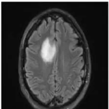
Let us consider the brain tumor image procured from MRI, containing the tumor in figure 1. Median filtering is implemented on the acquired images to get rid of the unwanted noises. The outcomes are displayed in the figure 2.
Fig. 1 Brain Tumor Image
Fig. 2 Median Filtering Outcome
ISSN(Online): 2319-8753 ISSN (Print) : 2347-6710
I
nternational
J
ournal of
I
nnovative
R
esearch in
S
cience,
E
ngineering and
T
echnology
(A High Impact Factor, Monthly, Peer Reviewed Journal)
Visit: www.ijirset.com
Vol. 8, Issue 5, May 2019
Fig. 3 K Means Clustering
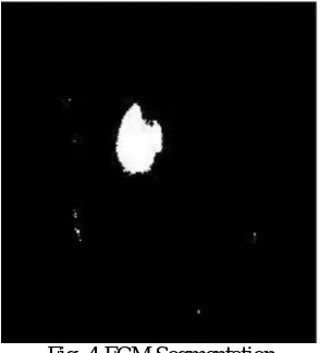
Once the K Means Clustering gets over, Fuzzy C-Means segmentation is eventually implemented on the resulted image procured from K Means segmentation. The region affected by ulcer is highlighted in this process. The outcome of the watershed segmentation is shown below in figure 4.
Fig. 4 FCM Segmentation
ISSN(Online): 2319-8753 ISSN (Print) : 2347-6710
I
nternational
J
ournal of
I
nnovative
R
esearch in
S
cience,
E
ngineering and
T
echnology
(A High Impact Factor, Monthly, Peer Reviewed Journal)
Visit: www.ijirset.com
Vol. 8, Issue 5, May 2019
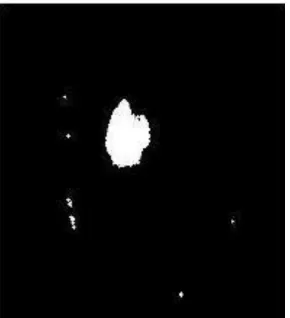
Fig. 5 Thresholding Segmentation
Fig. 6 Output Images of Tumor Area Estimation The output is displayed above in figure 6.
The proposed system is also very sensitive to the errors, because the small error will take the situation in ambiguous state which is not good for diagnosis of tumor. Again same FCM mean and k means algorithms are use to compare individual performance with the proposed method and the result of all are compare and we find that the proposed system having less errors in the system.
VII. CONCLUSION
Brain Tumor segmentation is carried out in this presented work. Firstly image pre-processing is done using median filter technique. If there is any noise are present in the MR image it is removed before the K-means process. The noise free image is given as an input to the k-means and tumor is extracted from the MRI image. And then segmentation using Fuzzy C means for accurate tumor shape extraction of malignant tumor and thresholding of output in feature extraction. Finally approximate reasoning step for calculating tumor area and position calculation and finally to identify stage of tumor from resultant area of tumor i.e. identifies stage of tumor which is easier, cost reducible and time savable.
REFERENCES
[1] Samir Kumar Bandhyopadhyay and TuhinUtsab Paul, “Automatic Segmentation of Brain Tumor from Multiple Images of Brain MRI” International Journal of Application or Innovation in Engineering & Management (IJAIEM),Volume 2, Issue 1, January 2013.
[2] A. Meena, “Spatial Fuzzy C-Means PET Image Segmentation of Neurodegenerative Disorder” , A. Meena et.al / Indian Journal of Computer Science and Engineering (IJCSE).
[3] SumanTatirajua and Avi Mehta, “Image Segmentation using k-means clustering EM and Normalized Cuts”.IEEE Trans. Parallel Diatribe. Syst., vol. 19, no. 5, pp. 710–720, May 2008.
ISSN(Online): 2319-8753 ISSN (Print) : 2347-6710
I
nternational
J
ournal of
I
nnovative
R
esearch in
S
cience,
E
ngineering and
T
echnology
(A High Impact Factor, Monthly, Peer Reviewed Journal)
Visit: www.ijirset.com
Vol. 8, Issue 5, May 2019
[5] Beshiba Wilson and Julia Punitha Malar Dhas, “ An Experimental Analysis of Fuzzy C-Means and K-Means Segmentation Algorithm for Iron Detection in Brain SWI using Matlab”, International Journal of Computer Applications (0975 – 8887) Volume 104 – No 15, October 2014
[6] M.H. FazelZarandia, M. Zarinbal and M. Izadi, “Systematic image processing for diagnosing brain tumors”, Department of Industrial
Engineering, Amirkabir University of Technology, P.O. Box 15875-4413, Tehran, Iran , journal homepage:www.elsevier.com/locate/asoc
[7] Samarjit Das, “Pattern Recognition using the Fuzzy c-means Technique”. International Journal of Energy, Information and Communications Vol. 4, Issue 1, February, 2013.
[8] Varnish Rajesh, BharathanVenkat, Vikesh Karan and M. Poonkodi, “Brain Tumor Segmentation and its Area Calculation in Brain MR Images Using K-Mean Clustering and Fuzzy C-Mean Algorithm”, Department of Computer Science and Engineering, SRM University
[9] Krishna Kant Singh1 and Akansha Singh, “A Study of Image Segmentation Algorithms for Different Types of Images”, IJCSI International Journal of Computer Science Issues, Vol. 7, Issue 5, September 2010.