INVESTIGATION
Activity-Dependent Human Brain
Coding/Noncoding Gene Regulatory Networks
Leonard Lipovich,* Fabien Dachet,†Juan Cai,†Shruti Bagla,† Karina Balan,†Hui Jia,* and Jeffrey A. Loeb*,†,1 *Center for Molecular Medicine and Genetics and†Department of Neurology, Wayne State University School of Medicine, Detroit, Michigan 48202
ABSTRACTWhile most gene transcription yields RNA transcripts that code for proteins, a sizable proportion of the genome generates RNA transcripts that do not code for proteins, but may have important regulatory functions. The brain-derived neurotrophic factor (BDNF) gene, a key regulator of neuronal activity, is overlapped by a primate-specific, antisense long noncoding RNA (lncRNA) called
BDNFOS. We demonstrate reciprocal patterns of BDNF and BDNFOS transcription in highly active regions of human neocortex removed as a treatment for intractable seizures. A genome-wide analysis of activity-dependent coding and noncoding human transcription using a custom lncRNA microarray identified 1288 differentially expressed lncRNAs, of which 26 had expression profiles that matched activity-dependent coding genes and an additional 8 were adjacent to or overlapping with differentially expressed protein-coding genes. The functions of most of these protein-coding partner genes, such asARC, include long-term potentiation, synaptic activity, and memory. The nuclear lncRNAs NEAT1, MALAT1, and RPPH1, composing an RNAse P-dependent lncRNA-maturation pathway, were also upregulated. As a means to replicate human neuronal activity, repeated depolarization of SY5Y cells resulted in sustained CREB activation and produced an inverse pattern of BDNF-BDNFOS co-expression that was not achieved with a single depolarization. RNAi-mediated knockdown of BDNFOS in human SY5Y cells increased BDNF expression, suggesting that BDNFOS directly downregulates BDNF. Temporal expression patterns of other lncRNA-messenger RNA pairs validated the effect of chronic neuronal activity on the transcriptome and implied various lncRNA regulatory mechanisms. lncRNAs, some of which are unique to primates, thus appear to have potentially important regulatory roles in activity-dependent human brain plasticity.
T
HE availability of mammalian genome sequences hasmade it possible to delineate the boundaries and struc-tures of all genes in a genome and has demonstrated an abundance of non-protein-coding transcriptional units that rivals the numbers of known protein-coding genes (reviewed in Carninci and Hayashizaki 2007). Complex and potentially functional regulatory relationships between protein-coding and noncoding genes, including noncoding RNA genes that are poorly conserved across different species, have recently
been delineated (Katayama et al. 2005; Engstrom et al.
2006). These long noncoding RNA (lncRNA) genes can be defined by four fundamental criteria: encoding transcripts
that lack any open reading frames (ORFs) .100 amino
acids or possessing protein database homologies (Dinger et al. 2008); being within the known range of lengths of mammalian mRNAs; support by transcript-to-genome align-ments from complementary DNA (cDNA) data; and absence of matches to any known noncoding-RNA classes. Function-ally, lncRNAs can have regulatory effects on coding mRNAs through a number of mechanisms, including those involving endogenous antisense lncRNA transcripts that repress their
sense-strand protein-coding partners (Katayamaet al.2005;
Yuet al. 2008).
Endogenous lncRNAs can also have catalytic roles, as exemplified by the TERC telomerase RNA, and by the RNAse P and MRP RNAs required for processing of other RNAs. lncRNAs essential to nuclear architecture include NEAT1 and NEAT2. Nuclear hormone receptors, homeobox tran-scription factors, tumor suppressors, and immune regulators are all endogenously modulated by lncRNAs (reviewed in
Lipovichet al.2010). Numerous lncRNAs are transcribed in
the vicinity of known protein-coding genes and regulate Copyright © 2012 by the Genetics Society of America
doi: 10.1534/genetics.112.145128
Manuscript received February 9, 2012; accepted for publication August 24, 2012 Supporting information is available online athttp://www.genetics.org/lookup/suppl/ doi:10.1534/genetics.112.145128/-/DC1.
those known genes through epigenetic mechanisms. Regu-lation of protein-coding genes by overlapping, or nearby encoded, lncRNAs is central in cancer, cell cycle, and
reprog-ramming (reviewed in Lipovich et al. 2010; Loewer et al.
2010; Oromet al.2010). lncRNAs encoded in an antisense
orientation to, and overlapping with, known protein-coding genes are particularly abundant, and the small number of antisense lncRNAs characterized to date is replete with novel functions. Endogenous antisense lncRNAs are
essential in mammalian X-inactivation (Tian et al. 2010);
can directly regulate tumor suppressors; function through dicer-independent mechanisms; and may be rapidly evolv-ing or not conserved, raisevolv-ing the potential for new
regula-tion of old genes over evoluregula-tionary time (Lipovich et al.
2010). RNA interference (RNAi) and overexpression of lncRNAs in cell lines generate reproducible phenotypes, as
we and others have shown (Bernard et al. 2010; Sheik
Mohamed et al.2010). Hundreds of human lncRNAs bind
the polycomb repressor complex 2 (PRC2), a key epigenetic
negative regulator (Khalil et al.2009). In addition to
high-throughput evidence of interactions with epigenetic factors, specific epigenetic roles of lncRNAs are beginning to be de-fined. Antisense lncRNAs actively and specifically modulate gene expression by serving as effectors of epigenetic changes
at target loci (Yu et al.2008). These changes include
anti-sense lncRNA-mediated epigenetic silencing of the anti- sense-strand protein-coding gene promoter; such silencing can be abrogated by Argonaute-2-dependent, small-RNA-mediated
suppression of the antisense lncRNA, resulting in“RNA
ac-tivation”of the sense gene (Morriset al.2008).
Promoter-overlapping antisense lncRNAs can also be targeted by ex-ogenous short RNAs that regulate sense gene expression,
also via Argonaute (Schwartz et al. 2008). Despite these
promising examples, a majority of the thousands of other lncRNAs evident in transcriptome data still remain devoid of assigned functions.
This abundance of lncRNAs, many of which are primate-specific, warrants a systematic assessment of whether they have functional, regulatory roles. Perhaps nowhere might this be more important than in the human brain that is composed of a diverse set of cell types connected through complex synaptic arrangements. The degree of synaptic activity in the brain can be translated into functional and structural changes through activity-dependent changes in gene expression (Katz and Shatz 1996). Although these changes can be effected through direct activation of synaptic genes, they can also be achieved through the release of neurotrophic factors such as brain-derived neurotrophic fac-tor (BDNF) that have direct effects on synaptic architecture and indirect effects by producing changes in gene expression
(Isacksonet al.1991; Binderet al.2001). BDNF, a member
of the nerve growth factor family, regulates the survival and differentiation of neuronal populations, axonal growth and pathfinding, and dendritic growth and morphology and has been linked to many human brain disorders (reviewed in Bibel and Barde 2000; Binder and Scharfman 2004; Hu
and Russek 2008). BDNF messenger (mRNA) and protein are upregulated by seizure activity in animal models of ep-ilepsy as well as in human brain tissues that display
in-creased epileptic activities (Ernfors et al. 1991; Lindvall
et al. 1994; Nibuya et al. 1995; Beaumont et al. 2012). The genomic locus encoding BDNF is structurally complex and also encodes BDNFOS, a primate-specific lncRNA that
is antisense to the coding BDNFgene (Liu et al.2006; Aid
et al. 2007; Pruunsild et al. 2007). BDNF and BDNFOS form double-stranded duplexes, suggesting a potential for BDNFOS to post-transcriptionally regulate BDNF (Pruunsild et al. 2007). Antisense knockdown of BDNFOS, in fact, has recently been shown to increase BDNF expression in HEK293
cells and promotes neuronal outgrowthin vitro(Modarresi
et al.2012)
BDNF binding to its receptors results in a diverse array of downstream signaling pathways including the activation of cyclic adenosine monophosphate response element binding protein (CREB), which, in turn, can also regulate BDNF by
binding to a cognate site within the BDNFgene (Taoet al.
1998; Spenceret al.2008). Activation of CREB by
phosphor-ylation at serine 106 as a result of neuronal activity leads to changes in gene expression that cause reinforcement and stabilization of more active neuronal circuits (reviewed in
Herdegen and Leah 1998; Kandel 2001; Matyniaet al.2002;
West et al.2002). Downstream from phosphorylated CREB (pCREB), immediate early genes have been shown to medi-ate long-lasting changes in neuronal structure and excitabil-ity. Upstream of CREB activation, several known signaling pathways are rapidly activated in response to neuronal
ac-tivity (Kandel 2001; reviewed in Westet al.2002), including
CaMKinase IV, protein kinase A, and MAPK. We have re-cently observed a pattern of transcriptional activation in human brain regions where seizures start that strongly implicates sustained MAPK/CREB activation and down-stream coding gene activations that could underlie layer-specific changes in synaptic architecture that makes these
regions prone to seizures (Rakhade et al.2005; Barkmeier
et al.2012; Beaumontet al.2012).
Given that human lncRNA genes tend to be less well-conserved than protein-coding genes, and can give rise to unique transcripts not found in other species, we sought out a uniquely human system to examine activity-dependent gene expression for both coding and noncoding RNAs using a pairwise comparison of human cortical regions displaying variable degrees of epileptic activities. These brain regions were removed as part of surgical treatment for intractable seizures. We show that regions of human neocortex that display increased activity and BDNF expression have re-duced BDNFOS expression and that BDNFOS directly
down-regulates BDNFin vitroin a neuronal cell line. We developed
a custom microarray platform to perform a transcriptome-wide discovery of other regulatory lncRNAs and matched these to nearby or overlapping, differentially expressed protein-coding
genes to develop a genome-wide list of lncRNA–mRNA gene
known to modulate activity-dependent gene expression in
the human brain, suggesting that these lncRNA–mRNA pairs
form a newly revealed regulatory network of human brain plasticity.
Materials and Methods
Human brain tissue
Informed consent was obtained from seven patients who underwent surgery for medically intractable epilepsy. Ex-treme care was taken to ensure that our study did not influence surgical decision making. All patients underwent presurgical evaluation and identification of epileptic and
control regions as previously described (Rakhadeet al.2005;
Beaumontet al.2012). To localize epileptic brain regions that
displayed both clinical seizures and interictal epileptiform discharges (spikes), a two-stage surgical approach using subdural electrodes with continuous brain-surface record-ings (electrocorticography) and video monitoring was
un-dertaken for 2–5 days. For these studies, paired tissue
samples from neocortex within each patient displaying high and low amounts of interictal (between seizures) spiking were used to compare differential gene expression as a func-tion of brain activity (Loeb 2010). To avoid introducing additional variables into the analysis, each block of tissue was examined histopathologically and demonstrated a nor-mal six-layered neocortical structure without lesions. The paired analysis of high- and low-spiking neocortex within each patient is also critical to isolate the variable under study, which is the degree of activity. Total RNA was pre-pared using a modification of the protocol described
previ-ously (Beaumontet al.2012). The difference was that only
gray matter was used by pooling two to three nearby strips of gray matter that extended from the pial surface to the white matter from each block of tissue corresponding to a given electrode location. This pooling method helps cor-rect for differences in dissections that could lead to over- or under-representation of specific cortical layers.
Cell cultures, transfections, and depolarizations
The SH-SY5Y cell line (ATCC) was maintained in Dulbecco’s
modified Eagle’s medium supplemented with 10% FBS and
used for experiments. Cells between 17 and 25 passages were transfected with BDNFOS-targeting and BC013641-targeting small interfering RNAs (siRNAs) by
electropora-tion according to the manufacturer’s instructions at80%
confluence (Neon electroporation system, Invitrogen). The electroporation conditions used for SH-SY5Y cell transfec-tion were the following: 1200 V; pulse width: 20 ms; and number of pulses: 2. Prior to the experiments, these condi-tions had been optimized using a condition matrix, a control
siRNA, and fluorescent reporters (data not shown). Single
and multiple depolarizations of cells were induced by add-ing 100 mM KCl (final concentration) to the medium at different time points as indicated in the Figure 5 legend.
Quantitative PCR, siRNAs, and primers
Total RNA from cultured SH-SY5Y cells was isolated with an
RNAeasy mini kit according to the manufacturer’s
instruc-tions (QIAgen). The first-strand cDNA was prepared using
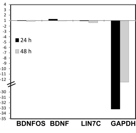
SuperScript First-Strand cDNA kit (Invitrogen), and mRNA and lncRNA expression levels were determined by Taqman quantitative real-time PCR (Taqman qPCR). BDNFOS siRNAs designated S1, S2, S3, and S4 were custom-designed and synthesized by Invitrogen. The BDNFOS Taqman primer/ probe combos were custom-designed by uploading FASTA-format sequences of preferred amplicon regions to the ABI Taqman custom-design website and were purchased from ABI/Life Technologies. This vendor does not release the ac-tual primer and probe sequences of custom-designed ampli-cons to the users. An siRNA against the housekeeping gene glyceraldehyde 3-phosphate dehydrogenase (GAPDH) was used to rule out nonspecific effects. While this siRNA knocked down GAPDH, it had no effects on BDNF, BDNFOS,
and Lin7C at 24 and 48 hr (see supporting information,
Figure S1andFile S4). Western blot analysis
Cell lysates were prepared with SDS sample buffer (Sigma) and subjected to Western blotting to measure CREB
phos-phorylation as described (Beaumont et al. 2012). Briefly,
proteins separated on 4–20% gradient sodium dodecyl
sulfate-polyacrylamide gel were electrically transferred onto nitro-cellulose membrane. After blocking with 5% (v/v) skim milk in TBS containing 0.05% Tween-20 for 1 hr at room temper-ature (RT), the membrane was incubated with rabbit poly-clonal antibody against pCREB (Cell Signaling) at a dilution of 1:1000 for 1 hr at RT and then with specific secondary antibody coupled with HRP (1:5000) for 1 hr at RT. pCREB was visualized with ECL detection system (Pierce). The mem-brane was then stripped and reprobed with CREB antibody (Cell Signaling) at (1:1000) to measure total CREB.
Custom microarrays
Seven 60-mer probes per gene, unambiguously mapping by BLAT (Kent 2002) to a single genomic location and free of interspersed and simple repeats, were designed using the Agilent Technologies OpenGenomics eArray interface for
5586 of the 6736 lncRNA genes from Jia et al. (2010).
The remaining lncRNA genes had been excluded because of eArray failure to yield seven probes per gene or because the eArray-designed probes failed our subsequent check for genomic uniqueness and absence of repeats. As a positive control, we also included seven probes each for 111 of the 137 previously determined protein-coding epileptic genes
(Beaumont et al. 2012) and for six housekeeping control
genes. The eArray Fill Array feature was used to randomly
select control protein-coding gene probes tofill all features
that would have otherwise remained vacant (,2% of total
features on a 44,000-feature, i.e., “44k,” array cell). The
entire probe set was printed in quadruplicate on each slide
microarray platform. Our custom lncRNA microarray
con-tained the combined resulting set of probes (File S2).
Our Agilent 60mer probes are longer than the 25mers used on the Affymetrix platform, and more importantly, we tested each probe (after the probe was proposed by the Agilent EArray design software) for genomic mapping uniqueness [by University of California at Santa Cruz (UCSC) BLAT] and for repetitive element overlaps (by RepeatMasker). Only repeat-free and uniquely-mapping 60mer probes were included. Specificity is assured by these sequence qualities of the probes as well as by our strategy of profiling each lncRNA gene with seven unique probe sequences (not seven replicates of the same probe).
A dye-flip quadruplicate two-color microarray experiment was performed on each within-patient pair of high-spiking and low-spiking surgically resected samples on both the Agilent human genome-wide array (G4112A) and our custom lncRNA array as described, but using a different
labeling method (Beaumontet al.2012). We used the
Epi-centre protocol to generate aminoallyl-amplified RNA (aRNA) for subsequent amplification and labeling with either cyanine or Alexa dyes. For our custom lncRNA arrays, we used label-ing with Alexa dyes (Alexa-647 and Alexa-555, Invitrogen)
within theflip-dye design, as described by the manufacturer
(SuperScript Indirect RNA Amplification System,
Invitro-gen) (Holloway et al.2008). For every patient, each of the
quadruplicates was hybridized on four separate slides. Four
slides of 4·44,000 Agilent arrays (4 arrays, each composed
of the same set of 44,000 probes) were used to screen
seven patients. All slides were scanned as described
previ-ously (Beaumontet al.2012).
Because our lncRNA custom microarray platform is new, we also used qPCR to validate a representative subset of differentially expressed lncRNAs. We considered which spe-cific probes were responsible for the differential expression of each coding and noncoding gene observed across all seven patients and used probes to target only the region of each transcript that was overlapped by the differentially expressed probes. Positive correlation coefficients were seen in all cases,
ranging from 0.61 to 0.96 (File S1) between the array and
qPCR results within each patient; all protein-coding gene dif-ferential expression results were from the G4112A or F cata-log protein-coding microarray, and all lncRNA results were
from our lncRNA custom microarray (Figure S4 inFile S4).
All lncRNA–mRNA overlaps in this work are in the
anti-sense orientation. For all lncRNA–mRNA neighbor pairs,
there is a spacer between the two genes along the genome, regardless of strand. All probes on our microarray are strand-specific and, therefore, even in the case of an
lncRNA–mRNA antisense pair, will exclusively profile either
only the lncRNA (on the custom array described in this article) or only the mRNA (on the Agilent catalog array).
Microarray statistical methods
To identify those differentially expressed lncRNAs that may be directly regulating their overlapping or neighboring
protein-coding genes, we integrated our custom lncRNA expression microarray data with our conventional mRNA
expression microarray data for thein vivohigh-/low-activity
cortical sample pairs from all seven patients analyzed with both array types. For each epilepsy patient, we had a within-patient sample pair of a high-spiking and a low-spiking re-gion. This within-patient sample pair was analyzed, using the same dye-flip quadruplicate strategy, for both the catalog coding (G4112A) and the custom lncRNA microarray. Dif-ferentially expressed genes were identified from both micro-array platforms but using the same strategy. Consistency
between arrays was first examined by correlating the fold
change of all protein-coding control genes common to both
arrays, which was possible because our 111“epileptic
tran-scriptome”genes from the prior protein-coding array work
(Beaumontet al.2012) were used as controls on the lncRNA
array. We used the average value of the seven probes corre-sponding to each control gene on the lncRNA custom array. For 140 catalog (Agilent G4112A) coding-array probes
cor-responding to these 111 genes, Pearson’s correlation
coeffi-cient was 0.90, attesting to very high reproducibility between the coding array and the noncoding custom array.
Scanned microarray images from coding and noncoding microarrays were analyzed by the software Agilent Feature Extraction (Agilent, V10.3.1) with the default protocol
GE2_107_Sep09. A fluorescent correction factor was
de-termined using both qRT-PCR and Agilent Spike-IN probes.
This correction factor was then applied on thefluorescence
intensity (fluorescence at exponent 1.125) and improved
the fold change prediction. The fluorescence distribution
inside each repetition of the microarray experiments was normalized by R V2.11 (R Development Core Team 2010)
using the library“limma”(Smyth and Speed 2003) in a
two-step process: (i) normalization of the intensity of
fluores-cence between dyes using a Loess correction (iterations:
50; span: 0.05) and (ii) independent scaling offluorescence
intensity on the same range across all the arrays for each dye using quantile normalization. The quality of the
normaliza-tion process of the microarray fluorescence was validated
using MA plot density and distribution analysis. Normality
was asserted using the Anderson–Darling test from the
li-brary Nortest (Gross 2006). For each array, the background
level was globally computed using the median of the
fluo-rescence intensity of the negative control probes and sub-tracted from the signal of each probe.
Once normalized, the microarrays were further analyzed using standard statistical methods (Kerr and Churchill
2001b; Wolfingeret al.2001). The differentially expressed
genes between high and low spiking were determined using
a two-step mixed model analysis of variance (Jinet al.2001)
with the library LME4 (Bateset al.2009). This mixed model
approach has been used to compute thefitted effect and the
random effects simultaneously (Littell et al. 1996). To
im-prove the sensibility of the analysis (Kerr et al. 2000; Jin
et al.2001), computation did not use the ratio but instead
(Tanaka et al. 2000) (RNA from the high-spiking area or RNA from the low-spiking area). The false discovery rate
(FDR) and corrected P-value for each gene was computed
with “R” using the library “fdrtool”(Strimmer 2009). The
differentially expressed genes were detected using fold change
and significance simultaneously (Tanakaet al.2000) and were
determined as significantly differentially expressed if their
fold change, for at least one probe per gene, was$1.4 and if
their FDR was#5%.
In addition to a number of custom approaches to identify
8 cis-acting coding lncRNA pairs, 26 trans-acting lncRNAs
were identified as significant and activity-dependent by their
tight correlation (Pearson’s correlation coefficient minimum of
0.9) to a well-known group of 13 activity-dependent
protein-coding genes (Rakhade et al. 2007; Beaumontet al. 2012),
which themselves had been co-expressed with a Pearson’s
correlation coefficient of 0.95. These results were displayed
graphically using Cytoscape (Smootet al.2011). To include
a trans-acting lncRNA in this group, at least one probe (of the seven available probes) representing the lncRNA gene had to meet this statistical requirement.
To study the genes represented both on the catalog array and on our custom array, we used genomic localization of all transcripts along the same human genome assembly, hg19, to find differentially expressed transcripts from the custom noncoding and the G4112A catalog coding array that were close to each other along the genome or overlapped in an antisense orientation within a genomic region. The genomic position, strand, and exon/intron location information for
each transcript is contained in the all_mrna BED file of the
UCSC Genome Database.
Results
Reciprocal patterns of BDNF and BDNFOS expression in electrically active human brain
Patients who fail to respond to medical management of their seizures can greatly benefit from a two-stage surgical
pro-cedure where long-termin vivobrain-surface recordings are
used to identify and remove epileptic brain regions. We have used this human system recently to identify a relatively
small group of genes, includingBDNFthat are differentially
expressed in regions of the human neocortex where seizures
start (Rakhade et al. 2005; Beaumont et al. 2012). While
removing seizure-onset regions is key to a good outcome for improved seizure control, seizures from these brain regions are relatively infrequent compared to the small, but
ex-tremely frequent “interictal,”epileptic discharges that can
occur almost constantly between seizures in some brain
regions (Staley et al. 2005). In fact, several of the genes
induced at seizure-onset zones correlate precisely with inter-ictal spiking rather than with seizure frequency (Rakhade et al. 2007), suggesting that interictal spiking may be the driving force behind this altered expression pattern. Consis-tently, an animal model of interictal spiking without seizures was sufficient to produce neuronal layer-specific changes in
these genes (Barkmeieret al.2012). Here we have focused
on brain regions with different levels of interictal spiking to identify the relationships between coding and noncoding
transcripts in thein vivohuman brain.
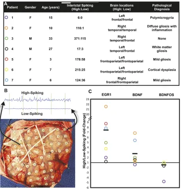
Figure 1A shows a table of seven patients used for the
present study, together with quantifiedin vivospike
frequen-cies, tissue locations, and pathological descriptions. Patients varied in both sex and age, but were chosen because of the availability of both high- and low-interictalspiking neocorti-cal brain samples from nearby brain regions for each patient that were removed as part of their seizure surgery treat-ment. Figure 1B shows how each of these pairs was selected with a short sample of the electroencephalogram recording that illustrates the large difference in interictal spiking. It is important to emphasize that, because of genetic differences, medication effects, and effects of tissue processing, our in-ternally controlled experimental design is crucial (Rakhade et al. 2005; Beaumont et al. 2012). Although patients are listed with different pathological diagnoses from multiple neocortical regions, only tissue samples that showed a nor-mal cortical architecture were used so as not to influence the major variable of interest: increased brain activity.
Because of the potential regulatory relationship of tran-scripts that code for BDNF with those that encode the
partially antisense BDNFOS, as afirst step we compared the
relative expression levels of BDNF and BDNFOS between paired high- and low-spiking regions of human neocortex using qPCR for each patient (Figure 1C). In most patients, BDNF expression was higher in more electrically active regions, whereas BDNFOS lncRNA levels were significantly reduced in the high-spiking regions. We used EGR1 expres-sion as a positive control for high-spiking human cortical brain regions as its expression has been shown to be directly
proportionate to interictal brain activity (Rakhade et al.
2007). These results raise the possibility that increased BDNF levels could in part be regulated by a decrease of the antisense BDNFOS RNA.
BDNFOS is a negative regulator of BDNF in an in vitro human cell culture system
The genomic antisense orientation of BDNF and BDNFOS is shown in Figure 2A, where both overlapping and non-overlapping regions are delineated. We have previously demonstrated that perturbation of lncRNA levels at multiple
cis-antisense lncRNA–mRNA pairs affects levels of the
cog-nate mRNAs (Katayamaet al.2005). To distinguish whether
the lncRNA BDNFOS directly regulates BDNF mRNA levels, we custom-designed three siRNAs targeting human BDNFOS (Figure 2A) and used qPCR to interrogate BDNFOS lncRNA and BDNF mRNA levels after the siRNA transfections. BDNFOS siRNAs were individually transfected into the human neuro-blastoma cell line SH-SY5Y by electroporation and caused re-producible BDNFOS knockdown at 24 hr (all three siRNAs) and at 48 hr (only S2). Two of the siRNAs led to knockdown of
BDNFOS by.70% (Figure 2B). BDNFOS knockdown by these
in BDNF mRNA levels (between 1.5- and 3.5-fold-change),
suggesting that the cis-antisense BDNFOS RNA functions as
a negative regulator of human BDNF (Figure 2B).
lncRNA genes in gene chains—loci where three or more
genes are joined through shared antisense overlaps and
bidirec-tional promoters—are a general property of the mammalian
genome (Engstrom et al. 2006). Human BDNFOS is part of
a three-gene genomic positional chain: it shares a putative
bi-directional promoter with the LIN7Cgene at its 59 end while
also encompassing an exonic cis-antisense overlap with BDNF
exonic sequences at its 39end. BDNFOS knockdown by the same
dsRNAs also increased the mRNA levels of LIN7C, suggesting that BDNFOS may negatively regulate other genes at its locus.
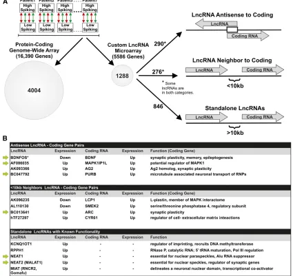
Transcriptome-wide profiling of all known human protein-coding and lncRNAs reveals activity-dependent regulatory pairs and networks
Our functional validation of the primate-specific BDNF/ BDNOS pair suggests the potential for many more coding/ noncoding regulatory relationships across the human ge-nome that may vary as a function of brain activity. Here we utilized these same paired RNA samples from the same seven patients to identify the activity-dependent coding/ noncoding interactome. To achieve this, we developed
a custom lncRNA microarray, which allowed us to compare transcriptional profiles of lncRNAs to coding RNAs from a commercial genome-wide coding array (Figure 3). This new custom array is based on our previously defined and characterized human lncRNA gene catalog from experimen-tal transcriptome data represented by cDNA and EST se-quences in GenBank, totaling 6736 lncRNA transcriptional
units (Jia et al.2010). Our human lncRNA gene catalog is
mostly nonredundant with respect to other recently
pub-lished human lncRNA collections (Figure S2 in File S4). In
contrast to our custom lncRNA array, current commercial microarray platforms do not adequately represent many genomically complex loci, including those encoding lncRNA
genes and sense–antisense pairs (Orlovet al.2007; Jiaet al.
2010).
Both platforms utilized a dye-flip (Kerr and Churchill 2001a) quadruplicate experimental design to obtain the most accurate statistical comparison of each pair of tissue
samples from each patient (Yaoet al.2004; Rakhade et al.
2005; Beaumont et al. 2012). Each within-patient sample
same strategy. Consistency between arrays was first exam-ined by correlating the fold-change of all differentially expressed protein-coding control genes common to both arrays, which was possible because our 111 epileptic tran-scriptome genes from the prior protein-coding array work were used as controls on the lncRNA array. We used the average value of the seven probes corresponding to each control gene on the lncRNA custom array. For 140 catalog (Agilent G4112A) coding-array probes corresponding to these
111 genes, Pearson’s correlation coefficient was 0.90,
attest-ing to very high reproducibility between the codattest-ing array and the noncoding custom array.
To define a gene as differentially expressed, we required at least one microarray oligonucleotide probe corresponding
to that gene to be $1.4-fold differentially expressed with
FDR #5% in a groupwise analysis of all seven patients.
These thresholds were selected based on a power analysis
using thisflip-dye quadruplicate design (Loeb and Beaumont
2009). Using this criterion, we identified 4004 protein-coding genes from the catalog array (1944 upregulated and 2060
downregulated in high-activity areas; File S1). On the
lncRNA arrays, 86 of the 111 positive control genes were upregulated, and 1288 lncRNA genes were differentially expressed between high-activity and low-activity neocortical
regions (698 upregulated lncRNA genes and 590
downregu-lated lncRNA genes in high-activity areas; File S2). BDNF
was represented on both the coding microarray and, as a brain-expressed known control gene, on the lncRNA mi-croarray. BDNF was upregulated in high-activity tissue from all seven patients according to both our array platforms: coding microarray, median 3.6-fold change; lncRNA micro-array, median 2.8-fold change.
To integrate the coding and noncoding transcriptomes of
the human neocortex (File S3), we then determined which
of the differentially expressed protein-coding genes were encoded by genomic loci overlapping, or adjacent to, the loci which also encoded differentially expressed lncRNA genes as outlined in Figure 3A. Here, we analyzed the entire extent of differential expression for lncRNAs that participate
in sense–antisense pairs and in non-overlapping gene
neigh-bor pairs such that one gene in the pair was protein-coding whereas the other gene encoded the lncRNA. Specifically, we
identified allcis-encoded gene pairs in which both a
protein-coding gene and a nonprotein-coding (lncRNA) gene were expressed
from the same locus. We refer to these pairs as coding–noncoding
gene pairs. We then separated the pairs into two categories—
antisense and neighbor—both of which carry the potential
for mRNA regulation by a paired lncRNA (Jia et al. 2010;
Lipovichet al.2010). We defined acis-antisense gene pair as
two genes transcribed from the opposite strands of the same locus in a configuration such that at least some sequence in at least one exon overlaps one exon of the other gene. We defined a neighbor-gene pair as any gene pair such that the nearest boundaries of two nearby, but
nonoverlap-ping, genes are ,10 kb away from one another. In this
study,“cis”therefore refers to any same-locus (not
neces-sarily same-allele) regulatory mechanisms, which include antisense-mediated regulation by lncRNAs of protein-coding
genes that are encoded in the same locus.“Trans”refers to
any regulation involving genes encoded at multiple distinct genomic loci.
Of our lncRNAs differentially expressed at high-activity
regions, 290 were members of sense–antisense gene pairs
(File S3). We define codifferential expression as a differen-tial expression profile of two genes such that the differendifferen-tial expression of one gene is either inversely or directly corre-lated with the differential expression of the other gene across multiple sample pairs, each of which originates from a different patient and all of which are statistically
signifi-cant. Only 4 of these 290 mRNA–lncRNAcis-antisense pairs
were codifferentially expressed in all seven patients (Figure 3B). Only 1 of the 4 pairs (BDNFOS/BDNF) featured an inverse differential expression profile. The other 3 pairs all had a positive, direct correlation. This is, in fact, not surpris-ing, given the prevalence of synergistic, as opposed to
in-verse, expression patterns in mammaliancis-antisense pairs
in response to a stimulus or to a knockdown (Katayamaet al.
2005). These 3 pairs featured lncRNAscis-antisense to
MAP-K1IP1L (MAP Kinase 1 Interacting Protein 1-like, potentially a modulator of MAP Kinase 1, whose role centers on the Figure 2 Downregulation of BDNFOS induces BDNF and LIN7C
CREB activation pathway upstream of brain activity-dependent gene expression); PURB (purine-rich element-binding pro-tein, a gene expression regulator); and C11ORF96, which we have shown by bioinformatic analysis to be a human
homolog of the ratAG2gene (Matsuoet al.2000), induced
as a consequence of sustained long-term potentiationin vivo
in rat hippocampus and therefore implicated in neuronal plasticity. In summary, the protein-coding genes at 3 of the
4 codifferentially expressed lncRNA–mRNA cis-antisense
pairs have known neuronal functions centered on synaptic
plasticity. The remaining 286 lncRNA–mRNA cis-antisense
pairs did not have any correlation between the mRNA and
the lncRNA, within each antisense gene pair, across the seven patients.
A total of 276 lncRNAs differentially expressed at high-activity brain regions resided at genomic loci where a protein-coding gene and an lncRNA gene were non-overlapping but
within 10 kb of each other along the genome (Jia et al.
2010). However, only four mRNA–lncRNA neighbor-gene
pairs were codifferentially expressed in the groupwise anal-ysis of the seven patients (Figure 3B). These four codiffer-entially expressed neighbor-gene pairs contained lncRNA
genes neighboring the protein-coding genes ARC
receptor endocytosis required for both synaptic plasticity
and long-term memory; L-plastin (LCP1), relevant to the
activity-dependent MAPK/CREB activation by its placement
within the human MAPK interactome (Bandyopadhyayet al.
2010);SMEK2, a regulatory subunit of Ser/Thr phosphatase
4; and CYR61, a secreted protein that associates with the
extracellular matrix and the cell surface, regulates Akt
acti-vation (Goodwinet al.2010), and is differentially expressed
in autism (Garbettet al.2008).
Of the 1288 lncRNA genes determined by our custom microarray to be differentially expressed at high-activity areas of the human neocortex, 846 remain largely refractory to functional interpretation as they were not genomically near, or antisense to, any known protein-coding genes. These include the lncRNA MALAT-1, originally discovered as a predictor of
metastasis and survival in lung cancer (Jiet al.2003) and now
known to be a regulator of several synaptic genes (Bernardet al.
2010; Lipovichet al.2010). Some are key components of specific
nuclear bodies, while other lncRNAs regulate imprinting genes
and still others perform essential catalytic roles (Bernardet al.
2010; reviewed in Lipovichet al.2010). Differential expression
of 5 of these known nuclear RNAs (Figure 3B, bottom) was significant. MIAT, the sole member of this group that was down-regulated in the more active areas delineates a novel neuronal
nuclear domain (Sone et al.2007) shown to be both a direct
target and a putative co-activator of the transcription factor Oct4
(Sheik Mohamedet al.2010). In contrast, the levels of the other
4 known nuclear lncRNAs were increased in the more active areas. These lncRNAs included: KCNQ1OT1, which may regu-late imprinting by recruiting the DNA methyltransferase DNMT1
to differentially methylated regions (Mohammad et al.2010);
RPPH1, the catalytic-RNA component of RNase P, essential for
tRNA 59 end maturation and for regulating Pol III-dependent
tRNA transcription (Jarrous and Reiner 2007); NEAT1, an es-sential component of nuclear paraspeckles that suppresses the nucleocytoplasmic export of Alu-containing RNAs; and NEAT2 (MALAT-1), an essential component of nuclear speckles and
a regulator of synaptic genes (Bernardet al.2010).
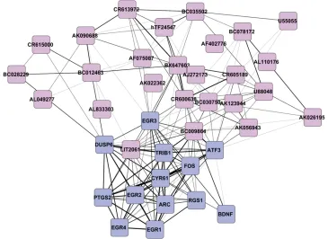
We also used a second unbiased approach to identify activity-dependent lncRNAs with potential importance in synaptic plasticity transcriptional regulatory networks. We have shown that a number of coding genes, including EGR1, EGR2, and FOS, are expressed in human brain in direct
relation to the degree of epileptic discharges (Rakhadeet al.
2007). Using co-expression clustering of protein-coding genes, we identified these and 8 additional genes (13 total) that have the same pattern of expression across the seven patients and then identified 26 lncRNAs whose pattern of expression correlated with this group of coding genes. Figure 4, constructed from our coding/noncoding transcrip-tome quantitation integration by Cytoscape software (Smoot et al. 2011), illustrates co-expression of these 26 differen-tially expressed lncRNA genes with the 13 differendifferen-tially expressed protein-coding genes. In Figure 4, genes that are closer together are more tightly linked. While it appears
that there are two linked clusters of coding vs. noncoding
is differentially expressed, although its parental geneIL8RB
and theRUFY4known gene that the pseudogene overlaps,
are not detectable above background in the same samples on our protein-coding gene arrays.
Time-dependent patterns of lncRNA–mRNA
codifferential expression with chronic depolarization of cultured human neuronal cells
To facilitate the study of primate-specific, activity-dependent,
coding–noncoding regulatory networks, we developed an
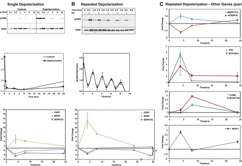
in vitro system of repeated depolarization using the human SH-SY5Y neuroblastoma cell line. Depolarization with supra-physiological concentrations of KCl has been used extensively as a means to study CREB activation and downstream
tran-scription in neuronal cells in culture (Sheng et al. 1990;
Connolly and Kingsbury 2010). Figure 5A shows that, while a single treatment of these cells with 100 mM KCl leads only
to transient CREB activation (CREB phosphorylation, detect-able by the pCREB Ser106 antibody), repeated 5-min expo-sures with KCl separated by 2-hr intervals lead to more sustained CREB activation, similar to that observed in highly
spiking human neocortex (Beaumontet al.2012) and in an
animal model of frequent interictal spiking (Barkmeieret al.
2012) (Figure 5B).
while BDNFOS remains downregulated at 24 hr. Impor-tantly, the 48-hr time point extends this trend, maintaining the sustained increase in BDNF expression and demonstrat-ing an even stronger downregulation of BDNFOS. While this is likely an oversimplification of a dynamic and complex set of regulatory interactions unrelated to BDNFOS, the chronic depolarization-induced reciprocal BDNFOS depletion and BDNF mRNA increase in culture parallel the inverse rela-tionship between BDNFOS and BDNF in high-activity areas of the human brain shown in Figure 1. Therefore, we ap-plied this cell culture system to interrogate the activity-dependent expression patterns of other lncRNAs and their
cis-encoded partner mRNAs from Figure 3.
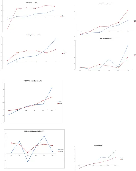
In Figure 5C, chronic depolarization of SH-SY5Y cells revealed a number of distinct time-dependent patterns.
Un-like the cis-antisense BDNF/BDNFOS pair, the
AF086035-MAPK1P1L cis-antisense pair had an opposite effect of
decreasing lncRNA and increasing mRNA levels early in the time course, although by 24 hr the lncRNA level showed a slight increase. The BC013641-ARC neighbor-gene pair displayed increased expression of both genes at the 4-hr time point in the depolarization-treated Sh-sy5y cells, mir-roring the increased expression of both genes in the high-activity human brain. At subsequent time points, ARC mRNA levels decreased back to the pretreatment levels although we observed a sustained elevated level of the BC013641
lncRNA encoded near theARCgene along the genome
(Fig-ure 5C). Since the BC013641 gene is located 6 kb from
ARC with a divergent genomic orientation relative to ARC
we designed two custom siRNA oligonucleotides targeting BC013641. Although both siRNAs knocked down BC013641, only one led to a 1.25-fold increase in the mRNA level of the
neighboring ARCgene, suggesting that reciprocal gene
ex-pression directionality at lncRNA–mRNA pairs may occur at
neighbor-gene loci such as BC013641-ARC, and not solely at
sense–antisense loci such as BDNFOS-BDNF (data not
shown). The BC047792-PURBcis-antisense pair showed
in-creased expression of both genes, which mirrored the co-ordinate increase observed in high-spiking brain regions; however, in contrast to BC013641-ARC, expression of both transcripts was maximal at the 8-hr time point and returned to baseline at 24 hr, showing no sustained increase in lncRNA expression. Finally, we looked at the time course of one stand-alone nuclear lncRNA, NEAT1, which has a po-tentially far-reaching regulatory role. NEAT1 goes up within 4 hr, returns to baseline at 8 hr, but shows some chronic elevated expression at 24 hr.
Discussion
A primate-specific lncRNA regulatory mechanism for BDNF
A striking feature of the BDNF/BDNFOS locus is the com-plexity of its genomic landscape, which is highly representa-tive of the genomic properties observed at lncRNA-encoding loci throughout mammalian genomes. In addition to residing
in a three-gene chain, with LIN7Csharing its bidirectional
promoter and BDNF overlapping its 39 end, BDNFOS may
have emerged in recent mammalian evolution after the
primate–rodent divergence. A possible recent origin for this
lncRNA gene is supported by two lines of evidence. First,
several splice sites ofBDNFOSare poorly conserved outside
of primates (data not shown), suggesting that the genomic
structure of BDNFOS either arose or was modified
specifi-cally in primate evolution. This is consistent with our recent finding that lncRNA genes may comprise a majority of
primate-specific genes (Tayet al. 2009). Second, there is no
evidence for a BDNFOS-like gene between the mouseLin7cand
Bdnfgenes in any public mouse cDNA and EST sequence data
represented by UCSC Genome Browser transcript-to-genome alignments (not shown); however, a recently identified non-orthologous postional equivalent Bdnf antisense tran-script was found in the mouse, suggesting an evolutionarily distinct, but similar, mechanism for lncRNA-dependent
reg-ulation of Bdnf (Engstrom et al. 2006; Modarresi et al.
2012). This genomic and evolutionary complexity of the BDNF/BDNFOS locus suggests that functional lncRNAs in the human brain may be characterized by interspecies non-conservation or high divergence of their gene structures. This is of particular interest because of the persistent inverse codifferential expression of the BDNF/BDNFOS gene pair as a function of human brain activity shown here together with the observed increase in BDNF mRNA levels following knockdown of BDNFOS. Our rescue of LIN7C by BDNFOS RNAi indicates that BDNFOS function may, in part, be nuclear and epigenetic. This would be consistent with the recent demonstration that an antisense lncRNA acts
epi-genetically by modulating target transcription (Tayet al.
2009). A possible explanation for the upregulation of both BDNF and LIN7C via BDNFOS RNAi might involve BDNFOS-mediated PRC2 recruitment to this locus. PRC2 association
with lncRNAs (Khalil et al. 2009) makes BDNFOS-PRC2
binding a distinct possibility. Knockdown of BDNFOS, pre-venting BDNFOS-mediated PRC2 targeting at this locus, would then result in increased LIN7C and BDNF mRNA lev-els, which is what we observed. BDNF is known to be under epigenetic control: it is activated by acetylation of multiple
H3 lysine residues in its promoter chromatin (Tian et al.
2010) and is repressed in vivo by H3K27Me2 (Tsankova
et al.2006), a direct PRC2-catalyzed modification (reviewed in Margueron and Reinberg 2011). Our model for BDNFOS-mediated PRC2 recruitment in LIN7C and BDNF suppression does not contradict the concurrent possibility of cytoplasmic, post-transcriptional BDNFOS-BDNF regulation; in fact, the efficiency of our RNAi knockdowns of BDNFOS implies cy-toplasmic localization, since RNA-induced silencing complex
(RISC) activity is cytoplasmic. Future work in this field
should also clarify whether BDNF mRNA has a reciprocal regulatory impact on BDNFOS.
part of the last exon and part of the last intron of BDNF (hg19::chr11:27,680,112-27,680,229). While there was little change in our ability to detect this amplicon with BDNFOS RNAi S1 and S2 treatment, there was an increase with S3, raising the possibility that the BDNFOS effect is at the level of new transcription (data not shown). However, more work is needed to address the question of whether BDNFOS directly regulates BDNF transcription.
In summary, our findings, which center on the poorly
conserved but functional lncRNA BDNFOS, provide a uniquely human view of activity-dependent transcriptional regulatory networks in the brain whose endogenous components cannot be modeled in rodents or other nonprimate species. A set of differentially expressed microRNAs, including miR-30a-5p, act as post-transcriptional inhibitors of BDNF in the prefrontal
cortex (Mellioset al.2008). Our demonstration of the
primate-specific lncRNA BDNFOS as an inhibitor of BDNF comple-ments this earlier miRNA work, suggesting that BDNF is targeted by multiple RNA-mediated regulatory mechanisms involving short and long, ancient as well as evolutionarily young, noncoding RNAs.
This is thefirst study where reciprocal lncRNA–mRNA
reg-ulation is inferred from thein vivohuman brain in a groupwise
analysis of multiple living patients and then validated by RNAi in a human neuronal cell line. Moreover, reciprocal
regulation in sense–antisense pairs is an exception rather
than the rule (Katayamaet al. 2005). Here, we pinpoint an
lncRNA, BDNFOS, which, through its ability to regulate BDNF, may be a key novel contributor to epileptogenesis in a locus where future mechanistic analysis is warranted.
Genome-wide integration of coding/noncoding RNAs as a function of human brain activity
Recently, we generated a stringently filtered catalog of
human lncRNAs and described the genomic positional relationships between these lncRNAs and protein-coding
genes, providing insights into lncRNA functions (Jia et al.
2010). Despite their prominence in the transcriptome, most lncRNAs remain poorly understood, although lncRNAs may contribute to the biological complexity of regulatory net-works (reviewed in Mattick and Makunin 2006). Because of their abundance, lncRNAs may be even more important than microRNAs. microRNAs function as post-transcriptional repressors, but lncRNAs have additional mechanisms to pos-itively and negatively regulate cotranscriptional and post-transcriptional alterations in gene expression. Here we have used these insights to develop a custom lncRNA microarray
to provide the first genome-wide analysis of human brain
lncRNA-based regulatory networks as a function of electrical brain activity. Several co-expressed lncRNA/coding gene pairs identified here have important roles in activity-dependent synaptic plasticity either directly, such as BDNF and others in-volved in the MAPK/CREB signaling, or indirectly through the expression of regulatory lncRNAs such as MALAT-1 (Bernard et al. 2010). Our focus on the relationship between cod-ing mRNAs and lncRNAs with respect to brain activity is
complementary to other human brain transcriptome stud-ies such as those focusing on developmental, regional, and
disease-related gene expression patterns (Johnson et al.
2009; Voineagu et al.2011), but significantly expands upon
those studies through our annotation of the human lncRNA transcriptome and its expression-level relationships with specific protein-coding genes.
This genome-wide lncRNA expression survey of electrically active human neocortex has uncovered hundreds of lncRNAs differentially expressed between more and less electrophys-iologically active areas of the human neocortex. Of these lncRNAs, 26 are expressed directly in proportion to known activity-dependent genes (Figure 4), and therefore these lncRNAs could represent biomarkers and drug targets for hu-man brain diseases, such as epilepsy (Loeb 2011). The co-expression clustering topology (Figure 4) suggests a network where mRNAs and lncRNAs are linked by previously unchar-acterized lncRNA nodes (such as BC009864) as hubs with spoke edges extending simultaneously to multiple mRNAs
and other lncRNAs. We also observed eight lncRNA–mRNA
cis-antisense and neighbor-gene pairs characterized by
coor-dinated differential expression of both genes in each coding–
noncoding pair, suggesting lncRNA-mediated regulation of protein-coding gene expression in the brain, and the even more intriguing reciprocal possibility that some mRNAs may function at the RNA level to regulate lncRNA expression or in
bidirectionally regulated feedback loops incis. Other lncRNAs
such as NEAT1 were detected only by the trans-regulation
analysis, where we searched for lncRNAs whose expression was highly correlated with protein-coding genes regardless of the genomic mapping location of those coding genes. Al-though the role of the NEAT2 (MALAT-1) lncRNA from nu-clear speckles in synaptic gene regulation is now known
(Bernard et al.2010), our study complements that work by
implicating NEAT1, the lncRNA from nuclear paraspeckles that is encoded near the NEAT2 locus, in regulatory interac-tions with activity-dependent genes in the brain. Our three lines of evidence for activity-dependent NEAT1 function in the neocortex are our detection of NEAT1 as a differentially expressed lncRNA on the custom microarray analysis of hu-man brain samples, our demonstration of activity-dependent NEAT1 expression in depolarized human SY5Y cell culture, and the assignment of NEAT1 as a central node to a co-expression cluster of specific coding and noncoding RNAs
(Figure 4). Our cis-regulation andtrans-regulation analyses
uncovered different, nonredundant sets of lncRNAs, suggest-ing that specific lncRNAs are involved in both types of regu-lation, which for any given lncRNA may be mutually exclusive.
These results represent thefirst functional evidence for a
re-markably diverse pattern of lncRNA expression in the hu-man brain.
Functionality of coding/noncoding RNA regulatory networks in the human brain
SH-SY5Y cell culture time course jointly represent thefirst demonstration that known lncRNAs are activity-dependent both in vivo and in cell culture. The complex, but similar,
pattern of lncRNA–mRNA expression in activated human
brain and in a chronically depolarized human neuronal cell line enables the temporal characterization of these regula-tory pathways and provides a new system in which to study these complex, primate-specific transcriptional regulatory networks. We previously performed time-course analysis of
mammalian cis-antisense pairs in cell culture subjected to
a specific stimulus, such as lipopolysaccharide induction of macrophages, revealing a wide diversity of temporal differ-ential expression patterns. These patterns, with concordant or synergistic regulation of the two paired genes, were
ob-served at most of the differentially expressed cis-antisense
pairs (similar to our results in Figure 5C). Less frequently, the patterns showed inverse or reciprocal regulation of the two paired genes at a locus, similar to our results in Figure
2B (Katayamaet al.2005).
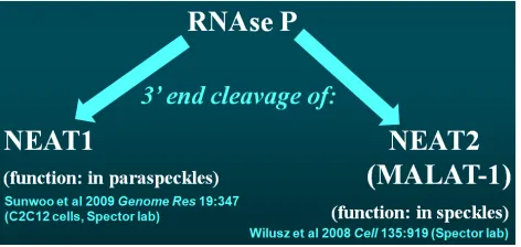
Fundamental functional roles have been previously estab-lished for a relatively few lncRNAs. We show upregulation of
three nuclear RNAs—RNase P (RPPH1), NEAT1, and
MALAT-1—in high-activity areas of the neocortex. The catalytic-RNA
component of RNAse P is essential for the 39-end cleavage of
both NEAT1 (Sunwoo et al. 2009) and NEAT2/MALAT-1
(Wilusz et al. 2008). Therefore, these three lncRNAs may
compose an lncRNA-mediated lncRNA maturation network
in highly active brain regions (Figure S3 in File S4). The
function of this induced network may be to modulate the expression of synaptic genes, such as those whose mRNA
levels are regulated by MALAT-1 (Bernardet al.2010). This
RNA-mediated regulatory network may function either inde-pendently from, or synergistically with, the MAPK/CREB pathway to regulate activity-dependent gene expression.
While BDNF regulation by BDNFOS bolsters previous precedents for reverse-genetic approaches to functional
validation of lncRNA genes (Sheik Mohamed et al. 2010),
further mechanistic studies of the novel regulatory lncRNAs will be needed to delineate the functions of these widely heterogeneous lncRNAs. For example, it is important to dis-tinguish nuclear epigenetic from post-transcriptional and cy-toplasmic regulatory mechanisms of lncRNAs. Such studies should be aided by structural insights into the mammalian lncRNAome, following in the footsteps of existing whole-transcriptome empirical RNA secondary structure
delinea-tion methodologies (Kerteszet al.2010). It has also become
increasingly evident that human lncRNAs perform diverse yet crucial functions, one of which is to regulate mRNA stability on a transcriptome-wide scale through repetitive elements embedded in exons of many lncRNAs (Gong and Maquat 2011). Ribonucleoprotein complexes that enable lncRNA function and complexes that facilitate
lncRNA-mediated regulation of mRNAs in sense–antisense pairs
can be identified by affinity columns and mass spectrometric analysis. This identification will allow therapeutic targeting
of lncRNA–mRNA regulatory relationships. Finally, integrated
differential expression analysis of the protein-coding and the long noncoding transcriptome represents only a limited en-try point into transcriptional regulatory networks underlying activity-dependent gene expression. A comprehensive as-sessment of this network is possible only if all transcript classes, including mRNAs, lncRNAs, microRNAs, and the
re-cently discovered endo-siRNAs (Smalheiseret al.2011), are
profiled jointly.
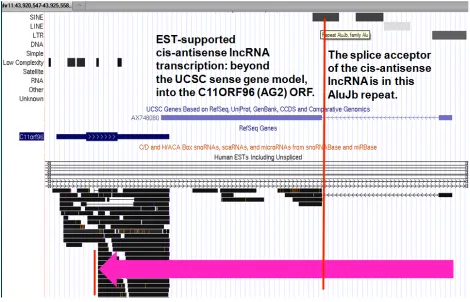
The genomic complexity of the human AK093366-AG2
lncRNA–mRNA cis-antisense pair (Figure S5 in File S4) is
reminiscent of that observed in the BDNFOS-BDNF lncRNA–
mRNAcis-antisense pair. Both theBDNFOSlncRNA gene and
the AK093366 lncRNA gene contained primate-specific
splice sites. The splice donor of AK093366’s sole intron is
primate-specific because it is harbored within an AluJb re-peat. Alu repeats are the best-known class of primate-specific interspersed repeats (Greally 2002), and therefore key gene structure elements, including splice sites, con-tained within Alu repeats provide direct evidence that the corresponding gene structures either arose or were modified after the mammalian radiation, specifically in the primate
lineage. Notably, EST-supported cis-antisense lncRNA
tran-scription of the Alu-containing AK093366 trantran-scriptional
unit extends substantially beyond the UCSC C11ORF96
(AG2) gene model and well into theC11ORF96 ORF. This
underscores the utility of EST data, much of which remains unincorporated into reference gene models and annotations in delineating the boundaries of lncRNA genes, including those involved in putative regulatory relationships with pro-tein-coding counterparts. While Alu-containing lncRNAs have
recently been implicated in the in trans post-transcriptional
regulation of gene expression via effecting mRNA decay (Gong
and Maquat 2011), our analysis suggests distinctcis-regulatory
roles in overlapping-gene regulation for certain Alu-containing
lncRNAs—specifically, AK093366. Two of the four
codiffer-entially expressed lncRNA–mRNAcis-antisense pairs in the
human neocortex, BDNFOS-BDNF and AK093366-AG2, thus feature primate-specific sequence at lncRNA gene splice
junctions. The protein-coding genes BDNF and AG2 are
overlapped by endogenous antisense lncRNAs containing exonic Alu repeats, and these gene pairs are codifferen-tially expressed in active areas of the human epileptic neocortex.
BDNFOS-mediated regulation of BDNF provides initial evidence that primate-specific regulation of conserved
protein-coding genes by cis-antisense lncRNAs takes place in
epi-lepsy, a complex human brain disorder. The co-expression of mRNAs and nonconserved lncRNAs at loci other than BDNF, including AG2, suggests that primate-specific regula-tion of conserved genes by nonconserved lncRNAs in the human brain may not be unique to the BDNF locus.
Acknowledgments
manu-script. This work was funded by National Institutes of Health (NIH)/National Institute of Neurological Disorders and Stroke grants R01NS045207 and R01NS058802 (J.A.L.) and by internal funds from Wayne State University (L.L.). Microarray development was supported by NIH/National Institute on Drug Abuse grant 1R03DA026021-01 (L.L.). Microarray scanning was performed by the Core Facility of the Department of Pediatrics, School of Medicine, Wayne State University.
Literature Cited
Aid, T., A. Kazantseva, M. Piirsoo, K. Palm, and T. Timmusk, 2007 Mouse and rat BDNF gene structure and expression re-visited. J. Neurosci. Res. 85: 525–535.
Bandyopadhyay, S., C. Y. Chiang, J. Srivastava, M. Gersten, S. White et al., 2010 A human MAP kinase interactome. Nat. Methods 7: 801–805.
Barkmeier, D. T., D. Senador, K. Leclercq, D. Pai, J. Hua et al., 2012 Electrical, molecular and behavioral effects of interictal spiking in the rat. Neurobiol. Dis. 47: 92–101.
Bates, D., M. Maechler, and B. Bolker, 2009 lme4: Linear mexed-ef-fects models using S4 classes. Available at:https://r-forge.r-project. org/R/?group_id=60. Accessed: September, 2012.
Beaumont, T., B. Yao, A. Shah, G. Pai, J. Kapatos, and J. A. Loeb, 2012 Layer-specific CREB target gene induction in human neocortical epilepsy. J. Neurosci. (in press).
Bernard, D., K. V. Prasanth, V. Tripathi, S. Colasse, T. Nakamura
et al., 2010 A long nuclear-retained non-coding RNA regulates synaptogenesis by modulating gene expression. EMBO J. 29: 3082–3093.
Bibel, M., and Y. A. Barde, 2000 Neurotrophins: key regulators of cell fate and cell shape in the vertebrate nervous system. Genes Dev. 14: 2919–2937.
Binder, D. K., and H. E. Scharfman, 2004 Brain-derived neuro-trophic factor. Growth Factors 22: 123–131.
Binder, D. K., S. D. Croll, C. M. Gall, and H. E. Scharfman, 2001 BDNF and epilepsy: Too much of a good thing? Trends Neurosci. 24: 47–53.
Carninci, P., and Y. Hayashizaki, 2007 Noncoding RNA transcription beyond annotated genes. Curr. Opin. Genet. Dev. 17: 139–144. Connolly, S., and T. J. Kingsbury, 2010 Caffeine modulates
CREB-dependent gene expression in developing cortical neurons. Bio-chem. Biophys. Res. Commun. 397: 152–156.
Dinger, M. E., K. C. Pang, T. R. Mercer, and J. S. Mattick, 2008 Differentiating protein-coding and noncoding RNA: chal-lenges and ambiguities. PLOS Comput. Biol. 4: e1000176. Engstrom, P. G., H. Suzuki, N. Ninomiya, A. Akalin, L. Sessaet al.,
2006 Complex loci in human and mouse genomes. PLoS Genet. 2: e47.
Ernfors, P., J. Bengzon, Z. Kokaia, H. Persson, and O. Lindvall, 1991 Increased levels of messenger RNAs for neurotrophic factors in the brain during kindling epileptogenesis. Neuron 7: 165–176.
Garbett, K., P. J. Ebert, A. Mitchell, C. Lintas, B. Manzi et al., 2008 Immune transcriptome alterations in the temporal cortex of subjects with autism. Neurobiol. Dis. 30: 303–311.
Gong, C., and L. E. Maquat, 2011 lncRNAs transactivate STAU1-mediated mRNA decay by duplexing with 39UTRs via Alu ele-ments. Nature 470: 284–288.
Goodwin, C. R., B. Lal, X. Zhou, S. Ho, S. Xiaet al., 2010 Cyr61 mediates hepatocyte growth factor-dependent tumor cell growth, migration, and Akt activation. Cancer Res. 70: 2932– 2941.
Greally, J. M., 2002 Short interspersed transposable elements (SINEs) are excluded from imprinted regions in the human ge-nome. Proc. Natl. Acad. Sci. USA 99: 327–332.
Gross, J., 2006 nortest: Tests for Normality. R package version 1.0. Available at:http://cran.r-project.org/web/packages/nortest/. Accessed: September, 2012.
Herdegen, T., and J. D. Leah, 1998 Inducible and constitutive transcription factors in the mammalian nervous system: control of gene expression by Jun, Fos and Krox, and CREB/ATF pro-teins. Brain Res. Brain Res. Rev. 28: 370–490.
Holloway, M. G., G. D. Miles, A. A. Dombkowski, and D. J. Waxman, 2008 Liver-specific hepatocyte nuclear factor-4alpha defi -ciency: greater impact on gene expression in male than in fe-male mouse liver. Mol. Endocrinol. 22: 1274–1286.
Hu, Y., and S. J. Russek, 2008 BDNF and the diseased nervous system: a delicate balance between adaptive and pathological processes of gene regulation. J. Neurochem. 105: 1–17. Isackson, P. J., M. M. Huntsman, K. D. Murray, and C. M. Gall,
1991 BDNF mRNA expression is increased in adult rat fore-brain after limbic seizures: temporal patterns of induction dis-tinct from NGF. Neuron 6: 937–948.
Jarrous, N., and R. Reiner, 2007 Human RNase P: a tRNA-process-ing enzyme and transcription factor. Nucleic Acids Res. 35: 3519–3524.
Ji, P., S. Diederichs, W. Wang, S. Boing, R. Metzger et al., 2003 MALAT-1, a novel noncoding RNA, and thymosin beta4 predict metastasis and survival in early-stage non-small cell lung cancer. Oncogene 22: 8031–8041.
Jia, H., M. Osak, G. K. Bogu, L. W. Stanton, R. Johnson et al., 2010 Genome-wide computational identification and manual annotation of human long noncoding RNA genes. RNA 16: 1478–1487.
Jin, W., R. M. Riley, R. D. Wolfinger, K. P. White, G. Passador-Gurgel et al., 2001 The contributions of sex, genotype and age to transcriptional variance in Drosophila melanogaster. Nat. Genet. 29: 389–395.
Johnson, M. B., Y. I. Kawasawa, C. E. Mason, Z. Krsnik, G. Coppola
et al., 2009 Functional and evolutionary insights into human brain development through global transcriptome analysis. Neu-ron 62: 494–509.
Kandel, E. R., 2001 The molecular biology of memory storage: a di-alogue between genes and synapses. Science 294: 1030–1038. Katayama, S., Y. Tomaru, T. Kasukawa, K. Waki, M. Nakanishi
et al., 2005 Antisense transcription in the mammalian tran-scriptome. Science 309: 1564–1566.
Katz, L. C., and C. J. Shatz, 1996 Synaptic activity and the con-struction of cortical circuits. Science 274: 1133–1138.
Kent, W. J., 2002 BLAT: the BLAST-like alignment tool. Genome Res. 12: 656–664.
Kerr, M. K., and G. A. Churchill, 2001a Experimental design for gene expression microarrays. Biostatistics 2: 183–201. Kerr, M. K., and G. A. Churchill, 2001b Statistical design and the
analysis of gene expression microarray data. Genet. Res. 77: 123–128.
Kerr, M. K., M. Martin, and G. A. Churchill, 2000 Analysis of variance for gene expression microarray data. J. Comput. Biol. 7: 819–837.
Kertesz, M., Y. Wan, E. Mazor, J. L. Rinn, R. C. Nutter et al., 2010 Genome-wide measurement of RNA secondary structure in yeast. Nature 467: 103–107.
Khalil, A. M., M. Guttman, M. Huarte, M. Garber, A. Raj et al., 2009 Many human large intergenic noncoding RNAs associate with chromatin-modifying complexes and affect gene expres-sion. Proc. Natl. Acad. Sci. USA 106: 11667–11672.
Lipovich, L., R. Johnson, and C. Y. Lin, 2010 MacroRNA under-dogs in a microRNA world: evolutionary, regulatory, and bio-medical significance of mammalian long non-protein-coding RNA. Biochim. Biophys. Acta 1799: 597–615.
Littell, R., G. Milliken, W. Stroup, and R. Wolfinger, 1996 SAS System for Mixed Models. SAS Institute, Carey, NC.
Liu, Q. R., L. Lu, X. G. Zhu, J. P. Gong, Y. Shaham et al., 2006 Rodent BDNF genes, novel promoters, novel splice var-iants, and regulation by cocaine. Brain Res. 1067: 1–12. Loeb, J. A., 2010 A human systems biology approach to discover
new drug targets in epilepsy. Epilepsia 51(Suppl. 3): 171–177. Loeb, J. A., 2011 Identifying targets for preventing epilepsy using
systems biology. Neurosci. Lett. 497: 205–212.
Loeb, J. A., and T. L. Beaumont, 2009 What goes in is what comes out: the design and analysis of microarray experiments, p. 213 inBioinformatics for Systems Biology, edited by S. Krawetz. Hu-mana Press, New York.
Loewer, S., M. N. Cabili, M. Guttman, Y. H. Loh, K. Thomaset al., 2010 Large intergenic non-coding RNA-RoR modulates re-programming of human induced pluripotent stem cells. Nat. Genet. 42: 1113–1117.
Margueron, R., and D. Reinberg, 2011 The Polycomb complex PRC2 and its mark in life. Nature 469: 343–349.
Matsuo, R., A. Murayama, Y. Saitoh, Y. Sakaki, and K. Inokuchi, 2000 Identification and cataloging of genes induced by long-lasting long-term potentiation in awake rats. J. Neurochem. 74: 2239–2249.
Mattick, J. S., and I. V. Makunin, 2006 Non-coding RNA. Hum. Mol. Genet. 15(Spec. No. 1): R17–R29.
Matynia, A., S. A. Kushner, and A. J. Silva, 2002 Genetic ap-proaches to molecular and cellular cognition: a focus on LTP and learning and memory. Annu. Rev. Genet. 36: 687–720. Mellios, N., H. S. Huang, A. Grigorenko, E. Rogaev, and S. Akbarian,
2008 A set of differentially expressed miRNAs, including miR-30a-5p, act as post-transcriptional inhibitors of BDNF in pre-frontal cortex. Hum. Mol. Genet. 17: 3030–3042.
Modarresi, F., M. A. Faghihi, M. A. Lopez-Toledano, R. P. Fatemi, M. Magistriet al., 2012 Inhibition of natural antisense tran-scripts in vivo results in gene-specific transcriptional upregula-tion. Nat. Biotechnol. 30: 453–459.
Mohammad, F., T. Mondal, N. Guseva, G. K. Pandey, and C. Kan-duri, 2010 Kcnq1ot1 noncoding RNA mediates transcriptional gene silencing by interacting with Dnmt1. Development 137: 2493–2499.
Morris, K. V., S. Santoso, A. M. Turner, C. Pastori, and P. G. Haw-kins, 2008 Bidirectional transcription directs both transcrip-tional gene activation and suppression in human cells. PLoS Genet. 4: e1000258.
Nibuya, M., S. Morinobu, and R. S. Duman, 1995 Regulation of BDNF and trkB mRNA in rat brain by chronic electroconvulsive seizure and antidepressant drug treatments. J. Neurosci. 15: 7539–7547.
Orlov, Y. L., J. Zhou, L. Lipovich, A. Shahab, and V. A. Kuznetsov, 2007 Quality assessment of the Affymetrix U133A&B probe-sets by target sequence mapping and expression data analysis. In Silico Biol. 7: 241–260.
Orom, U. A., T. Derrien, M. Beringer, K. Gumireddy, A. Gardini
et al., 2010 Long noncoding RNAs with enhancer-like function in human cells. Cell 143: 46–58.
Pruunsild, P., A. Kazantseva, T. Aid, K. Palm, and T. Timmusk, 2007 Dissecting the human BDNF locus: bidirectional tran-scription, complex splicing, and multiple promoters. Genomics 90: 397–406.
R Development Core Team, 2010 R: a language and environment for statistical computing. R: a language and environment for statistical computing. R Foundation for Statistical Computing,
Vienna. Available at:http://www.r-project.org/. Accessed: Sep-tember, 2012.
Rakhade, S. N., B. Yao, S. Ahmed, E. Asano, T. L. Beaumontet al., 2005 A common pattern of persistent gene activation in hu-man neocortical epileptic foci. Ann. Neurol. 58: 736–747. Rakhade, S. N., A. K. Shah, R. Agarwal, B. Yao, E. Asano et al.,
2007 Activity-dependent gene expression correlates with in-terictal spiking in human neocortical epilepsy. Epilepsia 48 (Suppl. 5): 86–95.
Schwartz, J. C., S. T. Younger, N. B. Nguyen, D. B. Hardy, B. P. Moniaet al., 2008 Antisense transcripts are targets for activat-ing small RNAs. Nat. Struct. Mol. Biol. 15: 842–848.
Sheik Mohamed, J., P. M. Gaughwin, B. Lim, P. Robson, and L. Lipovich, 2010 Conserved long noncoding RNAs transcription-ally regulated by Oct4 and Nanog modulate pluripotency in mouse embryonic stem cells. RNA 16: 324–337.
Sheng, M., G. McFadden, and M. E. Greenberg, 1990 Membrane depolarization and calcium induce c-fos transcription via phos-phorylation of transcription factor CREB. Neuron 4: 571–582. Smalheiser, N. R., G. Lugli, J. Thimmapuram, E. H. Cook, and J.
Larson, 2011 Endogenous siRNAs and noncoding RNA-de-rived small RNAs are expressed in adult mouse hippocampus and are up-regulated in olfactory discrimination training. RNA 17: 166–181.
Smoot, M. E., K. Ono, J. Ruscheinski, P. L. Wang, and T. Ideker, 2011 Cytoscape 2.8: new features for data integration and network visualization. Bioinformatics 27: 431–432.
Smyth, G. K., and T. Speed, 2003 Normalization of cDNA micro-array data. Methods 31: 265–273.
Sone, M., T. Hayashi, H. Tarui, K. Agata, M. Takeichi et al., 2007 The mRNA-like noncoding RNA Gomafu constitutes a novel nuclear domain in a subset of neurons. J. Cell Sci. 120: 2498–2506.
Spencer, T. K., W. Mellado, and M. T. Filbin, 2008 BDNF activates CaMKIV and PKA in parallel to block MAG-mediated inhibition of neurite outgrowth. Mol. Cell. Neurosci. 38: 110–116. Staley, K., J. L. Hellier, and F. E. Dudek, 2005 Do interictal spikes
drive epileptogenesis? Neuroscientist 11: 272–276.
Strimmer, K., 2009 fdrtool: estimation and control of (local) false discovery rates, R package version 1.2.6. Available at:http:// strimmerlab.org/software.html. Accessed: September, 2012. Sunwoo, H., M. E. Dinger, J. E. Wilusz, P. P. Amaral, J. S. Mattick
et al., 2009 MEN epsilon/beta nuclear-retained non-coding RNAs are up-regulated upon muscle differentiation and are es-sential components of paraspeckles. Genome Res. 19: 347–359. Tanaka, T. S., S. A. Jaradat, M. K. Lim, G. J. Kargul, X. Wanget al., 2000 Genome-wide expression profiling of mid-gestation pla-centa and embryo using a 15,000 mouse developmental cDNA microarray. Proc. Natl. Acad. Sci. USA 97: 9127–9132. Tao, X., S. Finkbeiner, D. B. Arnold, A. J. Shaywitz, and M. E.
Greenberg, 1998 Ca2+ influx regulates BDNF transcription by a CREB family transcription factor-dependent mechanism. Neuron 20: 709–726.
Tay, S. K., J. Blythe, and L. Lipovich, 2009 Global discovery of primate-specific genes in the human genome. Proc. Natl. Acad. Sci. USA 106: 12019–12024.
Tian, D., S. Sun, and J. T. Lee, 2010 The long noncoding RNA, Jpx, is a molecular switch for X chromosome inactivation. Cell 143: 390–403.
Tsankova, N. M., O. Berton, W. Renthal, A. Kumar, R. L. Neveet al., 2006 Sustained hippocampal chromatin regulation in a mouse model of depression and antidepressant action. Nat. Neurosci. 9: 519–525.